4850Pavel
1
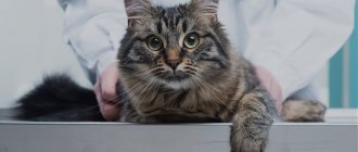
Ultrasound (ultrasound) is a widely used, accessible and painless method for diagnosing the internal organs of the abdominal cavity, both in humans and in cats and dogs. The technique is the use of ultrasonic waves to visualize the morphology, structure, and presence of pathological changes in the tissues of internal organs. The text will discuss how ultrasound is performed on cats and to whom the procedure is indicated.
An ultrasound machine is available in any modern veterinary clinic; it works on the principle of echolaction and emits high-frequency sound waves. These waves, hitting the organ, are deflected from it and returned back to the device, which interprets them as an image on the screen. However, these waves are difficult to pass through hollow organs and bones.
© shutterstock
Advantages of the method:
- Safe for the body.
- Completely painless.
- Non-invasive (no damage to tissues and organs).
- Has no contraindications.
- Informative and accurate.
- Shows the condition of internal organs using ultrasound waves: kidneys, spleen, liver, gall bladder, uterus and ovaries, stomach, pancreas, large vessels. Gives an image of the shape, size, structure, and the presence of pathological changes in organs.
- Shows pathological and functional changes in organs in the early stages of the disease, when clinical symptoms are not yet expressed.
- Allows you to monitor the results of treatment. You can compare images before and after treatment.
- Can be done repeatedly.
- Affordable price.
What diseases can be detected by ultrasound of the kidneys?
When performing an ultrasound of the kidneys, it is possible to determine the presence of a number of structural changes that may indicate various pathologies.
We can suspect signs of cystic kidney lesions, diffuse changes in the parenchyma, nephrolithiasis (kidney stones), we can see focal formations of the organ and suspect abscesses, hematomas, granulomas, neoplasia, signs of a decrease in the organ in size, suspected hypoplasia, aplasia, dysplasia of the kidney in a cat. We can see signs of acute kidney disease - nephritis (inflammation of the organ) or enlargement of the ureters, obstruction (blockage of the ureters due to a stone, clot or stricture) and so on.
To make a diagnosis, only the information obtained from ultrasound diagnostics is not enough. In some cases, a kidney biopsy may be necessary. Fine needle aspiration biopsy is a procedure that is performed under ultrasound guidance under general anesthesia. With a very thin needle, cells from the organ being examined are collected from the cat and sent for cytological examination to the laboratory.
As a rule, the kidneys are examined in conjunction with the entire genitourinary system, that is, the kidneys, ureters, bladder, urethra and genitals of cats and cats are examined. These organs are closely related to each other and the study of one of them is not very informative. An ultrasound scan of the kidneys is most often performed to monitor certain altered parameters (for example, in the presence of neoplasms).
When examining the bladder, you can detect suspension or uroliths (urinary stones), inflammation of the walls (cystitis), and neoplasms of the walls. The diagnosis of urolithiasis or cystitis in cats is usually made based on the results of an examination, medical history, ultrasound, and urinalysis.
Cats quite often have a problem with collecting urine for analysis - the easiest way to do this is to use an empty tray or a special kit with silica gel instead of litter, but not all cats agree to go into such a litter. Another disadvantage of such a collection will be the contamination of urine with bacteria from the external genital organs of the cat, the tray and the litter. It is also not always possible to immediately deliver the tests to the clinic, and for a correct study, no more than 4 hours should pass from the moment of urine collection (unless special tubes or refrigeration are used). Therefore, a routine procedure in veterinary medicine is cystocentesis (collection of urine by puncture directly from the bladder). It is the owners who are most afraid of this procedure, not the animals. The needle used is so thin that often the animals do not even feel the moment of puncture. This method is used in veterinary medicine due to the inability to collect urine from an animal in a sterile manner. And this is important for the doctor to understand whether the animal has a bacterial infection and whether it is necessary to use antibiotics, or inflammation of any other nature and a completely different therapy is needed.
Cats have very well developed compensatory mechanisms; this has developed evolutionarily and helped them survive in the wild. Therefore, unfortunately, our pets can hide diseases from us until the last stage. Very often, the only sign that something is hurting a cat is a decrease in activity, less active jumping, a change in sleeping position - this is often difficult to track, and such problems are often associated with age (the cat has matured, the cat has aged). To detect kidney disease at an early stage, it is recommended to undergo an annual medical examination after 4-5 years. Ultrasound of the kidneys is included in the standard set of studies during clinical examination in cats. Remember that cats are not people, and they cannot take revenge on us for anything when they go to the toilet in the wrong place. For a healthy cat, a clean, dry litter box is the optimal place to urinate; if she starts urinating on the sofa or folded things, in most cases it turns out that the cause is a bladder or kidney disease. If during this period you scold a cat that is already experiencing pain and discomfort, the problem can turn into a behavioral one and will be difficult to correct even after treatment.
Ultrasound of a cat's heart
An ultrasound examination of the cardiovascular system may be prescribed to a cat after identifying some characteristic symptoms associated with cardiac pathologies (shortness of breath, discoloration of mucous membranes, increased blood pressure). A cardiac examination is also mandatory before surgical interventions performed under anesthesia for such cat breeds as British, Scottish, Ragdoll, Persian, Maine Coon, due to a genetic predisposition to cardiac pathologies.
How to prepare for a kidney ultrasound?
As before an abdominal examination, it is advisable to fast for 10-12 hours before an ultrasound examination of the kidneys, since gas and intestinal contents will make visualization difficult. If your cat has contraindications to a starvation diet, and if it is necessary to perform an ultrasound on your kitten, consult your doctor. Since kidney examinations are rarely done without examining the bladder, it is important that the bladder is full before the ultrasound. Therefore, when preparing, remove the cat's litter box or close access to the toilet 4-5 hours in advance. If urination has been recent, the bladder will be empty, deflated, and it will be impossible to assess the condition of its walls, the presence or absence of urine.
Video with a visual diagnostic veterinarian about ultrasound of kidneys in cats:
When performing a kidney ultrasound, owners place the cat on its back on a special table and hold the front and back legs. The doctor shave the examination site, apply alcohol and acoustic gel to the skin. In large or obese animals, the doctor may apply pressure to the area being examined with a probe for better visualization due to the anatomical location of the kidneys. This may not be very comfortable for the animal, so restraint is important.
Kidney ultrasound is not the only method for diagnosing the disease. The attending physician makes a comprehensive diagnosis, based on the medical history, general condition of the animal, clinical symptoms, examination, ultrasound and laboratory diagnostic results.
Indications for ultrasound of a cat
Routine ultrasound examinations in cats most often include examinations of the urinary system, liver and gallbladder. They are often prescribed based on the results of blood tests.
For example, an increase in the level of urea and creatinine in the blood indicates damage to the kidney parenchyma, the level of bilirubin and liver transaminases - pathological processes in liver tissue, inflammation or blockage of the gallbladder and bile ducts. In cats with symptoms of hyperthyroidism and elevated levels of the hormone T4, an ultrasound examination of the thyroid gland is performed.
Ivanova Nadezhda Viktorovna Veterinarian. Specialization: therapy, reproduction
In an emergency situation, for example, a fall from a height, a car injury, or a state of shock, a veterinarian can conduct rapid screening examinations of the cat’s abdominal and thoracic cavities. In this case, an assessment and detailed description of each organ is not carried out. A quick study is aimed at searching for free fluid (blood, urine, ascites fluid) in cavities, which can appear due to internal bleeding, organ ruptures, and pathologies of the cardiovascular system. This method, along with radiography, allows the doctor to assess the need for emergency surgery and further treatment tactics at the first visit.
Another situation, typical mainly for cats, in which an ultrasound examination should be carried out immediately is acute urinary retention. If the owners notice that their cat has not been fully in the litter box for 24 hours, while he repeatedly tries to urinate, shows signs of anxiety, and screams, the condition of the wall of the bladder and urethra needs to be assessed using an ultrasound machine to eliminate the possibility of blockage with a urolith (stone). , as well as kidney pathologies.
Gastrointestinal pathologies determined by ultrasound
Below I would like to present some pathologies of the stomach and intestines that can be diagnosed in animals using ultrasound, as well as their main signs in the image.
- Chronic hypertrophy : layered walls are preserved, hypertrophy of the muscular or mucous layer, decreased motility.
- Gastritis : thickening of the wall, increasing its echogenicity, decreasing differentiation of layers, reducing the severity of folding.
- Gastric ulcers : local thickening of the wall, visualization of the ulcer crater, decreased differentiation of layers.
- Perforation of the stomach wall : signs of ulcerative lesions, increased echogenicity of the adjacent omentum, accumulation of gas/liquid near the affected area.
- Intussusception: with transverse scanning – a “target” picture, with a longitudinal scan – a “trident” picture, the outer segment is edematous, hyperechoic, enlarged mesenteric lymph nodes, decreased peristalsis, fluid accumulation proximal to the affected area.
Intussusception of the small intestine. - Intestinal obstruction. There are two types: mechanical (foreign bodies, scar changes, neoplasia) and functional (acute gastroenteritis, impaired innervation).
Linear foreign body in the small intestine. - Enteritis and colitis: thickening of the wall, decreased differentiation of layers, changes in the echogenicity of the wall, increased intestinal lumen, decreased peristalsis, increased echogenicity of the adjacent omentum, increased mesenteric lymph nodes, thickening of the spleen, decreased echogenicity.
Inflammation of the small intestine (enteritis).
Thus, you can see that with various pathologies, the condition of the stomach and intestines will differ from the norm. That is why ultrasound examination of the gastrointestinal tract is important in diagnosing an animal.
Find out prices for ultrasound of animals
Preparation
An abdominal ultrasound is one of those types of examinations for which you need to prepare. This preparation is necessary and includes, for example, a certain diet and a number of other measures: all rules play an important role in the informativeness of the study. Before an abdominal ultrasound, important requirements and recommendations must be observed.
Three days before ultrasound
3 days before the scheduled procedure, you need to change your diet and switch to split meals: eat small portions every 3 hours. Be sure to drink water or tea about 1.5 liters per day. What should you not eat before the test? Completely exclude from the menu products that enhance gas formation processes. Which? These are, first of all, yeast-containing products, fruits, peas and beans, soda, fatty protein dishes, alcoholic drinks, milk, juices, cabbage and grapes.
What can you eat before an abdominal ultrasound? Boiled, baked or steamed vegetables, cereals, and soups are allowed for consumption. Lean meat, lean fish, light cheese, boiled egg are allowed, no more than 1 per day. If there is a tendency to increased gas formation, it is better to drink adsorbents (“Espumizan”, “Simethicone”, “Enterosgel”, activated carbon: how to take them can be found in the instructions for the drugs).
To prevent flatulence, the doctor may recommend enzyme preparations: “Festal”, “Mezim” or “Pancreatin”.
Evening before the test
How long do you not eat before the ultrasound? A light dinner on the eve of the examination should be completed before 20.00. You should not include fish and meat dishes, even low-fat ones. If the patient is prone to constipation, a single use of a laxative is recommended (take Senade, Fitolax, Microlax, Fortrans and others). If laxatives are ineffective, the day before the ultrasound the patient is prescribed a cleansing enema.
Day of procedure
If an ultrasound is performed in the morning, it is better to do it on an empty stomach. If the procedure is planned in the afternoon, then breakfast should be light and should be completed before 11.00. A couple of hours before the planned test, you need to take Simethicone (or Espumisan) emulsion or 2 capsules of the drug. You can replace it with 6-10 carbon tablets or Filtrum. In the morning before the procedure, an additional enema may be needed if a tendency to flatulence has already been noted.
Get expert advice on any issue
Leave your number for advice or call
Ultrasound examination is based on the principle of echolocation. Ultrasound waves sent by the sensor pass through the body's tissues, are reflected and deciphered by the device and converted into an image that allows the veterinarian to see the condition of the internal organs, tissues and cavities. The doctor can accurately confirm or exclude the diagnosis, prescribe or adjust treatment.
Causes of intestinal obstruction in cats
Intestinal obstruction occurs due to blockage of some part of the intestine, as a result of which gastric juice, feces and gases are not able to escape. This is a very dangerous condition that can lead to the death of the animal within a couple of days.
Juices in a cat's stomach are constantly produced. If there is a blockage in the intestines, they begin to accumulate in the stomach, which leads to vomiting. Many useful substances come out with the liquid, which leads to rapid dehydration of the body. The animal loses strength before our eyes.
Most often, the main cause of intestinal blockage is the ingestion of any large objects. However, obstruction can also develop against the background of other diseases.
The main causes of mechanical intestinal obstruction:
- Swallowing large objects . Cats are very curious, and they often try different objects to their teeth. However, not all of them are able to leave the body naturally. The most dangerous are: cellophane bags, rustling paper wrappers, New Year's tinsel, etc.
- Hairballs in the stomach . Cats lick themselves, and all the hair that falls out, especially during the molting period, ends up in the stomach. In most cases, the fur is easily removed on its own - this is what is associated with periodic vomiting in cats. There are times when a hairball begins to move further along the esophagus and stops the intestines.
- Infection with helminths . Although all stray cats suffer from worms to one degree or another, sometimes the situation can get significantly worse. Helminths multiply very quickly. They often stop various internal organs of the animal. The resulting lump of helminths can clog the intestines.
- Neoplasms in the intestines . A tumor does not have to be malignant to cause the death of an animal. Often the tumor grows slowly, and therefore the cat first begins to suffer from partial intestinal obstruction. The tumor will grow until it completely blocks the esophagus.
- Volvulus . One of the sections of the intestine is pinched. The situation is aggravated by the fact that necrosis of dying tissue is added to the obstruction.
- Hernia . In some cases, part of the intestine may prolapse into the abdominal cavity. It is compressed by tissues, which can lead to tissue necrosis and intestinal obstruction.
- Constipation . Poor nutrition leads to stool becoming increasingly dry. They have difficulty passing through the intestines. This can lead first to constipation, and then to the formation of fecal impaction.
In addition to mechanical, there is also dynamic intestinal obstruction. It is not associated with mechanical blockage of the intestines, but with a violation of intestinal contractions.
The main causes of dynamic intestinal obstruction:
- Abdominal surgery performed.
- Other serious diseases: ascites, peritonitis, lead to paralysis of the intestinal walls.
- Spinal injury.
- Poor circulation in the abdominal area.
- A strong spasm leads to compression of the intestinal muscle. In this case, the animal feels very severe pain. Spasm can be caused by inflammatory diseases (enteritis, enterocolitis), or severe mechanical trauma to the abdominal cavity.
You might be interested in: What causes cats' gums to turn red?
Ultrasound of the abdominal cavity of a cat includes examination of the condition of such internal organs as
- Pancreas
- Liver
- Spleen
- Kidneys
- Intestines and stomach
- Bladder
- Reproductive organs
When performing an abdominal ultrasound in our clinic for a cat, the price will be 1,600 rubles. Examination of this list of organs can reveal a number of diseases that cannot be detected with the naked eye.
Preparation for the procedure scheduled for the first or second half of the day
— If a person has been given a referral for the morning, then it is most convenient to come to the hospital on an empty stomach.
In this case, the last meal should be before 18.00. Dishes should be light and easily digestible. This rule is mandatory for both men and women. Therefore, it is important to adhere to these recommendations so that the ultrasound scan is successful and the doctor is able to identify the problem. It is necessary to come to the procedure only after the person has drunk about 1 liter of water
— Preparing for an ultrasound of the kidneys in women and men in the afternoon involves an early breakfast. One hour after breakfast you need to take activated or any other sorbent. And do not forget that before coming to the procedure (1 hour before it), you need to drink about 1 liter of liquid.
Can you do an ultrasound at home?
If for some reason a visit to the hospital is not possible, you can carry out diagnostics at home by pre-ordering the appropriate service.
This method has its advantages and disadvantages. It is important to consider that there are many factors at home that can influence the results of the study. Therefore, it is not always possible to accurately determine whether a cat’s health is normal or if there are abnormalities. In emergency situations, it is recommended to take the animal to a clinic that is open 24 hours a day. Diagnostic services, including ultrasound, are provided by the Zoovet veterinary clinic, whose specialists will help you quickly and accurately establish a diagnosis and prescribe adequate treatment.
Indications for ultrasound of the abdominal cavity in a cat:
- Pain in your pet's abdomen
- Presence of blood in the urine or unusual color of urine
- Frequent vomiting
- Indigestion, diarrhea, blood or mucus in stool
- Fever
- Obvious enlargement of the spleen and liver
If palpation of the abdominal wall, urine and blood tests indicate problems in the cat’s body, ultrasound scanning is used. This allows us to most accurately identify tumor processes, determine the nature of the pathology, as well as inflammatory processes in the digestive system, urinary and reproductive organs.