Anatomy of the structure of a cat's penis
The cat's reproductive system consists of:
- testicles;
- seminal ducts;
- urogenital canal;
- testicular appendages;
- penis.
A cat's testicles produce sperm that are ready for fertilization with cat cells.
Sperm are constantly produced in the testicles, which indicates that the pet is ready to conceive. When an animal mates, sperm enter the urogenital canal, and then into the pet’s vagina. Active cat cells enter the cat's reproductive organs, after which fertilization occurs.
In males, the testicles are hidden in the scrotum (skin pouch), which is located below the anus.
Reproductive system of a cat and cat
Screams at night, marking of housing, a sharp and unpleasant smell of urine - the effect of testosterone on the pet’s body.
Reproduction
The cat is a polycyclic person. This means that readiness to conceive offspring appears at a certain time. They are called sexual cycles. They are characterized by physiological changes throughout the body caused by hormones. In cats, such a seasonal dependence is not observed. He is always ready for fertilization. Although in the spring they have a high concentration of sperm, which increases their activity. If a cat mates with several males, the litter will contain kittens from different fathers. The female becomes pliable, restless, walks slowly with her tail raised, and rubs near her legs. The cat is ready to be accepted for mating. Soon, estrus occurs, which is the release of mucus from the vagina.
Predisposing behavior of a cat towards a cat arises as a result of irritation of centers in the brain and marks sexual heat.
Sexual arousal in a cat is manifested by “concerts”, running away from the cat. Therefore, mating of fluffy ones is recommended to be carried out on the territory of the male. Full readiness for mating is signaled by the female's stance with her rear raised and her tail moved to the side. The cat covers the cat, holding it by the withers. The act lasts about 5 seconds. The end is marked by the cat screaming and the male jumping away. The cat, lying on its back, sways. Ovulation occurs one day after coitus. If conception does not occur, a period of rest begins until the next estrus. The intervals between them are variable and depend on many factors.
Anatomy of the structure of the feline genital organs in a cat
The reproductive system of a pet includes:
- external genitalia;
- uterus;
- ovaries;
- mammary glands (some consider them to be part of the pet’s reproductive system).
Urine is removed from the pet’s body thanks to the female’s external organs and vagina.
They are released at the moment of mating with the male, and conception occurs in the oviducts. In the uterus, the fetus continues to grow and develop.
Due to the physiological structure of the cat's body, the cervix is in a closed state - this protects the pet from foreign infections. The cervix opens only when the female goes into heat. During mating, sperm penetrate at this moment.
The horn of the uterus serves for the development of the fetus. Due to the fact that it stretches, the female is able to bear from one to eight kittens.
Cat reproductive system
The female, along with her urine, secretes hormones - estrogens, thanks to which the male receives a signal to mate. Mammary glands are necessary for feeding offspring. Four pairs of nipples are considered the norm; they are located symmetrically along the body, from the groin to the chest.
Females begin to walk one to two months after winter. During this period of time, the cat's body produces melatonin, which is responsible for the reproductive process in cats.
Cats are much more careful in lapping than dogs.
© www.wodumedia.com
Instead of simply scooping up water with its tongue, as a dog does, a cat, when lapping milk or other liquid, touches the surface with the rough tip of its tongue and draws it into the mouth along with the resulting column of liquid, closing its jaws to prevent moisture from escaping.
A cat can lap at a rate of four times per second, receiving about 0.1 ml of liquid each time - so it can drink about 5 tablespoons (24 ml) per minute.
Genital organs in cats
The genital organs in cats are organs that produce germ cells, which excrete sexual products and are the site of fertilization and development of the embryo.
The genitals form the reproductive or reproductive system of cats.
Cats, like all mammals, have two sexes - male and female, whose genital organs are different.
Hello student
Foreskin
The foreskin, or prepuce, praeputium, of a male dog is adjacent to the ventral abdominal wall and directed towards the navel. The end of the foreskin, covering the narrow prenucial opening, ostium praeputiale, is free along the entire circumference. This makes it easier to retract the foreskin when inserting the penis. The preputial sac is covered with a mucous inner layer. In the area of the bottom, fundus praeputii, this inner leaf turns into a hollow leaf. The transition site is at the top of the bulb of the head. The internal and penile layers, lamina interna praeputii and lamina penis praeputii contain a large number of lymph nodes; purulent inflammation often occurs. The system of thin vessels of the mucous membrane and arteriovenous anastomoses are described by Ninomiya Nakamura (1981 a, b).
Veins run parallel to arteries and bear the same names; dorsal vein of the penis, V. dorsalis penis, vein of the bulb of the penis, v. bulbi penis, deep vein of the nenis, v. profunda penis. All of them, connecting in the vein of the penis, flow into the internal pudendal vein, v. Pudenda interna. At the same time, the outflow of blood from the glans penis occurs through the external pudendal vein, v. Pudenda externa. All veins have reliable valves. In addition, during the erection process, the outflow of blood is blocked due to squeezing of blood vessels and due to contraction of the muscles of the penis. For a more detailed description of the mechanism of erection, see Christensen (1954)
Rice. 15. Pars libera penis and foreskin, transverse section (according to Schummer, 1987)
a os penis; b urethra in sulcus urethralis, surrounded by the corpus spongiosum of the penis; with pars longa glandis, cavernous tissue; d lamina penis praeputii; e lamina interna praeputii
Cranial preputial muscle, m. praeputialis cranialis, a loop covers the preputial foramen. During an erection, the outflow of blood from the penis into the external pudendal vein is blocked by it. Caudal preputial muscle, m. praeputialis caudalis, is not always present.
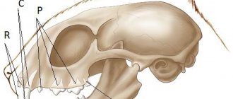
The penis, the penile part of the urethra and the foreskin of a cat
The penis of a cat is relatively short and, unlike males of other domestic animals, is directed caudally. Since the designation of the parts of the penis is determined by the principles of comparative anatomy, it therefore turns out that the dorsum of the penis, dorsum penis, is located ventro-cranially, and the penile part of the urethra is adjacent to the body of the penis from the dorso-caudal side. The structure of the cat's penis is described by Jackson (1902), Wagner (1909) and Redlich (1963), its corpora cavernosa - by Koniget al. (1979)
The cavernous body of the penis, corpus cavemosum penis, has the shape of a harpoon tip. Its legs, crura penis, are attached to the ischial arch. They are approximately 7 mm long and meet each other at an obtuse angle. The length of the trunk, truncus penis, is approximately 12 mm. It gradually narrows and passes into the bone of the penis, os penis. At the wide base of the bone of the penis there is a semblance of a urethral groove; the length of the bone of the penis is 3.5 -5.5 mm. In kittens, the bone of the penis begins to form after 3 months of life.
The tunica albuginea, tunica albuginea, of the corpora cavernosa of the penis consists of two layers; the outer layer of longitudinally oriented fibers is absent on the legs and in the urethral groove. On the back of the trunk, the apical ligament of the penis, ligamentum apicale penis, is formed from bundles of longitudinal fibers, which is attached to the bone of the penis and during erection passively directs the head of the penis cranially (Redlich, 1963).
Rice. 16. Diagram with the nomenclature of the cat’s penis
Left: parts of the corpus cavernosum of the penis, right: parts of the spongy bodies of the urethra and glans penis, middle: parts of the penis as a whole.
Rice. 17. The principle of the structure of the tunica albuginea of the cavernous body of the cat’s penis (according to Redlich, 1963) a ligamentum apicalc penis; b outer wall layer, c inner wall layer; d transverse and horizontal trabeculae; e trabeculae of the septum
Trabeculae extend inward from the tunica albuginea, trabeculae corporum cavemosorum, which in the form of transverse bridges are located in the cavity of the caverns or participate in the formation of the septum, which in the cat is not continuous; The trabeculae of the septum are round in cross section. Between the connective tissue-muscular trabeculae there are caverns, cavernae corporum cavernosorum, surrounded by elastic fibers. Large caverns are oriented along the longitudinal axis and communicate with the caverns of the opposite side. Small cavities completely fill the legs and peripheral areas of the trunk, especially its distal half. In the distal part of the cavernous bodies of the penis, the main structural element, as Jackson (1902) established, is adipose tissue, so that a kind of “articular part” is formed here (Redlich, 1963), with the help of which the penis bends cranially during erection.
The spongy body of the penis, corpus spongiosum penis, is covered with a thinner tunica albuginea. The paired thickened bulbs of the penis, bulbi penis, closely reach the bulbourethral gland. The spongy part emerging from the pelvic part of the urethra passes under the bulbs and further through the spongy body of the penis. Thus, the spongy layer communicates with the spongy body of the urethra. It covers the urethra up to the head of the penis, becoming increasingly thinner towards the head.
Rice. 18. Pars libera penis of a cat, dorsal view (after Schummer, 1987)
a glans penis; b lamina penis praeputii; with fundus praeputii; d lamina interna praeputii, here covers the extended body of the penis 1 trenulum praeputii
Rice. 19. Schematic representation of the normal (A) and erect (B) position of the cat’s penis, view from the lateral side (according to experimental studies by Redlich, 1963) a crus penis; b truncus penis; with os penis
1 so-called articular part in the corpus cavernosum penis: 2 ligamentum apicale penis; 3 m. retractor penis
The spongy body of the glans penis, corpus spongiosum glandis, consists of two or three layers of coarse cavernous tissue, forming a cap, which at the apex is pierced by the external opening of the urethra. In the internal space of the cap there is the final part of the spongy body of the penis, as well as the bone of the penis. Under the bone of the penis, that is, from the back, the cap is split. In general, the corpus spongiosum forms the urethral and lateral surfaces of the glans penis.
The foreskin covering the head bears approximately 120 keratinized denticles arranged in rows. Wagner (1909) reports specific terminal nerve bodies at the base of the denticles. On the non-erect penis, the tips of the denticles are adjacent to the head; during erection, they rise as a result of the enlargement of the head. After castration, their involution occurs. The erection process was experimentally studied by Redlich (1963). In this case, the shaft lengthens by 1 1/2 times, the diameter of the penis increases by 2 times. The direction of the axis passively changes as the apical ligament of the penis pulls the glans cranially and the body of the penis bends at the so-called articular part.
A cat's thickened foreskin is located under the scrotum. The prenucial foramen is directed caudally. The short inner leaf at the bottom of the prepuce passes into the genital leaf, equipped with denticles. When the penis extends, the inner leaf covers the body of the penis.
The blood vessels of the cat's copulatory organ belong to the system of the internal pudendal artery, which, after the vessels branch off from it to the urethra and bulbourethral gland, continues as the artery of the penis and is divided into three branches, similar to the circulatory system of the male penis. The smallest branch is the dorsal penile artery, a. dorsalis penis, in a cat it does not participate in the blood supply to the penis, but stretches to the foreskin. Artery of the penile bulb, a. bulbi penis, goes into the bulb, supplies it and the corpus spongiosum of the penis with blood, then reaches to the head and also vascularizes the corpus spongiosum of the glans. Deep artery of the penis, a. profunda penis, is responsible for the blood supply to the corpus cavernosum (for more details, see Konig et al., 1979). Dorsal vein of the penis, v. dorsalis penis, drains blood not only from the foreskin, but also from the corpus spongiosum of the glans and the distal part of the corpus spongiosum of the penis. Otherwise, the veins run the same way as the arteries of the same name.
Rice. 20. Lymph nodes and lymphatic vessels of the urinary and genital tract of a male dog in situ (but Baum, 1918)
1 lymphonodi iliaei mediales; 2, 2′ lymphonodi lumbales aortici; 3 lymphonodi sacrales; 4 lymphonodi scrotales; 5, 6 lymphatic vessels of the inner layer of the penis; 7 lymphonodi scrotales, vasa efferentia; 8 lymphatic vessels of the kidney capsule, going, like the lymphatic vessels of the testis, to the lymphonodi lumbales aortici a os ilii (sawed off); b ventral abdominal wall; from the bottom of the pelvis; d lumbar muscles; e aorta, f v. cava caudalis; g a. iliaca externa sinistra; h a. iliaca interna dextra; i vesica urinaria; k prostate; l urethra; m ligamentum vesicae laterale; n ureter; o m. coccygeus (cut off); p slice m. adductor; q m. bulbospongiosus; r m. ischiocavernosus; s penis; v praeputium; v' skin of the scrotum; w testis; x epididymis; in funiculus spermaticub, y' ductus deferens; z ren sinister
Ischio-cavernous muscle, m. ischiocavernosus, bulbospongiosus muscle, m. bulbospongiosus, and the muscle that retracts the penis, m. retractor penis, in principle, pass and act in the same way as in a male dog. No special studies have been carried out on the muscles of the cat’s copulatory organ.
Features of innervation and lymphatic drainage in the genital organs of dogs and cats: the dorsal nerve of the penis, the dorsalis penis, extending from the pudendal nerve, contains sensitive fibers of the penis, including the head and foreskin, as well as vegetative fibers of the blood vessels involved in erection. The regional lymph nodes for the testes and testicular appendages are the aortic lumbar lymph nodes, for the pelvic part of the urethra and accessory sex glands - the medial iliac and sacral lymph nodes, for the penis, foreskin and scrotum - the scrotal lymph nodes.
Literature used: Anatomy of a dog and a cat (Coll, authors) / Transl. with him. E. Boldyreva, I. Kravets. - M.: “AQUARIUM BUK”, 2003. 580 pp., ill. color on
Download abstract: You do not have access to download files from our server. HOW TO DOWNLOAD HERE
Cat genitals
• The scrotum is a cavity of the sac-like protrusion of the abdominal wall, which ensures the maintenance of the temperature necessary for the maturation of sperm (several tenths of a degree lower than in the abdominal cavity). In cats, the muscles of the scrotum can pull it towards the groin if it is too cold outside. The scrotum is located between the penis and the anus.
• The spermatic cord is a fold of the peritoneum that contains blood vessels, nerves leading to the testis, as well as lymphatic vessels and the vas deferens emerging from it.
• The testes are paired, egg-shaped organs located in the scrotum. The diameter of each testis in cats is 1-1.5 cm. In the tubules of the testes, sperm are produced by a special epithelium.
Cat genitals
• The ovaries are a paired, bean-shaped organ located in the lumbar region of the cat. The ovaries are connected to the uterus by the fallopian tubes, where eggs mature. The ovaries consist of follicles - sacs containing eggs and fluid. When the follicle matures, it bursts and the egg with follicular fluid enters the fallopian tube (ovulation occurs). And in place of the follicle, a corpus luteum is formed, suppressing the development of other follicles. In cats, multiple follicles burst at the same time, resulting in multiple pregnancies.
• Fallopian tube (oviduct) is a narrow, convoluted tube that connects the ovary to the uterus. Fertilization occurs in the fallopian tube, after which the egg moves to the uterus.
• The uterus is a hollow, membranous organ in which the fetus develops. In cats, the uterus is bicornuate, that is, it has the shape of the Latin letter Y. Each horn (3 mm in diameter and 10 mm in length) is connected to its fallopian tube, and closer to the vagina the horns merge, forming the body of the uterus (2 cm in length). The body of the uterus gradually narrows, in turn forming the cervix, which opens into the vagina. Fruit ripening occurs in the horns of the uterus. The uterus is located in the pelvic and abdominal cavity.
• The vagina is a tubular organ designed for copulation and located between the cervix and the urogenital opening.
• Vulva – a set of external genitourinary organs (vestibule of the vagina + external opening of the urethra). Located below the anus and separated from it by a small perineum).
• The vaginal vestibule is the common part of the genitourinary tract located behind the urethral opening.
• The labia are folds of skin that protect the vestibule of the vagina.
• The genital fissure is the entrance to the vagina.
• The clitoris is a sexual organ, a rudiment of the penis in females, representing an erogenous zone. In cats, it is located in the genital cleft, and its stimulation ensures ovulation.
The genital organs of cats are similar to the genital organs of other mammals, but they have a number of features characteristic exclusively of felines.
Digestive system
The cat's digestive system consists of:
- esophagus;
- stomach;
- small intestine;
- duodenum;
- jejunum;
- liver;
- large intestine.
The esophagus has a hose-like shape of a relatively small size, and connects the animal’s mouth and its stomach.
The esophagus originates from the inner base of the mouth, extends through the neck and chest, passes close to the heart, extends through the muscles of the diaphragm, and connects to the stomach. It is important to note that the esophagus is equipped with special muscles that push food into the stomach, producing synchronous movements similar to waves. The esophagus is one of the most difficult organs in terms of surgical treatment, as it is difficult to access and extremely difficult to heal.
Cats control you
© Salvo Bombara
This fact will be confirmed by all cat owners without exception - pets do not just meow and purr, but in this way try to establish communication with the owner. In the process of evolution and living side by side with people, these cunning creatures have developed a cry of a special timbre, which a person perceives as unbearably shrill and disgusting. Of course, cats most often use this method of training people when they want to get something edible and, as a rule, achieve the desired result.
Circulatory system
The circulatory system in cats works the same way as in other mammals: the heart pushes blood through arteries that have elastic walls and rhythmically carry out contraction and relaxation movements. It is thanks to such movements that arteries located close to the skin can be felt, and this is called the pulse. The cat's pulse is easiest to detect on the inside of the thigh, and in a healthy animal it should range between 100-150 beats per minute.
The cat's brain absorbs 15-20% of the blood, the muscular system absorbs up to 40% of the total blood, and about 25-30% of the blood goes to the internal organs. During physical activity, muscles can absorb up to 90% of the blood, which is why cats get tired so quickly, but can focus maximum strength for a short period of time.
The animal's heart is a hollow organ located in the chest, just behind the breastbone. An important nuance is the fact that the weight of a cat’s heart depends on their weight, and does not have clearly established standards. Most often, the heart of an animal weighs 0.6% of the total body weight. The cat's heart consists of 2 ventricles and 2 atria.
Cats live for 10 minutes
© www.followthewabbit.com
Cats are able to remember any obstacle in their environment for only about 10 minutes - this is the conclusion made by experts based on the results of a study conducted in 2007.
During the experiment, scientists created artificial barriers that furry subjects had to overcome. It turned out that if you stop a cat after it has stepped over an obstacle with its front paws and do not allow it to continue on its way for 10 minutes, the animal forgets that it is necessary to step over the barrier with its hind paws too, although if you stop it for a short time, the barrier will be “taken” practically straightaway.
The visual memory of cats is much shorter than the muscular memory - if you distract the pet from an obstacle before it begins to overcome it, the cat forgets about the unsolved problem.
Endocrine system
The endocrine system is primarily responsible for hormones and their production in the corresponding organs. Thus, the cat’s brain produces antidiuretic hormone, oxytocin, corticotropic hormone, adrenocorticotropic hormone, cortisol and growth hormone.
The adrenal glands produce a lot of other hormones, the main purpose of which is to regulate metabolism, and are also responsible for behavioral characteristics. The adrenal glands also produce cortisol, a small portion of testosterone, as well as epinephrine and norepinephrine.
There are a number of other glands of external and internal secretion, the principle of operation of which is common to all mammals.
Nervous system
The nervous system of cats is divided into central and peripheral. Each of these systems in a cat performs functions that are standard for most mammals.
The central nervous system consists of the brain, brain stem and the so-called spinal cord. The central nervous system is the most important in the body of any living creature, and simple and complex reactions, as well as some reflexes, depend on it. In addition, the central nervous system interacts with the peripheral and autonomic ones, ensuring their functioning and control.
The peripheral nervous system is responsible for the cat's conscious motor abilities. So, thanks to this system, a cat can move its paws, extend its claws, run, and generally lead the lifestyle that it leads. Also, the peripheral nervous system transmits pain impulses to the central nervous system from any part of the body where peripheral nerve endings are present.
Cat parasites can turn you neurotic and kill you
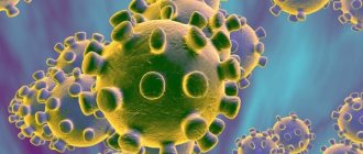
Toxoplasma gondii
Cats are capable of not only controlling people, but also infecting them with parasites that directly affect the human personality. Microorganisms of the species Toxoplasma gondii are transmitted to cats from rats and mice, which, under the influence of this parasite, behave very recklessly and themselves look for places where they can encounter cats. If a furry pet manages to eat an infected mouse, the microbes will most likely appear in its owner, which is fraught with the development of toxoplasmosis , a disease that can noticeably change a person’s behavior.
A study conducted in 2006 found that after infection with Toxoplasma gondii, people's anxiety levels increase, their reactions slow down, neurotic traits appear, and for a person with a weak immune system, toxoplasmosis can even become fatal.