Uveitis in cats is a pathology that is dangerous for the animal’s vision.
The system of small vessels and capillary network in the eye provides blood supply to the visual organ and nourishes the organ.
The blood supply system to the eyeball is called the uveal tract. With the development of dangerous changes in the vascular network of the eye, serious pathologies arise, one of which is uveitis. The pathological process is an inflammation, which has a sharply negative effect on the vascular network, causing not only pain and discomfort, but also increases the risk of loss of vision in animals. Uveitis is diagnosed in domestic cats not as an independent disease, but as a complication of conjunctivitis and keratitis not treated in a timely manner.
Causes of the disease
The inflammatory process in the area of the vessels supplying blood to the eyeball is called uveitis. The vascular network is located in the middle part of the eye and is the median membrane.
The uveal tract, where inflammation occurs, is represented not only by vessels, but also by other structures - the iris, ciliary body and choroid (the choroid itself).
In the anterior part of the uveal tract is the iris and ciliary body. In the posterior part there is only the vascular network.
Depending on which part of the uveal tract is affected, several types of uveitis are distinguished in veterinary medicine. The following types of inflammation of the choroid are distinguished:
- The anterior form of uveitis is characterized by an inflammatory process in the area of the iris and ciliary body. Its scientific name is iridoccilitis.
- The posterior form of the pathology is characterized by inflammation in the area of the vascular network, called choroiditis.
- Panuveitis is a disease manifested by inflammation of all structures of the uveal tract.
A dense network of vessels, forming the membrane, is located between the fibrous membrane and the retina. The fibrous membrane is based on the corneal layer and sclera, and the retina is formed by internal structures. As the pathology develops, inflammation may not be localized in one place, but may spread to nearby tissues of the eyeball. In this case, the following pathologies develop:
- Endophthalmitis is an inflammatory pathology affecting the anterior part of the eye, the vitreous body and nearby tissues of the visual organ.
- Panophthalmitis is an inflammation diagnosed in the case of an advanced course of a certain pathology. Panophthalmitis is characterized by the involvement of all tissue structures in the inflammatory process - the choroid, fibrous membrane and retina.
There are several factors that provoke the development of uveitis in cats. The main ones are:
- chronic diseases of the organ of vision - cataracts, high blood pressure in the eye, ulcerative lesions of the corneal layer;
- mechanical eye lesions – bruises;
- systemic pathologies - various infectious factors, pathologies of the endocrine system, lead to inflammation in the visual apparatus.
In domestic cats, systemic pathologies play an important role in the development of uveitis. The risk of developing eye disease increases if the animal has viral leukemia, toxoplasmosis, viral peritonitis or herpes virus. Feline viral immunodeficiency plays a special role in the development of inflammation of the vascular system of the visual apparatus.
Separately, it is worth noting fungal pathologies. Infection of a cat with coccidomiasis, asperigillosis or blastomycosis provokes systemic changes in the body.
Differential diagnosis is based on the clinical picture of manifestations and anamnesis. It is noted that with blastomycosis, cryptococcosis, and coccidioidomycosis, the posterior part of the uveal tract is affected.
With fungal diseases, eyelid lesions occur. There is a constant discharge of specific exudate from the eyes.
https://www.youtube.com/watch?v=nJbs89TFFYQ
The following types of pathologies can cause damage to the uveal tract and provoke uveitis in pets:
- bacterial damage (hemobartonellosis);
- helminthiasis – damage to the body by dirophyllaria (heartworms) and toxocara (inflammation is provoked by the migration of larvae through the body of some worms);
- damage by protozoan microorganisms (for example, toxoplasmosis).
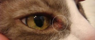
Among the reasons for the development of inflammation of the vessels of the visual apparatus, metabolic disorders are distinguished. One of the most common is systemic hypertension. Uveitis in a cat can be provoked by tumor processes - primary tumors in the optic organ or systemic neoplasms - metastases, lymphosarcoma, hemangiosarcoma.
Symptoms and diagnosis of uveitis in cats
It is not difficult to notice a change in your pet’s condition. With the development of uveitis, the animal experiences pain, increased secretion of tear secretion from the eyes, and fear of light. The pet hides in secluded places, actively scratches its eyes, the pupil is constricted, and swelling of the cornea is noted. There may be blood stasis in the front of the eye and cloudiness.
In advanced cases, the pet begins to see poorly and blindness develops. The development of pathology can be almost lightning fast - over the course of several days. There is also a long course of the disease - for 2-3 months.
Complications of untreated uveitis in a cat include increased intraocular pressure, retinal detachment, and even atrophy of the eye muscles.
The main signs of uveitis in a pet are:
- severe lacrimation and pain;
- photophobia (fear of bright light);
- constriction of the pupil and constant itching, as a result of which the animal squints the affected eye or tries to scratch it;
- swelling of the cornea, clouding of the intraocular fluid.
Against the background of characteristic changes in the visual apparatus due to inflammation of the blood vessels of the eye, the animal begins to lose orientation in space, bumps into objects, and tries to avoid jumping on any surface.
Due to the fact that uveitis can develop rapidly, symptoms increase quickly. Lack of timely treatment leads to the development of complications - retinal detachment, cataracts or glaucoma, up to complete loss of the ability to see.
When visiting a veterinary clinic, a specialist will prescribe a series of tests to make a diagnosis. A comprehensive diagnosis is carried out, including a number of studies. Mandatory are:
- tonometry – important for determining pressure;
- ophthalmoscopy;
- study of eye structures - biomicroscopy;
- assessment of the condition of the anterior chamber of the eye - the presence of blood accumulations, turbidity of the intraocular fluid (gonioscopy);
- study of the condition of the posterior chambers of the eye - fundoscopy.
In some cases, an ultrasound examination of the eye is necessary to assess the condition of the retina or vitreous.
A number of laboratory tests are carried out - general clinical blood test, biochemical panel, blood serology to identify possible systemic infectious pathologies.
Based on the data obtained, the doctor will be able to draw up a treatment plan based on the clinical picture and factors that provoked the development of uveitis.
Treatment and prevention
Therapy of the inflammatory process inside the visual apparatus requires an integrated approach. Specific treatment is prescribed depending on the factors that provoked the disease. Specific therapy includes taking antifungal drugs and antibiotics. If uveitis is of viral origin, immunoglobulins are prescribed.
Symptomatic treatment is based on the use of nonspecific agents. The veterinarian’s task is to relieve the inflammatory process and eliminate discomfort.
For these purposes, cycloplegic drugs are used to reduce discomfort and corticosteroids are necessary to relieve inflammation. Elimination of pain is achieved by administering non-steroidal anti-inflammatory drugs.
It is important to follow your doctor's instructions and prescriptions to prevent overdose.
In the chronic form of diagnosed inflammation in the visual apparatus, long-term treatment is carried out. The disease develops slowly, but constantly progresses, which inevitably leads to complete loss of vision. With reduced intraocular pressure, a process such as phthisis or wrinkling of the eyeball gradually develops.
In order to prevent the development of uveitis in domestic cats, it is recommended to adhere to a number of simple rules. First of all, it is necessary to provide the animal with proper care and diet.
Don’t forget about regular checks at the veterinary clinic for the purpose of early diagnosis of possible diseases.
It is necessary to avoid infecting your pet with various infections - fungal, viral, bacterial.
Source: https://zen.yandex.ru/media/ivethelp/uveit-u-koshek-patologiia-opasnaia-dlia-zreniia-jivotnogo-5ed52c88cbfac70930dac0af
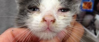
Acute catarrhal conjunctivitis
The disease usually occurs in an acute form. It is associated with inflammation of the mucous membrane of the eye.
Symptoms
When acute catarrhal conjunctivitis appears, swelling and redness appear around the eye. The cat will experience pain, which will affect its behavior. Serous-mucous discharge will begin to accumulate in the corner of the animal’s eye and lacrimation will increase.
Causes
This disease can develop as a result of mechanical trauma. If it has not healed well and dirt has gotten into it, then conjunctivitis may appear. Any infectious disease is also considered another cause. And finally, a lack of vitamin A in the body (hypovitaminosis A) very often causes such inflammation.
Giving help
For proper treatment, it is first necessary to establish the cause of acute catarrhal conjunctivitis. If damage is detected, you must immediately treat the eye with a 0.25% novocaine solution. This procedure must be repeated regularly, and a solution of 0.25% chloramphenicol should be instilled several times a day. For other causes of this disease, after washing, a 0.25% solution of chloramphenicol or a 1% solution of kanamycin should be instilled into the eye. Sofradex and a 10-30% solution of sodium sulfacyl are well suited for the same purposes. This procedure should be carried out up to 5 times a day, instilling 2-3 drops of medicine. If the disease is advanced, then in addition to this treatment it is necessary to lubricate the eyelid with antibiotic ointments several times a day or apply bandages with these ointments.
Separation of the retina in animals, dogs and cats
| > Eye diseases > Retinal detachment When the retina of the eye is detached in dogs, cats and other animals, vision decreases to the point of complete blindness (dilated pupils during the day, blurred vision), and in advanced cases leads to the death of the eye. Therefore, this pathology is an emergency condition and requires immediate contact with a veterinary ophthalmologist. Below is the work of our specialist on this topic. Komarov Sergey Vitalievich, Ph.D. MGAVMiB im. K.I. Scriabin, bmdg.ru Center for Emergency Veterinary Ophthalmology and Microsurgery, okovet.ru Veterinarian-ophthalmologist, microsurgeon, Moscow. IntroductionRetinal detachment in cats and dogs is a serious emergency condition in which the neuroretina (NR) separates from the pigment epithelium (RPE). This leads to disruption of the interaction of these layers, which results in visual impairment. The main method of treatment is surgical. The principle of treatment is to bring the neuroretina closer to the PE and localize the rupture with foci of chorioretinal inflammation. A long-term state of OS causes irreversible changes and death of neurons. This always leads to decreased vision. StructureThe retina includes 10 layers, all of them extend deep into the eyeball.
nerve fibers; The retina contains neurons located in 3 levels: The RPE is a monolayer of neuroepithelium and performs very important functions: Photoreception mechanismWhen light is absorbed, the structure of rhodopsin changes. This leads (through a series of intermediate events) to the closure of Na+ channels in the plasma membrane. Therefore, the transmembrane potential increases. Excitation of light-sensitive neurons does not lead to depolarization (as usual), but to hyperpolarization of the membrane. Hyperpolarization extends to the area of synaptic contact and causes excitation of associative neurons. Development factorsTypes of diseaseAccording to the mechanism of formation, four types are distinguished: Reasons for developmentRhegmatogenous OS – with a rupture of the retinal plane. Causes: Types of breaks: Traction OS The presence of vitreoretinal adhesions (proliferative vitreoretinopathy) can develop with proliferative diabetic retinopathy, hemorrhage, thrombosis of the retinal veins, etc. Membranes are formed by PE cells; Deformation and tension of the retina due to the formation of fibrous tissue leads to elevation of the neurosensory retina above the underlying pigment layer - OS. Exudative OS Occurs when the integrity of the vascular wall is violated, which causes plasma to escape into the subretinal space (hypertension, central vein thrombosis, vasculitis, optic disc edema, infusion therapy errors) Often without rupture, total bladder-like OS, sometimes with subretinal hemorrhages DiagnosticsAvailable objective diagnostic methods for dogs and cats. |
| Total V-shaped OS. | Total OS with a gigantic gap in the AP. |
| High traction | Tractions during PVR. No OS yet. |
Drug treatment
Pneumoretinopexy
Surgical methods of treatment
Endovitreal intervention
Material base for surgical treatment
Source: https://www.okovet.ru/otsloyka-setchatki.php
Corneal ulcer
All eye ulcers are divided into two types: purulent and perforated.
Symptoms
Firstly, this is severe pain, so the cat will somehow express its feelings. She develops photophobia and eyelid spasms. Mucopurulent fluid is released from the conjunctival sac. Various defects appear on the cornea. The edges of the ulcer become swollen and vascularization of the cornea appears.
If an animal has a perforated ulcer, then the iris prolapses in the central part of the eye. Part of the cornea takes on a gray-blue color, swells, and mixed vascularization develops in it. Subsequently, after a corneal ulcer, scar formation occurs.
Causes
This disease is the result of a superficial or deep wound, infection in the eye, or destruction of eye tissue after purulent keratitis.
Giving help
You should immediately begin washing your eyes with an antiseptic solution of 3% boric acid, 1% solution of rivanol or a weak solution of furatsilin. Additionally, it is necessary to do intramuscular injections of antibiotics and dressings with kanamycin or sulfapyridazine in combination with dexamethasone. When the ulcers are perforated, the prolapsed part of the iris is removed and sutures are placed on the edges of the wound. But this can only be done in a veterinary hospital.
Hereditary pathologies of the retina in cats - IVC MBA
Boyarinov S.A. – veterinary ophthalmologist IVC MBA, microsurgeon
The retina is a unique organ, which is determined by the complexity of its structure and subtle functioning mechanisms. Any pathological changes occurring in the retina are reflected in the visual functions of animals, and, accordingly, on the perception of the surrounding world.
Figure 1. Fundus of healthy cats under ophthalmoscopy.
Retinal pathologies in cats occupy one of the leading places in the development of low vision and blindness.
Among these pathologies, a special place is occupied by hereditary diseases of the retina of cats, which can be transmitted from generation to generation and lead to irreversible blindness.
A distinctive feature of hereditary retinal pathologies in cats is a strict breed predisposition. This means that only cat breeds such as the Abyssinian or Persian, as well as a few others, can have such serious diseases.
It is worth noting that, unlike dogs, this pathology is much less common in cats and affects young animals.
Pet owners may notice the following symptoms of disease:
Figure 2. Assessment of chromatic pupillary responses.
The diagnosis of hereditary retinal disease in cats is made only on the basis of a comprehensive ophthalmological examination. Mandatory and key research methods are examination of the fundus (ophthalmoscopy and fundoscopy), as well as examination of retinal activity (electroretinography and assessment of chromatic pupillary reactions).
Hereditary early rod-cone dysplasia of the retina (early-onset rod–cone dysplasia (Rdy)
The disease occurs in Abyssinian cats. It develops already at a young age in kittens and affects both eyes.
Rod-cone dysplasia is a hereditary disease and develops in an autosomal dominant pattern in Abyssinian cats.
The common clinical symptom characterizing early rod-cone retinal dysplasia is decreased pupillary reflex, mydriasis, nystagmus and vision loss.
With fundus ophthalmoscopy, the first pathological changes can be visualized in kittens from 2-3 months of age. These changes include:
- tapetal hyperreflexia (reflectivity);
- loss of pigmentation;
- thinning and/or disappearance of retinal vessels.
With electroretinography, changes in the functional state of rods and cones are recorded already on the 14th day of life of kittens. The pathological process begins, as a rule, in the central zone of the retina and spreads to the periphery, ultimately affecting the entire retina.
There is no treatment for hereditary rod-cone retinal dysplasia, so it is necessary to carry out selection work aimed at excluding from breeding animals that carry the mutant gene.
Figure 3 . Fundus of cats with hereditary retinal pathologies.
Recessively-inherited rod–cone degeneration (rdAc)
The disease occurs in cats of the Abyssinian and Somali breeds . The initial stage of the pathology begins at 1.5-2 years of age and over the next 2-4 years progresses to complete loss of vision in both eyes.
Hereditary rod-cone retinal degeneration is characterized by development in several stages.
Symptoms of the disease include: discoloration of the peripapillary zone in the precursor stage, discoloration of the tapetum and weakening of the vasculature in the early stage, the appearance of local hyperreflexia and disappearance of retinal vessels in the moderately advanced stage, and generalized hyperreflexia and complete absence of retinal vessels in the advanced stage.
With electroretinography, changes in the functional state of rods and cones are recorded already from 8-12 weeks of age in cats, which can be used for early diagnosis of the disease, when ophthalmoscopic changes are not yet observed.
There is no treatment for hereditary rod-cone retinal degeneration, so it is necessary to carry out selection work aimed at excluding sick animals from breeding.
Hereditary early progressive retinal atrophy in Persian cats (PRA)
The disease occurs in Persian and is hereditary, developing in an autosomal recessive manner.
The pathology affects both eyes and is clinically manifested at 2-3 weeks of age by a decrease in the pupillary reflex. Complete blindness occurs at 16-17 weeks of age.
Ophthalmoscopic changes are observed from 2-4 weeks of age and include weakening of the vasculature, in particular the central veins and retinal arteries. From 6 weeks of age, there is an increase in the reflectivity of the tapetum and the development of hyperreflexia. From 9 weeks of age, hyperpigmentation and atrophy of the optic nerve head are recorded.
Figure 4. Fundus of cats with hereditary retinal pathologies.
Electroretinography also notes a decrease in the activity of the rod-cone response at the early stage of the disease, which speaks of this pathology as a disease with an early onset.
There is no treatment for hereditary early retinal atrophy in Persian cats. Since this pathology is hereditary and is passed on from generation to generation, it is necessary to exclude from breeding animals that carry the defective gene.
Hereditary early progressive retinal atrophy in Bengal cats (PRA)
The disease occurs in Bengal cats and is hereditary, developing in an autosomal recessive manner with an early onset. This pathology has no connection with a similar pathology in Persian cats.
Retinal lesions develop symmetrically in both eyes in kittens from 8 weeks of age and progress to complete blindness by 6 months.
Figure 5 . Fundus of cats with hereditary retinal pathologies.
Unlike early hereditary retinal atrophies in Persian and Abyssinian cats, which are clinically manifested by a decrease in the pupillary reflex in the initial stages, visual clinical symptoms of this pathology are observed closer to 1 year of the animal’s life, which significantly complicates the ability to detect retinal atrophy in the early stages.
Pathological changes in the fundus of the eye with progressive retinal atrophy in Bengal cats can be visualized starting at 8 weeks of age and include a generalized increase in granularity, an increase in the reflectivity of the tapetum, weakening of the vasculature, and a decrease in the diameter of veins and arteries. By 20-25 weeks of age, the changes become more pronounced, and by 40-60 weeks, total retinal degeneration is observed: generalized hyperreflexia, disappearance of central and peripheral vessels.
There is no treatment for hereditary early retinal atrophy in Bengal cats. Since this pathology is hereditary, it is necessary to exclude from breeding animals that carry the defective gene.
In conclusion, it is worth noting that hereditary retinal pathologies are a serious eye disease that irreversibly leads to blindness. Unfortunately, no treatment for these pathologies has been developed. However, when making a diagnosis, it is necessary to exclude animals from breeding to prevent the spread of pathology among cats.
Return to list
Source: https://vetacademy.ru/obuchenie/stati/nasledstvennye-patologii-setchatki-u-koshek/
Vitreoretinal intervention. Jack Russell, 3G, single eyeHD after retinal straightening. | In 3 weeks. |
Purulent conjunctivitis
This disease occurs as a result of various infections, mainly pyogenic microbes, entering the eye.
Symptoms
The skin of the eyelid swells and turns pale. Spasms of the eyelid and purulent discharge appear in the corner of the eye. The eyeball goes deep into the orbit, and the third eyelid becomes half closed.
Giving help
If this disease occurs, it is necessary to wash the eyes 2-3 times a day with a weak solution of furatsilin or rivanol. Then lubricate the conjunctiva with ointments such as tetracycline, oletethrin or erythromycin. It is useful to apply eye patches with kanamycin and dexamethasone. In addition to these procedures, for more effective treatment, it is advisable to administer antibiotics to the cat and give sulfa drugs.
Glaucoma
There are three types of glaucoma: congenital, open-angle and closed-angle . As a result of this disease, atrophy of the optic nerve nipple occurs as a result of a constant or periodic increase in intraocular pressure.
Symptoms
With glaucoma, the pupil dilates, and congestive hyperemia forms on the mucous membrane. The eyeball increases in size, and upon palpation you can feel its hardening. The vessels of the mucous membrane of the eye acquire a dark red color, are greatly dilated and have a tortuous structure. Open-angle glaucoma is characterized by the fact that the cornea of the eye becomes colorless and may have slight opacities. In this case, its sensitivity is noticeably reduced. When angle-closure glaucoma develops, ring-shaped opacification and vascularization are noticeable in the cornea of the eye.
Cause of occurrence
The cause of congenital or open-angle glaucoma may be degenerative changes in the trabecular apparatus of the anterior chamber angle. Angle-closure glaucoma develops as a result of lens luxation or swelling, as well as deep purulent keratitis or hemorrhage.
Giving help
For glaucoma, it is necessary to instill medications into the eyes that dilate the pupil. This is a 1% solution of pilocarpine, 0.5% solution of physostigmine or 0.135% solution of phosphacol. If medications cannot reduce eye pressure, you should contact your veterinarian. Surgery is required.
Treatment in our clinic
We will conduct a comprehensive examination of your pet’s eyes and prescribe optimal, modern and high-quality treatment.
Center for Emergency Veterinary Ophthalmology and Microsurgery (CNVOiM), Moscow. Site materials are prohibited from being used on other resources (495) 362-20-38 Directions
Eye diseases entail many negative consequences for the entire body. A disease such as retinal detachment can lead to blindness, which will affect the pet’s quality of life. To avoid this, when the first symptoms appear, you should contact a veterinary ophthalmologist who can prescribe adequate treatment.
Types of disease
According to the mechanism of formation, four types are distinguished:
Symptoms of the disease
Usually the disease occurs in a chronic form (duration is a year or even more) with periods of exacerbation. The onset of the disease is characterized by a sudden loss of appetite, a sharp increase in body temperature and weakness of all limbs. Then both eyes of the cat are affected by conjunctivitis.
The conjunctiva becomes red and swollen, and tears, as well as serous-mucous and even purulent discharge, are released from the eyes profusely. Photophobia is sometimes noted.
With pneumonia, a prolonged and painful cough, moist rales, rapid and difficult breathing (shortness of breath) are recorded. Death from this disease in adult cats is extremely rare, but kittens die in most cases. It should be noted that chlamydia often appears sporadically, that is, as an isolated case, and not as an epidemic.
Causes
Retinal detachment can occur in any dog, regardless of breed or age.
However, it is noted that
older animals are most often susceptible to the disease
. In some dogs, detachment occurs due to congenital defects. If retinal detachment occurs in both eyes, this may be a sign of a serious disease, such as glaucoma. A possible cause of the disease can also be exposure of your pet to toxic substances.
High blood pressure (hypertension) is considered one of the risk factors.
Other metabolic causes of retinal detachment may be
hyperthyroidism
, characterized by increased activity of the thyroid gland,
hyperproteinemia - increased protein levels in the blood,
and hypoxia, that is, reduced oxygen content in the body's tissues. In addition to these conditions, possible causes include eye trauma, ocular neoplasia (intraocular tumor), and inflammation of the blood vessels inside and outside the eye.