Cosmetics
- The coprogram is important for diagnosing diseases of the digestive system and assessing the results of their treatment. Includes physical and chemical indicators and microscopic examination data.
CONSISTENCY
Normally, it is dense and well-shaped.
Dense, shaped, except in normal cases - in case of insufficiency of gastric digestion. Ointment-like - reasons: 2. Lack of bile flow. Liquid - reasons: 1. Insufficiency of digestion in the small intestine (putrefactive dyspepsia or accelerated evacuation); 2. Insufficiency of digestion in the large intestine (colitis with ulcerations, increased secretory function). Pasty - causes: 1. Fermentative dyspepsia; 2. Colitis with diarrhea and accelerated evacuation from the colon. Foamy - for fermentative dyspepsia. Very dense (“sheep”) - for colitis with constipation. COLOR
Normally brown. Black (tarry) - for gastrointestinal bleeding. Dark brown - reasons: 1. Insufficiency of gastric digestion; 2. Putrid dyspepsia; 3. Colitis with constipation; 4. Colitis with ulcerations;
6. Constipation. Light brown - with accelerated evacuation from the colon. Reddish - with colitis with ulcerations. Yellow - reasons: 1. Digestive insufficiency in the small intestine; 2. Fermentative dyspepsia. Light yellow - with pancreatic insufficiency. Grayish-white - when bile does not enter the intestines.
SMELL
Normally - fecal, unsharp, specific. Putrefactive - causes: 1. Insufficiency of gastric digestion; 2. Putrid dyspepsia; 3. Colitis with constipation; 4. Intestinal movement disorders. Fetid - causes: 1. Violation of pancreatic secretion; 2. Lack of bile flow; 3. Increased secretory function of the colon. Weak - reasons: 1. Insufficiency of digestion in the large intestine; 2. Constipation; 3. Accelerated evacuation from the colon. Sharp - for colitis with ulcerations. Sour - for fermentative dyspepsia. Butyric acid - with accelerated evacuation from the colon.
REACTION
Normally, it is neutral, slightly acidic. Weak basic - with insufficient digestion in the small intestine. The main reasons are: 1. Insufficiency of gastric digestion;
3. Colitis with constipation; 4. Colitis with ulcerations; 5. Increased secretory function of the colon; 6. Constipation. Sharply basic - for putrefactive dyspepsia. Strongly acidic - for fermentative dyspepsia.
STERKOBILIN
Normally present. Reasons for the decrease: 1. Parenchymal hepatitis; 2. Cholangitis. Increases in hemolytic anemia.
BILIRUBIN
Normally absent. Reasons for appearance: 1. Increased peristalsis; 2. Accelerated evacuation from the intestine; 3. Long-term use of antibiotics and sulfonamides (suppression of intestinal microflora - in case of dysbacteriosis).
SOLUBLE PROTEIN
Normally absent. Causes of appearance: 1. Putrefactive dyspepsia; 2. Colitis with ulcerations; 3. Increased secretory function of the colon; 4. Bleeding; 5. Inflammatory processes in the gastrointestinal tract.
MUSCLE FIBERS
Normally absent or detected in small quantities. Causes of appearance: 1. Insufficiency of gastric digestion; 2. Violation of pancreatic secretion; 3. Violation of absorption processes in the intestines; The presence of muscle fibers in feces is accompanied by a picture of putrefactive dyspepsia.
CONNECTIVE WOVEN FIBERS
Normally none. Causes of appearance: 1. Insufficiency of gastric digestion; 2. Functional pancreatic insufficiency.
NEUTRAL FAT
Normally absent.
The cause of its appearance is insufficiency of the secretory function of the pancreas. FATTY ACIDS
Normally absent. Reasons for detection: 1. Lack of bile supply; 2. Insufficient digestion in the small intestine; 3. Accelerated evacuation from the small intestine; 4. Fermentative dyspepsia; 5. Insufficiency of pancreatic secretion; 6. Accelerated evacuation from the colon.
SOAP
Normally present in small quantities. Appear in the feces in excess amounts in all conditions listed for fatty acids, with a tendency to constipation.
STARCH
Normally undetectable. Reasons for the determination: 1. Violation of the secretory function of the pancreas; 2. Insufficiency of digestion in the small intestine; 3. Fermentative dyspepsia; 4. Accelerated evacuation from the colon; 5. Insufficiency of gastric digestion.
IODOPHILIC FLORA
Normally undetectable. Causes of appearance: 1. Insufficient digestion in the small intestine; 2. Accelerated evacuation from the colon; 3. Fermentative dyspepsia; 4. Violation of pancreatic secretion.
DIGESTIBLE FIBER
Normally absent. Causes of appearance: 1. Insufficiency of gastric digestion; 2. Putrid dyspepsia; 3. Lack of bile flow; 4. Insufficiency of digestion in the small intestine; 5. Accelerated evacuation from the colon; 6. Fermentative dyspepsia; 7. Insufficiency of pancreatic secretion; 8. Colitis and ulcerations.
SLIME
Normally absent. Reasons for the definition: 1. Colitis with constipation; 2. Putrid dyspepsia; 3. Increased secretory function of the colon; 4. Constipation.
erythrocytes
Normally none. Causes of manifestation: 1. Colitis with ulcerations; 2. Dysentery; 3. Polyps and neoplasms of the colon.
HIDDEN BLOOD
Occult blood is blood that does not change the color of stool and is not detectable macroscopically or microscopically. Normally, less than 2 ml of blood is excreted in the feces (2 mg of hemoglobin per 1 g of feces). Normally, it is not detected if the sick animal is properly prepared. Proper preparation of the animal for stool testing for occult blood: 3 days before the test, meat feed is excluded from the diet, ascorbic acid, iron supplements, and non-steroidal anti-inflammatory drugs are discontinued. The reaction to occult blood can be weakly positive (+), positive (++ and +++) and strongly positive (++++), depending on the speed of occurrence of the reaction (staining) and the intensity of the coloring. Reasons for a false positive reaction: 1. Improper preparation of the animal for stool testing for occult blood; 2. Excessively high sensitivity of the reagents. The reasons for a false negative reaction are the low sensitivity of the reagents. Reasons for a positive reaction: 1. Ulcers of the stomach and duodenum; 2. Primary and metastatic tumors of the esophagus, stomach, intestines; 3. Idiopathic chronic ulcerative colitis, eosinophilic ulcerative colitis; 4. Infestation by helminths that injure the intestinal wall; 5. If blood enters the digestive tract from the mouth, larynx, or swallows blood during nosebleeds.
LEUCOCYTES
Normally none. Causes of appearance: 1. Colitis with ulcerations; 2. Disintegration of intestinal neoplasm.
CALCIUM OXALATE CRYSTALS
Normally none. The cause of its appearance is insufficiency of gastric digestion.
CHARCOTT-LEIDEN CRYSTALS
Normally none.
Appear when eosinophilic granulocytes enter the stool. Reasons: 1. Allergy; 2. Helminthic infestation; 3. Eosinophilic colitis or enterocolitis. HEMOSIDERIN CRYSTALS
are normally absent. The cause is intestinal bleeding.
HELMINTH EGGS
Normally they do not occur. Identified in various helminthiases.
PROTOZOTS
Pathogenic protozoa are not normally found. Pathogenic protozoa are detected in the formed stool in the form of cysts. To detect vegetative forms, it is necessary to examine the feces while still warm. It should be taken into account that cola may contain non-pathogenic protozoa, which must be differentiated from pathogenic ones. Of the pathogenic protozoa, the following can be found: 1. Coccidia of the Isospora family - Cystoisospora canis and Cystoisospora ohioensis - oocysts are found (dogs); 2. Giardia canis (dogs) and Giardia spp. (cats) - oocysts in feces or mobile trophozoids are detected during microscopy of fresh fecal smears; 3. Сryptosporidium sp. (dogs and cats) - oocysts are detected in the feces.
Analysis of cat feces allows you to diagnose diseases of the digestive tract in an animal, as well as evaluate the results of treatment, therefore, in CityVet veterinary clinics, laboratory examination of feces is of great importance. The presence of advanced equipment allows you to obtain accurate data in a short time, and the correct interpretation of fecal analysis in cats allows you to identify abnormalities in your pet’s body at an early stage, promptly begin treatment, and, therefore, increase its effectiveness.
In CityVet laboratories, fecal analysis in cats consists of two parts: scatological examination and analysis for helminth eggs. The owner can order both studies at once, or only one.
Coprological examination and interpretation of cat feces analysis
In deciphering the analysis of feces in cats, you can find such indicators as:
- The presence of digested and semi-digested muscle fibers. Deviation from the norm indicates a disturbance in the digestion of proteins, which may indicate achylia, enteritis, or a violation of the enzymatic activity of the pancreas.
- Presence of neutral fats, fatty acids and soap A healthy cat's body almost completely absorbs fat and fatty acids from food, so normally it is not detected when analyzing feces in cats. If neutral fats are detected, this indicates disturbances in the gastrointestinal tract. The reasons for the inability of these elements to be absorbed may be insufficient secretion of bile (problems with its entry into the gastrointestinal tract), disturbances in the functioning of the pancreas. This may also be a symptom of enteritis.
- Starch Like fats, starch is practically not found in the feces of a healthy animal. If it is found in quantities exceeding the norm, this indicates diarrhea.
- Fiber (indigestible and digestible) Interpreting the analysis of cats for the presence of digestible fiber can also indicate accelerated evacuation of feces.
- Blood and mucus The content of mucus and blood in the animal’s feces (including hidden) indicates damage to the gastrointestinal tract tissues - for example, the presence of ulcers or bleeding. There may also be other reasons, which should be clarified by an experienced veterinarian.
A scatological study involves analyzing feces for physical and mechanical properties (consistency, color, smell), which can provide the specialist with no less information about the health status of the four-legged patient. But at the same time, it is better to entrust the interpretation of fecal analysis in cats to a veterinarian: a qualified employee can assess not only the degree of deviation from the norm of all indicators, but also, based on these data, obtain an accurate clinical picture, diagnose a disease or evaluate the effectiveness of the chosen course of treatment.
The accuracy of the result may be affected by the correct collection of stool. To conduct a scatological examination, veterinarians recommend taking the freshest feces.
When a visit to the vet should not be postponed
Urgent veterinary care is needed for your pet when the following additional symptoms occur:
- stomach ache;
- plaintive meow;
- temperature above 40 °C;
- diarrhea;
- vomit;
- refusal to eat;
- blood clots in excrement.
Be sure to read:
Urinary incontinence in a cat: causes and treatment, symptoms, medication and alternative treatment
The most dangerous sign is the appearance of melena - feces with a tarry consistency, indicating large-scale bleeding.
Cost of fecal analysis for cats
At CityVet veterinary clinics, the price of fecal analysis for cats remains affordable for a wide range of caring owners.
Qualified veterinarians; A healthy and happy pet is the main goal of the specialists at CityVet veterinary clinics, and we have everything necessary for this:
- Well-equipped laboratories;
- A wide range of services, including interpretation of fecal analysis for cats.
To make an appointment or get additional information on questions of interest, you can call the contact numbers listed on the website.
Fecal analysis in dogs is of primary importance in diagnosing various diseases in the animal.
Material for fecal analysis in dogs is collected during a walk; it can be taken from the tray. The stool is placed in a sterile container, purchased in advance at any pharmacy. Within a day after collection, the stool must be submitted to the laboratory.
Decoding a dog's stool analysis
Laboratory research consists of macroscopic, biochemical and microscopic research methods
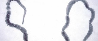
Visual examination of feces allows you to identify adult helminths, segments of tapeworms, the feeding pattern of the animal, and the presence of dangerous components such as bone fragments, polyethylene, and ropes. The main indicators of macroscopic examination of stool are color, smell, and consistency.
Water, fiber, mucus and fat determine the consistency of stool. With pathology of pancreatic secretion, stool has an ointment-like consistency. Liquid feces occur due to insufficient digestion in the small intestine with enteritis, colitis with ulcerations. With fermentative dyspepsia, the stool becomes mushy. The foamy character indicates fermentative colitis, dysbiosis, dysbacteriosis. Feces become ribbon-like and pencil-shaped in the presence of hemorrhoids, anal fissure, or rectal tumor.
Brown feces are normal; they are normally yellow with a milk diet and dark brown with a meat diet. Black stool indicates bleeding in the anterior gastrointestinal tract.
With pathology of gastric digestion, as well as colitis of various forms and hyperfunction of the large intestine, the stool turns dark brown. Red stool indicates bleeding in the gastrointestinal tract, in the posterior sections. Yellow stool appears due to pathology in the small intestine. With pancreatic insufficiency, the stool becomes gray in color. White feces are caused by pathology of the bile duct or intrahepatic stagnation.
The smell of feces comes from the breakdown of food proteins. The putrid smell of feces indicates poor functioning of gastric digestion, possibly ulcerative colitis. The unpleasant smell of rancid oil appears when there is insufficient secretion of lipase by the pancreas. A sour smell appears due to the fermentation process in the large intestine and impaired absorption of fatty acids in the small intestine.
Microscopy reveals protozoa and their cysts, helminth eggs, muscle fibers, fatty feed components, leukocytes, erythrocytes, epithelial and tumor cells, and crystals in feces. Sometimes they find fleas and ticks that came from outside.
Biochemical indicators of feces are reflected in crosses, being qualitative, not quantitative indicators.
The reaction to protein is positive if undigested food protein is present in the stool. It is detected when the stomach is damaged; pathology of the duodenum is also possible.
They make a reaction to occult blood, which is not detected during macroscopic examination. If it is positive, then this indicates bleeding from all parts of the digestive tract. There is also a false positive reaction that is given by peroxidases from bacteria, fungi, and medications containing iron.
Timely analysis of feces in dogs allows you to promptly identify and prevent unwanted pathologies of internal organs in your pet. Give health and a happy life to your faithful and dear dog. If you suspect any illness or illness, take your animal's stool for analysis to a veterinary clinic. I recommend that all dog breeders do a stool test, which will prolong the life of your pet.
Other characteristics
According to its characteristics, feces in an adult are directly related to lifestyle and nutrition. What causes an unpleasant odor? Pay attention to what you've been eating more frequently lately. A foul odor is also associated with taking certain medications and can manifest itself as a symptom of some kind of inflammatory process. In cases of food absorption disorders (Crohn's disease, cystic fibrosis, celiac disease), this symptom also appears.
Floating stool in itself should not be a cause for concern. If the floating stool has a very unpleasant odor or contains a lot of fat, this is a symptom of poor absorption of nutrients in the intestine. In this case, body weight is quickly lost.
Dog stool analysis: form
Very often, cat owners turn to veterinary clinics with the problem of stool problems in their animals.
This could be diarrhea, feces with blood, mucus, or vice versa - constipation. As a rule, owners initially try to cope with this problem on their own, but most often home treatment does not give a positive result.
When contacting the clinic, the veterinarian will first conduct a physical examination of the animal and collect anamnesis (diet, deworming, vaccination, nuances of keeping the animal). Based on this data, the veterinarian will order a stool test for the cat. Stool analysis can help assess how nutrients are digested by liver and pancreatic enzymes, and can also help determine the composition of the intestinal microflora. It will allow you to find out about the presence of helminths and protozoan cysts, which can cause discomfort to your pet. The veterinarian will be able to obtain information, for example, about the insufficiency of the exocrine activity of the pancreas and severe infectious diseases, such as feline panleukopenia, coronavirus infection, toxoplasmosis, which is a zooanthroponosis.
Based on the results of the stool analysis, the attending physician will prescribe the appropriate treatment or preventive measures for the cat and recommend the correct feeding diet specifically for your pet.
To obtain an informative stool coprogram (general stool analysis), you need to follow some rules when preparing biomaterial, then the analysis will be accurate:
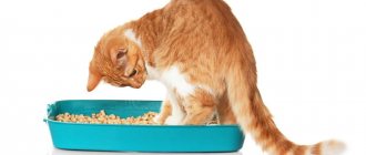
Make sure that 2-3 days before the analysis the cat was not given laxatives and adsorbents (for example, activated carbon), as well as drugs that color feces (iron, bismuth). Feces collected after an enema or an X-ray of the digestive tract, during which the animal was given radiocontrast agents, will not be suitable for research. The stool sample must not contain particles of cat litter.
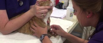
Three days before the test, stop giving your cat meat, fish, and green plants, as these foods can give a false positive result and show the presence of occult blood in the feces. If the cat consumed these products the day before taking the material, notify your veterinarian about this.
The optimal container for collecting and storing feces is a plastic container with an airtight lid.
In order for a laboratory technician to detect vegetative forms of protozoa in feces, it must be delivered to the laboratory immediately after defecation (15-20 minutes). Detection of cysts is possible if stool is delivered within twenty-four hours.
The correct diagnosis - dysbiosis can be made to a cat when feces for analysis are collected from the central part of the mass.
Fecal analysis in cats is an important link in the diagnosis of many diseases. Do not neglect it, because a correct diagnosis is the key to successful treatment of the disease.
A stool analysis, if performed correctly, can tell the doctor about many disorders and pathological conditions:
- helps detect inflammatory processes in the gastrointestinal tract in the early stages,
- indicates the degree of digestibility of the feed,
- shows the activity of the pancreas and bile pigments,
- identifies frequently occurring groups of microorganisms responsible for dysbiosis and intestinal infections,
- detects protozoa.
Bristol scale
English doctors at the Royal Hospital in Bristol have developed a simple but unique scale that characterizes all the main types of feces. Its creation was the result of the fact that experts were faced with the problem that people are reluctant to open up about this topic; embarrassment prevents them from talking in detail about their stool.
Based on the developed drawings, it became very easy to independently characterize your own bowel movements without any embarrassment or awkwardness. Currently, the Bristol Stool Shape Scale is used throughout the world to assess the functioning of the digestive system. For many, printing a table (types of feces) on the wall in your own toilet is nothing more than a way to monitor your health.
1st type. Sheep feces
It is called so because it is shaped like hard balls and resembles sheep feces. If for animals this is a normal result of intestinal function, then for humans such stool is an alarm signal. Sheep pellets are a sign of constipation and dysbacteriosis. Hard feces can cause hemorrhoids, damage to the anus, and even lead to intoxication of the body.
2nd type. Thick sausage
What does the appearance of stool indicate? This is also a sign of constipation. Only in this case are bacteria and fibers present in the mass. It takes several days to form such a sausage. Its thickness exceeds the width of the anus, so emptying is difficult and can lead to cracks and tears, hemorrhoids. It is not recommended to self-prescribe laxatives, as sudden release of feces can be very painful.
3rd type. Sausage with cracks
Very often people consider such stools to be normal, because they pass easily. But make no mistake. Hard sausage is also a sign of constipation. When defecating, you have to strain, which means there is a possibility of anal fissures. In this case, irritable bowel syndrome may be present.
4th type. Ideal chair
The diameter of the sausage or snake is 1-2 cm, the feces are smooth, soft, and easily amenable to pressure. Regular bowel movements once a day.
5th type. Soft balls
This type is even better than the previous one. A few soft pieces form and come out gently. Usually occurs with a large meal. Stool several times a day.
6th type. Unshaped chair
The feces come out in pieces, but unformed, with torn edges. It comes out easily without hurting the anus. This is not diarrhea yet, but it is already a condition close to it. The causes of this type of stool can be laxative medications, increased blood pressure, excessive consumption of spices, and mineral water.
7th type. Loose stools
Watery stools that do not include any particles. Diarrhea requiring identification of causes and treatment. This is an abnormal condition of the body that needs treatment. There can be many reasons: fungi, infections, allergies, poisoning, liver and stomach diseases, poor diet, helminths and even stress. In this case, you should not postpone your visit to the doctor.
How to correctly collect a stool sample from an animal?
You can collect stool samples from a tray or during a walk into laboratory containers, or into a clean container (plastic or glass). Special requirements for dishes are imposed only when sowing is prescribed and, in this case, are separately discussed by the veterinarian. With formal stool
The middle part of the feces is taken, and if the animal is small, the feces can be taken as a whole.
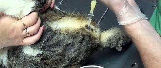
With loose stools,
collection with a syringe is possible. If the stool is visually changed - there is mucus or jelly-like inclusions, then all types of changed stool are collected. Important: if an animal has loose stools, there is no need to wait until they become formed. The contents are collected in the form in which they exist, because altered loose stools are not a contraindication for testing, but on the contrary can be even more informative. This is especially true for intestinal lesions such as giardiasis or infection with other protozoa.
Fecal analysis of dogs and cats in our veterinary clinic
Our veterinary laboratory uses a unique phase-contrast microscopy method
, thanks to which you get the most accurate results in the shortest possible time. This method allows you to accurately determine the slightest deviations from the norm. In addition, it makes it possible to accurately detect a number of diseases that are quite difficult to detect using standard methods (for example, giardiasis, which is a common cause of indigestion).
The results of the analysis will be available to you on the same day and can be sent to the email address you provided.
We conduct a detailed analysis of stool for dysbacteriosis, determine the state of the digestive organs, as well as the correct digestion of feed. Stool analysis is a very informative study and provides direction for further diagnostic studies. The condition of the skin, ears, eyes and general vigor of the animal is a mirror of the condition of the digestive tract. By analyzing stool, we can immediately roughly determine the affected organs of the digestive system and further research will be highly specific, which will significantly save time, money for the owner and the nerves of the animal in the process of making a diagnosis. In our laboratory, stool analysis can be done daily from 10 a.m. to 7 p.m. For the cost of laboratory tests, see our