5267Pavel
Alkaline phosphatase enzyme activity occurs mostly in the liver, but also in the intestines and to a small extent in bone tissue. In a healthy animal, the total ALP activity in the blood consists of bone and liver isoenzymes. The second reason for increased levels of alkaline phosphatase in the blood of animals is cholestasis or cholangitis. This is a disease in which the secretion of bile slows down and the production of the enzyme increases. Diagnosis of this pathology is problematic, since alkaline phosphatase is present in the blood of cats for only a few hours and then disintegrates. Therefore, veterinarians do not consider ALP as a marker of cholestatic disease.
The second subtype of hepatic isoenzyme rises as a result of the cat taking corticosteroid substances during drug therapy. Another reason is the presence of hyperadrenocorticosis.
© shutterstock
The isoenzyme that is responsible for the activity of alkaline phosphatase in the intestinal tract is located in the thin section. Little research has been carried out in this direction, therefore, when the level of alkaline phosphatase in the intestines changes, they speak with caution about gastrointestinal diseases, and additional research is being carried out.
Increased phosphoric acid metabolism in a cat may indicate hyperthyroidism. This is a hormonal disease that causes an increase in thyroid function. Other liver enzymes are tested to make a diagnosis.
Alkaline phosphatase is elevated in liver diseases, including cirrhosis, necrosis of liver tissue, cancer, infectious and toxic hepatitis, drug-induced hepatitis, and tuberculosis. The activity of the enzyme increases with infarction of the kidneys and lungs, Cushing's syndrome, taking antibiotics, vitamin C, sulfonamide, and bacterial gastrointestinal infections.
In cats, ALP is active in the third trimester of pregnancy, when the fetus and placenta are actively growing. This is a physiological change in the enzyme that does not require treatment.
Causes
One of the main reasons for an increase in the enzyme is diseases such as cholestasis and cholangitis. Pathologies are characterized by a decrease in the volume of bile produced.
ATTENTION! In practice, it is quite difficult to use the values of ALP in the biochemical composition of blood to determine diseases, since the latter decomposes quite quickly, within a few hours.
The second reason is long-term use of corticosteroid drugs during rehabilitation after surgery. Kittens are characterized by an increase in alkaline phosphatase during the period of strengthening bone tissue. In older cats, the level of the enzyme changes depending on the orthopedic diseases they have suffered.
The isoenzyme that regulates the activity of alkaline phosphatase is produced in the small intestine. But, in general, its activity has not been sufficiently studied, so changes in the enzyme level alone cannot be used. A comprehensive analysis of all blood components is required.
Let us note the main diseases of the gastrointestinal tract that provoke an increase in alkaline phosphatase:
- Cirrhosis and necrosis of the liver.
- Hepatitis.
- Infectious poisoning.
Enzyme activity provokes cancer and tuberculosis. ALP levels in cats are affected by taking certain antibiotics, vitamin C, and kidney problems.
For cats during pregnancy, changes in enzyme levels are normal and should not cause concern. Alkaline phosphatase is involved in the formation of the fetus and placenta.
Activity increases in the last weeks of pregnancy (3rd trimester).
High concentrations of ALP in children
Alkaline phosphatase in the blood of children is several times higher than in adults. This situation is explained by the active growth and development of babies. The highest concentration will remain until the transition to adolescence. It is at this time that the body produces hormones that affect all metabolic processes.
Despite the high level, when the baby gets sick, alkaline phosphatase can increase several times more for pathological reasons, for example:
- Liver diseases in combination with stagnation of bile or its excretion into the duodenum in insufficient quantities,
- Rickets. Thanks to ALP, rickets can be detected long before symptoms appear,
- Infectious mononucleosis is a disease that can cause serious complications.
- Diseases affecting bone tissue
- Intestinal infection.
More complete information in our article Alkaline phosphatase in children: normal, increased, decreased.
Cholestasis
The disease is characterized by disruption of the liver and gallbladder. At the first stage, bile ceases to be excreted into the duodenum.
Of course, not immediately, but every day more and more secretions remain in the gallbladder. Stagnation of bile occurs and it is gradually absorbed into the blood. Internal intoxication of the body occurs. Cholemia develops, which is fraught with the most tragic consequences for the cat. Let us note the main symptoms that will help owners identify impending cholestasis:
- Partial or complete refusal to eat, the animal does not even touch its favorite treats.
- The animal sleeps more often and avoids contact with family members.
- The mucous membranes turn yellow;
- Urine acquires a sharp, unpleasant odor and becomes significantly darker.
- Vomit.
- The feces are mostly liquid, with a white coating, mostly light in color.
- Kidney failure develops.
The veterinarian chooses the treatment independently. It depends on the cause of the disease. Benign tumors that can form during stagnation are removed surgically . Pathogenic microorganisms are destroyed by antibiotics. Drugs are often used in parallel to thin the bile.
What to do if alkaline phosphatase is elevated?
Proper treatment and care for the animal will help get rid of the disease.
It is clear that when a disease occurs, it must be treated. After undergoing a clinical examination and identifying pathology in your pet, you need to find out more information about this disease and learn how to properly care for the animal during treatment.
Cholestasis
The animal needs immediate care and treatment. The consequences may be irreversible!
A disease associated with dysfunction of the liver and gallbladder is cholestasis.
The mechanism for the appearance of pathology is the impossibility of removing bile into the lumen of the duodenum, as a result of which it stagnates in the bladder itself and its pathways and is subsequently absorbed into the blood, thereby giving rise to a difficult process - cholemia. As a result of severe toxicity of bile, severe poisoning of the entire body occurs. If action is not taken immediately, the cat will die.
Symptoms
Externally, a sick cat does not differ from its healthy counterparts.
The signs of this disease are very extensive and contradictory, due to the variety of causes of its occurrence.
- The cat may be overly sleepy, refuse to eat, or, conversely, experience insatiability.
Excessive sleepiness of the pet should alert the owner. - Vomiting and severe yellowness of the mucous membranes are often present.
- The total body weight decreases, the urine changes color and becomes dark orange.
A cat's weight loss can also be an alarming symptom! - Feces have a fuzzy consistency, pale, almost white.
- Liver failure develops.
Treatment
Treatment depends on the causes of the disease.
- Neoplasms and echinococcal blisters are eliminated surgically.
Sometimes surgery is an inevitable way to treat a cat with cholestasis. - Parasites are destroyed by antiparasitic agents.
- Medicines are recommended to help thin out bile.
Hyperparathyroidism
The disease occurs as a result of excess hormones produced by the parathyroid glands. Directly depends on the lack of calcium and excess phosphorus in the cat’s menu.
Causes, diagnosis and symptoms
The main reasons that provoke the development of pathology are related to the cat’s diet:
- improperly selected diet, rich in food rich in phosphorus and containing insufficient amounts of calcium;
An improper diet poses a real threat to the health of cats. - genetic predisposition;
- impaired absorption of calcium;
- accelerated kitten growth.
The cat's rapid growth can also trigger the development of the disease.
Hyperparathyroidism is diagnosed by radiography.
Laboratory tests of urine and blood are usually normal. The only exceptions are severe forms. X-rays will show decreased bone density. The kitten's bones will look thin, and the difference between muscle mass and bones will be almost invisible. You will also see abnormal bone structure as a result of deformation.
It is recommended to take an x-ray of the entire skeleton as a whole. The main symptom of this disease will be lameness and inability to move freely, bone pain.
Hyperparathyroidism
Problems arise when the thyroid gland begins to produce too much hormone. An imbalance is created in the body, the functioning of many organs is disrupted. Among the main reasons, experts note feeding with cheap food, fried, spicy and any food from the human table. Oddly enough, excessive consumption of fish will also lead to hyperparathyroidism.
ATTENTION! For prevention purposes, it is worth balancing your cat’s diet. Eliminate foods rich in phosphorus as much as possible and add foods high in calcium (eggs or a little milk) to your diet.
Other reasons include a genetic predisposition to calcium absorption disorders or an overly active kitten's mouth. For diagnostic purposes, the radiography method is used . A comprehensive blood and urine test is performed. As you know, a lack of calcium leads to problems with bone tissue. For better results, the entire animal is examined.
The test results show areas where bone tissue is difficult to distinguish from muscle tissue. X-rays can help identify areas where bones have become thinner and their overall density has been reduced.
External signs include:
- low mobility;
- unsteadiness of gait;
- frequent problems with bone fractures.
As mentioned above, it is difficult to judge problems only by deviations of alkaline phosphatase in one direction or another. Additional information about bilirubin and gamma-glutamine stransferase levels is often required. Judging by the norm of the enzyme, its indicators range from 39 to 55 units.
Symptoms
Pyoderma
Rickets
Haemorrhoids
At the initial stage of the disease, the cat becomes lethargic and apathetic, constantly lies in one place, gets nervous, and tries to run away when picked up.
Over time, the animal develops severe pain in the bones, the pet becomes aggressive, and when trying to pet or pick it up, it hisses and may bite.
When the disease enters the active phase, its symptoms become noticeable even to the most inattentive owners. The cat experiences pain when walking and limps. With sudden movements (for example, jumping from a chair), fractures and microcracks form in fragile bones. The pet's gait changes: it moves slowly, on widely spaced paws.
There are also the following symptoms:
- Deformation of the pelvic bones and sternum.
- Impaired tooth growth, tooth loss.
- Problems with urination and bowel movements (retention or incontinence).
- Development of complete or partial paralysis of the paws.
- Abdominal bloating.
- Curvature of paws.
Progression of the disease and lack of treatment leads to the development of arthritis caused by pathological changes in bone tissue.
Prevention and treatment
Many owners who are faced with the problem of enzyme production are interested in under what circumstances should the level of SF be examined in a biochemical blood test? Changing enzyme levels act as a litmus test for the following diseases:
- kidney and bone cancer;
- problems with the liver and biliary tract;
- osteodystrophy;
- metastases in bone tissue.
During the analysis, it is imperative to warn the veterinarian about the medications that the pet has taken. The analysis results are distorted when exposed to large doses of ascorbic acid, interferon, non-steroidal anti-inflammatory drugs, levamisole, penicillin and a number of other drugs. In any case, it is useful to check the list with your veterinarian.
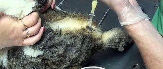
As with most tests, blood is drawn from a vein on an empty stomach. A competent specialist checks the level of alkaline phosphatase throughout the course of treatment in order to respond in time to possible deviations.
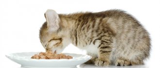
IMPORTANT! During the rehabilitation period at home, a proper diet is important. In most cases, experts recommend specialized foods.
In case of obvious fractures, the pet's mobility should be limited. Give the cat a new home in a carrier or specially organize a place in the apartment for this. In extreme cases, a regular box with high sides will do.
The main thing is to ensure a comfortable temperature in the apartment, quick access to water and food. The average period of forced isolation is about 2 months. Much depends on the level of alkaline phosphatase and other enzymes. In extreme cases, droppers or intravenous injections of calcium solution are possible.
the cat has chronic renal failure and a highly elevated alkaline phosphatase level
Rostov region. The cat is 17 years old. Weight 2.6 kg. 12 years ago my uterus was removed. He sits at home, sometimes goes out into the yard for a walk. No vaccinations were given. I give it regularly for worms. Almost all my life I ate domestic canned food like “Night Hunter”, as well as canned food “Whiskas”. Two years ago I switched to Royal dry food for sensitive digestion. About a year ago, vomiting appeared - 30-40 minutes after eating, but not every day. I thought it was from the wool. I started giving Maltpaste regularly. Did not help. The pharmacy recommended Mezim (the cat is old - there are not enough enzymes). Got better. After some time, she appeared again. vomit. Moved to Hills. Instead of Mezim I gave Pancreatin and the vomiting stopped. In 2011 I had cystitis. We successfully treated him. Since February 23, the cat did not pee for 2 days, but she was feeling as usual: she ate and drank water. The temperature was 38 degrees. On the third day, she urinated. I collected urine and went to the doctor. The clinic did biochemistry and ultrasound. A general blood test was apparently considered unnecessary. Gamavit, Kantaren and KotErwin were prescribed. The ultrasound did not find any stones or sand. Here are the test results. Biochemical blood test 02.25.14. Indicator Unit change Normal Result Total protein g/l (57-80) - 70.7 Albumin g/l (24-38) - 23.4 Glucose Mol/l (3.4-6.9) - 6.69 α-Amylase Units ./l (371-1193) - 1889.5 Bilirubin µmol/l (1.2-7.9) - 10.5 Alkaline Phosphatase U./l (12-65) - 114.5 AlAt U./l ( 8.3-53) - 261.9 AsAt U/l (9.2-40) - 216.9 Creatinine µmol/l (49-165) - 249.1 Urea mmol/l (5.5-11, 1) — 12.34 General urine analysis Indicator Unit. change Norm Result quantity ml (-) - 120 Color (Yellow) - Yellow transparency - (Full) - Not complete Density - (1.015-1.060) - 1.025 pH kg/m3 (5.5-7.5) - 5.5 Ascorbic acid units (absent) - 15 Glucose mg/100 ml (absent) - Absent Nitrites mg/100 ml (absent) - Absent Protein presence (absent) - Absent Ketones mg/100 ml (absent) - Absent Urobilinogen mg/100 ml (0. 1-1.0) - 1.0 Bilirubin mg/100 ml (Absent) - 0.5 Leukocytes presence (1-3 in x) - 1-3 in x Red blood cells Presence (1-3 in x) - Absent Salts Presence ( - ) - Uric acid Renal epithelium Presence (Absent) - Absent Flat epithelium Presence (0-2 in x) - 0-4 in x Cylinders Presence (Absent) - Absent Bacteria Presence (Absent) - Cocci (+) rods (+ +) Mucus Presence (absent) - Absent Transitional epithelium (Units) - Absent Ultrasound 02/25/14 bladder - empty; Left kidney - uneven edges, size. – 2.58 by 2.06 cm, parenchyma 0.51 cm, heterogeneous structure, increased echogenicity, pelvis not dilated, no stones identified. Right kidney – uneven edges, size. – 2.93 by 1.98 cm, parenchyma – 0.46 cm, heterogeneous structure with increased echogenicity, the pelvis is not dilated, no stones were identified. Conclusion – structural changes in the renal parenchyma. After receiving such tests, they told me that everything was very bad and offered to put her to sleep and not torment her with treatment, because it was impossible to force the kidneys to work. I took tests and went to another doctor. Another doctor, upon palpation, discovered uneven contours of the kidneys and tubercles on the peritoneum. Due to increased alkaline phosphatase, she suggested oncology. According to the urine test, she suggested injecting ceftriaxone for 3 days, cantaren, gamavit, Essentiale and Creon capsules (1/4 cap x 3 rubles per day). She said that the cat is 90 years old by human standards and there is no need to torture her, but if you want, you can put IVs. The cat’s state of health practically did not change, she just lay more, but she met and saw me off from work, ate less than usual, but did not urinate and became constipated. After reading veterinary forums on the Internet, I decided to prescribe IVs for the cat myself, because she stopped drinking, although I gave her kidney herbs, but this was not enough. I went to the clinic again and asked for a cannula for IVs. There they considered it necessary to instill saline solution 70 ml, glucose 70 ml, Maniiit 15 ml, cytoflavin 10 ml. On the third dropper, I removed Cytoflavin and added Essentiale IV 1 ml. In addition to herbs, I also poured polysorb into my mouth. The cat's condition did not improve. She was bothered by the cannula and began to have trouble eating. She had no stool for 5 days. Therefore, after 5 days of droppers. I decided to remove the catheter and do repeated tests. The cat’s weight increased by 300 g (why?). At my request, they added calcium, phosphorus and potassium indicators and a sugar test to the biochemistry (they immediately said it was normal). Biochemical blood test 03/6/14 Indicator Unit. change Normal Result Total protein g/l (57-80) - 63 Albumin g/l (24-38) - 19.9 Glucose Mol/l (3.4-6.9) - 6.82 α-Amylase Unit/ l (371-1193) - 1579.6 Bilirubin µmol/l (1.2-7.9) - 15.6 Alkaline Phosphatase U./l (12-65) - 190 AlAt U./l (8.3- 53) - 389.5 AsAt Units/l (9.2-40) - 156.2 Creatinine µmol/l (49-165) - 173.5 Urea mmol/l (5.5-11.1) - 7 .58 Calcium mmol/l (2.0-2.7) - 3.37 Phosphorus mmol/l (1.3-2.4) - 1.26 Potassium µmol/l (3.6-5, - 7. 6 General blood test Red blood cells million/µl (6.6-9.4) - 6.64 Hematocrit % (24-45) - 31.4 Hemoglobin g/l (80-150) - 95 Leukocytes thousand/µl (5. 5-19.5) — 4.1 Basophils % (0-1) — — Myelocytes % (-) — — Young % (-) —- — Lymphocytes % (36-54) — 36 Monocytes % (1-5) — 0 Eosinophils % (2- — 4 Bands % (3-9) — 2 Segientonuclear % (40-45) — 58 Thrombocytes thousand/μl (300-800) — 61 General urine test 03/6/14 Protein is normal Sugar -norm Acetone-norm Urobilin-norm Leukocytes 0-1vx Bile pigments -norm Indican-norm Hyaline cylinders -2 Unchanged red blood cells -norm Granular cylinders-norm Waxy cylinders -norm Cylinders -norm Released red blood cells -norm Kidney epithelium -norm Transitional epithelium 0- 1vx According to the test results, it turns out that urea, creatinine, AST, AlAt, alpha-amylase decreased, but alkaline phosphatase increased. I continue Gamavit 2 ml, Kantaren 2 ml, Essentiale 13 capsules x 3 times a day, Liarsin 1 t. x 3 rubles, Renal advanced 2 scoops per day. The cat began to eat and drink better, but pees once a day. Please give me advice after how long to resume IV drips and what to do with the alkaline phosphatase level?
Blood chemistry
For the owner, it is important, if not to understand, then at least to navigate the abundance of enzymes and substances that will tell you about the current state of the animal. Of course, a detailed diagnosis will be carried out by a specialist, but an owner who cares about the health of the pet should show interest in the treatment process.
Let us note the main indicators that are taken into account in the blood test:
- Bilirubin. One of the main substances that make up bile. The substance is also involved in the coloring of blood. Its excess leads to changes in skin color and says a lot about liver disease (hepatitis) or problems with the bile ducts. Deficiency leads to bone marrow damage. The norm for an adult animal is 3-12 units.
- Urea. The external factor of many diseases is excessive urination, change in color and odor. An abundance of urea indicates blockage of the urinary tract and poor kidney function. But it is important to understand that excess discharge can be associated with diet. Problems arise when feeding foods rich in animal protein. The norm for an adult animal is 5-12 units.
- Glucose. Excess sugar in the body is the cause of a variety of diseases. For example, diabetes mellitus, the release of adrenaline into the blood during frequent, excessive exercise or stress. Experts also note problems with the kidneys, liver, and pancreas. Glucose levels drop during poisoning, prolonged fasting or excess alcohol. The norm for an adult animal is 3-6 units.
- Phosphorus. Increased levels are typical for animals with leukemia or bone tumors. Other causes include excess vitamin D and kidney failure. What is interesting is that long-term diarrhea leads to a decrease in phosphorus levels. The norm for an adult animal is 1-2.3 units.
- Calcium. The microelement increases due to severe dehydration, damage to bone tissue by cancer and excess vitamin D. Problems with kidneys, pancreatitis and vitamin D deficiency, calcium levels drop rapidly. The norm for an adult animal is 2-2.7 units.
- Total protein. Excessive vomiting and prolonged diarrhea contribute to increased levels of the substance. This also includes dehydration. A sharp decline in total protein is observed with intestinal diseases, cirrhosis, hepatitis, renal failure and prolonged fasting. The norm for an adult animal is 54-77 units.
As you can see, it is not only the indicators of alkaline phosphatase or amylase that you should pay attention to. Only a comprehensive analysis and competent processing of results will make it possible to prescribe an effective course of treatment.
Liver tests for liver cirrhosis
In liver diseases of various origins, blood counts can be both higher and lower than normal. A common condition is cirrhosis. With this pathology, the liver gradually loses its functional tissue - parenchyma - it is replaced by fibrous connective tissue, the cells of which can no longer perform the functions assigned to the organ. Tests for liver cirrhosis, such as determining the concentration of specific enzymes and bilirubin, in most cases show liver test levels above normal. However, ALT and AST may be at the upper limit of normal values. Bilirubin is almost always elevated.
But at the terminal stages of the disease, when there is multiple liver damage, the level of ALT and AST begins to rapidly decrease. The reason for such changes lies in the fact that the process of necrosis reaches the stage when liver cells become almost unable to synthesize these enzymes.
In addition to the fact that free bilirubin is one of the main markers of liver pathology, it is also a very toxic substance. First of all, it acts on the “energy stations” of the cell - mitochondria. Indirect bilirubin has the ability to interrupt the respiratory chain. Thanks to this chain of reactions, our body receives energy. When a disruption occurs in the activity of this mechanism, it becomes more and more difficult for cells to function, which can ultimately lead to their death.
Another dangerous effect of bilirubin is that with the bloodstream it can reach the blood-brain barrier, and at certain concentrations it begins to penetrate the central nervous system. Here its toxic effect can lead to encephalopathy or even coma.