Rules for operations to remove the eyeball
- Use of general anesthesia
- Compliance with the rules of asepsis and antiseptics. The operation is performed in a sterile operating room. Before the procedure, the hair around the eye is shaved and disinfected with available isoseptic agents.
- Using local anesthesia to prevent pain in the cat.
Before the operation, owners are prescribed antibiotic therapy and the use of hemostatic agents 5 days before the intended operation. Convenient if it was planned. During unscheduled surgical interventions during eye surgery, drugs are used in the postoperative period.
- Use of general anesthesia
- Compliance with the rules of asepsis and antiseptics. The operation is performed in a sterile operating room. Before the procedure, the hair around the eye is shaved and disinfected with available isoseptic agents.
- Using local anesthesia to prevent pain in the cat.
When performing an operation to remove an eye from a cat or dog, veterinarians must follow certain rules that can prevent dangerous consequences for the health of the animal.
When performing this procedure in a clinic, specialists must fulfill the following requirements:
- During the operation, the doctor must cause minimal harm to the cat’s health;
- The doctor must perform the operation correctly in compliance with all necessary measures, this will avoid complications after surgery;
- The procedure should be done using anesthesia so that the pet cannot feel all the unpleasant feelings. In addition, so that the cat does not feel pain after removal of the eye, it is necessary to inject special painkillers;
- The anesthetic during surgery should be used in small quantities so as not to cause serious harm to the health of the animal.
Types of operations
There are three types of surgical intervention. Each of them is prescribed depending on the indications.
- Enucleation is the removal of the eye with all its contents, without affecting the structural components of the orbit - muscles and ligaments. Possible consequences include cosmetic deformation, chronic entropion of the eyelids and conjunctivitis.
- Evisceration – removal of the eyeball and ligaments.
- Exenteration is the removal of the eyeball along with muscles and ligaments. Recommended in case of panophthalmitis or cancerous tumors affecting surrounding tissues. Gives a good cosmetic result without complications.
During the operation, it is possible to install an implant that will replace the removed eye in the orbit. This procedure is carried out at the owner’s request, regardless of the type of surgical intervention.
When performing surgical interventions on the eyes, it is better to give preference to specialized veterinary ophthalmologists.
For surgical interventions on the eyes, it is better to give preference to specialized veterinary ophthalmologists
Why in some cases do they resort to removing the eyeball?
In case of some diseases in animals, surgery to remove the eye becomes necessary, since the eyeball becomes a source of suffering for the animal, and the pathological processes that develop in it can threaten the health and life of the animal. The procedure is performed when the intraocular structures are severely damaged and visual functions cannot be restored.
Such diseases include: buphthalmos (terminal glaucoma), penetrating wounds into the eyeball, blunt injuries and contusions, which are combined with damage to the choroid, with extensive hemorrhages and disorders of the retina and vitreous body. Surgery to remove the eyeball is necessary in cases where therapeutic treatment is pointless.
Pathological processes that rapidly develop in an animal’s eye can only be stopped with the help of surgical intervention to relieve the patient of pain and return him to normal life. Operations can be planned or emergency. At the Antistress clinic at the SQ-lap veterinary hospital, they are carried out after a preoperative examination of the animal, which includes ultrasound, ECG, General and Biochemical Blood Tests, and an 8-hour diet is required.
Before the operation, the doctor and the owner draw up a document on informed consent for the operation, after all the risks of these activities are explained to the owner. It is very important that the owner brings a warm blanket to the operation; it will be needed to warm the animal after the operation, disposable absorbent diapers, and napkins.
Evisceration
- Antibiotic therapy for 5-7 days
- Treatment of the seam 2 times a day for two weeks
- Use of non-steroidal anti-inflammatory drugs for a week.
- Use a protective collar if the cat is bothered by a stitch in the eye area.
- Limit the cat from contact with the street and other animals.
- Provide her with a source of heat during the recovery period.
Properly selected diet therapy plays a special role in a cat’s recovery.
The diet should include a large amount of protein, which will be the foundation for the restoration and healing of damaged tissue.
In the postoperative period, it is important to follow all the veterinarian’s instructions to prevent the development of secondary infections in the cavity of the eye orbit.
Removing the eyeball is a simple operation that does not pose a danger to the animal’s life. The absence of one eye does not affect the pet's quality of life.
Eye damage in cats is a serious and dangerous traumatic injury. During examination, the veterinarian can provide assistance, but it is not always possible to save this organ. In these cases, it may be necessary to remove the cat's eye. But in order for this procedure to be successful, you need to know the main features of its implementation.
This is a surgical procedure in which the eyeball itself and part of its auxiliary apparatus are removed while preserving the muscular-ligamentous apparatus of the organ. In this case, there are several approaches to performing the operation - transconjunctival and transpalpebral. The latter is recognized as the cleanest method.
The advantage of this method is the extraction of the orbicularis eyelid muscle and a significant reduction in skin prominence deep into the orbit in the postoperative period. However, the intervention is accompanied by significant bleeding, so in some cases there is a need for ligation of blood vessels. Enucleation gives a stable result and a low percentage of postoperative complications.
The operation assumes that it is necessary to completely remove the contents of the eyeball and all intraocular structures: the retina, lens, vitreous body, choroid. Despite the scale of the work, this method is low-traumatic and does not require the use of special equipment. After the blood clot dissolves, a fibrin framework remains in the cavity. After a few years, it resolves and the eye completely atrophies.
The disadvantages of evisceration are an unsatisfactory aesthetic result and the possibility of developing complications in the postoperative period: inversion and eversion of the upper and lower eyelids, which provoke injury to the conjunctiva, an increase in lacrimal and mucous secretions, and the appearance of chronic conjunctivitis in the dog. Prolapse of the third eyelid gland may occur.
Bleeding occurs after ophthalmic surgery. Among the complications that can develop in the postoperative period in dogs, it is worth noting emphysema, fistula, purulent inflammation of the orbital cavity, damage to the chiasm as a result of tension on the optic nerve, etc. Therefore, it is so important to provide the pet with proper care and not neglect regular examinations by a veterinarian, who will control the healing process.
After surgery, the four-legged patient is prescribed a course of painkillers and anti-inflammatory drugs. To avoid the risk of wound infection, systemic antibacterial therapy is carried out. Local treatment of the seam is required once or twice a day. If necessary, a special protective collar is placed around the dog's neck. After 10–14 weeks, the stitches are usually removed. After this, the doctor will prescribe a histological examination of the eyeball.
We suggest you read: Hip dysplasia in cats
Make an appointment for your dog to see the doctor. To do this, fill out the online application form. If you have questions, you can ask them to a TIM clinic specialist by phone.
Severe injuries with destruction of the eye;
Purulent inflammatory processes (panophthalmitis, endophthalmitis);
Painful absolute glaucoma;
Malignant tumor processes in the eye area;
Threat of sympathetic ophthalmia;
Long-term inflammation in the blind eye;
Atrophy and subatrophy of the eyeball;
Removal of an eye for cosmetic purposes.
Enucleation of the eye is a very aggressive procedure that requires deep anesthesia. Very often the operation is accompanied by significant blood loss, and changes the appearance of the animal far from for the better. This circumstance is extremely painful for animal owners, especially those with an exhibition animal.
Enucleation, execution technique
First of all, the conjunctiva is separated from the limbus, then the eye muscles are grabbed with a hook, stitched (except for the oblique muscles) and cut off. Special scissors are carefully placed behind the eye and used to cut off the optic nerve (after moving the eye anteriorly). Stopping bleeding is an important point; it is carried out using tamponing with a solution of 3% hydrogen peroxide.
Enucleation of the eyeball in dogs, as well as in cats, takes place under conditions of absolute sterility using the most modern methods, instruments and drugs used in veterinary medicine.
In recent years, a method of cosmetically restoring the appearance of animals has been developed and successfully implemented so that owners do not perceive the visual defect of a pet too much.
Ocular prosthetics are used after enucleation and are produced to restore the cosmetic appearance of dogs and cats.
Maintains normal shape and size of the eyeball;
Ensures the anatomically correct location of the eyeball in the orbit and adherence of the eyelids (and third eyelid) to the eyeball;
Preserves the normal functioning of the extraocular muscles;
Provides an excellent cosmetic effect, the animal looks almost the same as in normal condition;
The operation to install the implant does not last longer than half an hour and is performed under general anesthesia. But, it is contraindicated for intraorbital and intraocular neoplasia, purulent inflammatory processes in the eye, ulcerative and degenerative changes and lesions of the cornea, which are accompanied by its thinning.
After enucleation, the animal must wear a special protective collar for at least a month to prevent damage to the suture. Local use of antibiotics in solution and keratoprotectors is indicated (2-3 times a day, for a month). Systemic therapy, nonsteroidal anti-inflammatory drugs, and antibiotics are used. The use of absorbable suture material allows the sutures not to be removed; they gradually dissolve on their own.
Evisceration of the eye in animals
Evisceration - consists of the complete removal of all intraocular structures, after which only the fibrous membrane remains.
Technique of the operation
After the conjunctiva is separated from the limbus and sclera, the cornea is removed with special scissors, capturing a small strip of the sclera. The contents of the eye are removed with a surgical spoon, leaving only the fibrous membrane. The cavity is washed using hemostasis lavage and treated with antiseptic solutions. Evisceration of the eye is performed using 4 incisions on the sclera, and the conjunctiva is sutured with a continuous suture, and a drainage is inserted into the eye cavity.
There is evisceration of the eyeball with excision of the posterior pole and neurotomy.
Frequently asked questions to the doctor.
How difficult is the recovery period for a dog after eye enucleation?
It all depends on the severity of the pathological process and the traumatic mechanism that preceded the operation. As a rule, the recovery period takes place in at least a month.
Is it necessary for the animal to wear a surgical collar after surgery?
Yes, it is desirable, since the animal will definitely “try” to wipe the newly operated eye with its paw. As a result, the sutures will be removed, and pathogenic microorganisms will inevitably enter the fresh wound.
To what extent does evisceration and enucleation spoil the appearance of an animal?
It all depends on the perception of others and on the extent of the owner’s love for his four-legged pet. Of course, one cannot expect the same appearance, but in this case the question arises not about a cosmetic effect, but about saving the life of the animal.
Veterinary
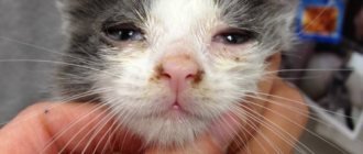
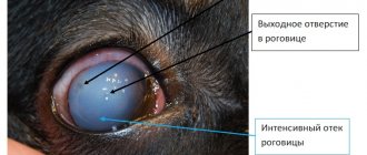
Eye injury in cats is a common problem. The injury can be blunt, the eye injury can be penetrating or non-penetrating, but in any case, this condition requires the help of an ophthalmologist to assess the damage and preserve vision.
Blunt eye injury in cats occurs when falling from a height, car injury, or fights (Fig. 1). The following clinical signs are noted: blepharospasm, miosis (constriction of the pupil), fibrin in the anterior chamber of the eye, hyphema (hemorrhage in the anterior chamber of the eye), hyperemia of the vessels of the iris, hemorrhage under the conjunctiva of the eyeball. These signs may be mild and may be partially present in cases of minor injury.
After an ophthalmological examination, a veterinary ophthalmologist assesses the severity of the eye condition and prescribes symptomatic treatment (anti-inflammatory drugs); in case of minor injury, the eye condition stabilizes after a few days, the eyeball can perform its functions normally.
In cases of severe blunt trauma to the eye in cats, proptosis (prolapse of the eyeball), rupture of the sclera with severe intraocular hemorrhage may occur. Proptosis of the eyeball in cats is a rare pathology because the cat's orbit securely anchors the eye, unlike some breeds of dogs that are predisposed to proptosis, which means that trauma must be very serious to cause this condition in a cat.
Postoperative care for animals
- Antibiotic therapy for 5-7 days
- Treatment of the seam 2 times a day for two weeks
- Use of non-steroidal anti-inflammatory drugs for a week.
- Use a protective collar if the cat is bothered by a stitch in the eye area.
- Limit the cat from contact with the street and other animals.
- Provide her with a source of heat during the recovery period.
Properly selected diet therapy plays a special role in a cat’s recovery.
The diet should include a large amount of protein, which will be the foundation for the restoration and healing of damaged tissue.
In the postoperative period, it is important to follow all the veterinarian’s instructions to prevent the development of secondary infections in the cavity of the eye orbit.
Removing the eyeball is a simple operation that does not pose a danger to the animal’s life. The absence of one eye does not affect the pet's quality of life.
Eye damage in cats is a serious and dangerous traumatic injury. During examination, the veterinarian can provide assistance, but it is not always possible to save this organ. In these cases, it may be necessary to remove the cat's eye. But in order for this procedure to be successful, you need to know the main features of its implementation.
For what diseases does a cat need eye removal?
Surgery to remove an eye in cats, as well as dogs, may be required in the presence of a serious pathological process. But first you need to carry out a series of therapeutic procedures that can save at least part of your vision. If the situation is very serious, the veterinarian often recommends removing the cat’s eye.
We invite you to familiarize yourself with: Spotted cats color features Description of spotted cats
Surgery to remove a cat's eye may be required for the following pathological processes:
- the presence of internal infectious lesions that can spread to other organs;
- damage to the back of the eyeball, but within the orbit;
- congenital deformations of the visual organs;
- oncological tumors;
- infectious and inflammatory processes of the eyeballs (superficial and internal), which can only be cured using surgery;
- glaucoma with a severe course;
- the presence of severe physical injuries from which it is impossible to recover independently.
Many cat owners often have a question: why remove the eyeball from an animal? Enucleation is required to reduce pain in animals, as well as to eliminate all suffering. In addition, the damage can often affect other internal organs in addition to the eye.
The main indications for surgical removal of the eyeball are diseases that can cause irreparable harm to internal organs and the brain, and also cause unbearable pain to cats.
List of diseases:
- Panophthalmitis is a purulent inflammation of the cavity of the eyeball. The entire cavity is filled with purulent exudate, and, unfortunately, the inflammation cannot be treated therapeutically. Without removing the affected gas, inflammation can spread to surrounding tissues and even to the brain, which leads to the death of the pet.
- Glaucoma with increased intraocular pressure without therapeutic effect - increased intraocular pressure has a negative effect on the optic nerve and causes blindness. The eyeball is bursting and it “falls out” a little from the orbit. If the animal does not respond to therapeutic treatment, it is recommended to remove the organ to prevent functional and structural damage to the optic nerve and brain.
- Severe injuries to the eyeball - these can be various wounds that cannot be healed with medication. Without surgery, the cavity of the eyeball becomes clogged with pus and panophthalmitis develops.
- Loss of the visual organ with changes in the tissue structure - changes in the color of the retina, rupture of blood vessels and the optic nerve.
- Malignant tumors of the eye
- Scleral rupture
The operation is carried out only when diagnostic measures are carried out that confirm the presence of the above diseases.
Panophthalmitis is a common condition that requires removal of a cat's eye
Ocular evisceration is an attractive alternative to enucleation.
The procedure, like enucleation, is performed on completely diseased eyes. This operation eliminates the need to use local and systemic medications and provides a good cosmetic effect.
Indications for surgery (evisceration):
- The end stage of primary glaucoma, in which the response to drug therapy disappears, the eyeball becomes enlarged and painful, and the visual functions of the eye are lost.
- Severe eye injuries without the development of purulent inflammation.
- Traumatic and endogenous uveitis with vision loss when drug treatment is ineffective.
The surgical technique is evisceration of the eye with placement of an intraocular prosthesis in dogs and cats.
The essence of the method is to remove the contents of the eye through a scleral incision, leaving only the fibrous membrane. A sterile silicone bead is inserted into this corneoscleral membrane and the scleral wound is sutured. The usual result is a painless, mobile and cosmetically good-looking eye that does not require drug therapy. There are no visible seams; the seams are located under the upper eyelid.
We recommend reading: Cleaning Your Dog's Mouth
What is a prosthetic eye for dogs and cats?
The use of ophthalmic prostheses in animals has been of interest to veterinary scientists since the 19th century.
Previously, veterinarians tried to place prosthetic eyes in the conjunctival space or in the orbit. As a result, we realized that these prostheses require daily care; orbital and conjunctival tissues undergo contracture over time, which leads to the prosthesis being pushed out.
In recent decades, scientists have decided that it is best to use silicone prostheses and place them inside the fibrous membrane of the eye.
These prostheses do not cause pain or discomfort, look good, are non-toxic, do not have antigenic activity, are easy to install, are not expensive and fill the intraocular volume.
Our network of veterinary clinics “Vasilyok” uses prosthetics from the German company Acrivet. They come in different diameters (from 12 to 26 mm) for dogs and cats.
During the preoperative examination, our veterinarian ophthalmologist takes measurements and determines the required diameter of the intraocular prosthesis for a given animal.
How long does the operation - evisceration of the eye with placement of an intraocular prosthesis - take and is postoperative care difficult?
This surgery is performed under general anesthesia, the dog does not experience pain. The duration of the operation is no more than an hour.
After this operation, the animal needs to wear a protective collar for 2-3 weeks, the owner needs to treat the sutures on the skin and instill drops into the eye. Two stitches in the skin are the only visible stitches and are removed after 2 weeks.
At our veterinary clinic “Vasilyok”, ophthalmologists try to save the eyes “to the last.” If it is impossible to save the eye, then our veterinary ophthalmologists, if possible, prefer to perform evisceration of the eye with the installation of an intraocular prosthesis rather than enucleation.
Make an appointment with a veterinarian ophthalmologist: 8 (499) 110-0105
Endoprosthetics of the eye. A case from the practice of a veterinary ophthalmologist.
The owners of a Siberian Husky puppy contacted a veterinary ophthalmologist at our clinic. The owners recently acquired a puppy and did not immediately notice that he kept his left eye closed. The owners did not have dogs before, they wanted this particular breed, quickly chose a puppy on the Internet, quickly went to pick him up, and quickly picked him up. As if there was no time to “look” at the baby. The next day it was noticed that there was “something wrong” with the left eye - the puppy was squinting it. And a day later the puppy was taken to an appointment with a veterinary ophthalmologist.
A little about the patient: a Siberian Husky puppy; nickname Holmes; age 2.5 months; general condition is satisfactory, nutrition is average, appetite and thirst are preserved, temperature is normal. Exterior, character, temperament correspond to the breed.
An ophthalmological examination revealed increased intraocular pressure and swelling of the cornea of the left eye. When measured with an applanation tonometer, the intraocular pressure of the left eye was 35 mmHg. Art., which is twice the norm.
When examining the anterior chamber angle using a gonioscopic lens, it was found that the iridocorneal angle was closed.
A veterinary ophthalmologist diagnosed primary angle-closure glaucoma - goniodysplasia . Congenital glaucoma, unfortunately, occurs in Siberian Huskies and is considered a hereditary disease.
A husky puppy has primary angle-closure glaucoma - goniodysplasia.
With this type of glaucoma, the structures of the eye are damaged - the pectineal ligaments, through which most of the aqueous humor flows out. Intraocular pressure increases.
The puppy experienced severe pain in the eye, he had buphthalmos (severe enlargement of the eye in size), corneal edema, cracks in the endothelial layer of the cornea, and injection of the deep vessels of the conjunctiva.
The pupil in the affected eye was slightly dilated and did not react to light.
The veterinary ophthalmologist warned the owners about the seriousness of the disease. The prognosis is “unfavorable”; nothing more comforting can be said for this disease. With a congenital disorder of the anatomical structure of the eye, it is very difficult to normalize its functional state. In addition, increased intraocular pressure has an extremely negative effect on the retina. With increased intraocular pressure up to 30-40 mm Hg. Art. Within a few days, retinal atrophy occurs, and blindness occurs in the affected eye.
The puppy was prescribed therapeutic treatment, but in this case it did not lead to improvement. It was possible to relieve corneal edema, but it was not possible to reduce intraocular pressure.
14 days after the start of treatment, the intraocular pressure of the left eye was 42 mmHg. Art.
Before surgery, intraocular pressure was 42 mm. rt. Art.
Together with the owners, a decision was made to replace the left eye with prosthetics.
This operation has several advantages compared to complete removal of the eye - enucleation. This, of course, is the aesthetic side, which is of great importance to the owners. In addition, endoprosthetics is a much less traumatic operation that is done faster than removing an eye. But endoprosthetics requires highly qualified veterinary ophthalmologist.
The veterinary ophthalmologist selected the size of the puppy's eye prosthesis. Considering that the puppy will grow, the prosthesis corresponded to the size of the eye of an adult husky. Operation was successfully completed. The postoperative period was without complications.
1.5 months after intraocular prosthetics
Holmes easily adapted to seeing with only one eye. He is very cheerful and active, which is typical for the breed. The fact that the left eye is “a little different” is noticed only by “super-experienced dog lovers” if they look at Holmes “point-blank.” Most people don't notice anything because Huskies often have different colored eyes.
We are pleased with the result of the operation. The dog is practically healthy and feels great. The owners are pleased with the dog's appearance.
Holsm 2.5 months after intraocular prosthetics
Discounts on veterinary ophthalmologist services
Don’t miss the opportunity to take advantage of our seasonal discounts on veterinary services, as well as receive a veterinary discount card PLUS VIP status as a regular customer: Discounts on veterinary services >>
We recommend reading: Nicknames for Calm Cats
Veterinarians will answer your questions: 8 (499) 110-01015
A veterinary ophthalmologist diagnosed primary angle-closure glaucoma - goniodysplasia . Congenital glaucoma, unfortunately, occurs in Siberian Huskies and is considered a hereditary disease.
Eye removal technique
Having retreated from the edge of the eyelid about 1-1.5 cm (more is possible depending on the free volume of the skin), the skin is cut parallel to it. The tissue is then separated and advanced to the eyeball in such a way as to enclose the eyelids, the orbicularis pallid muscle, the entire conjunctiva, the third eyelid, the lacrimal gland of the third eyelid, and the main lacrimal gland as a single block.
The result of this manipulation at this stage should be the following: the entire auxiliary apparatus of the eyeball, with the exception of the extraocular muscles, should be removed as a single block, but at the same time it remains connected to the eyeball by the conjunctiva. Thanks to this, the eyeball is removed in a “package” and the contents of the eyeball and conjunctival sac are not in contact with the orbit.
The next stage is the separation of the eyeball from the extraocular muscles. The muscles are cut directly at the eyeball itself. After separation of the extraocular muscles, the eyeball becomes more mobile and it becomes possible to visualize the optic nerve, which must also be cut off from the eyeball.
In this way, the eyeball and all its supporting apparatus are removed. If possible, ligation of the vessels is carried out. Then the wound is sutured using absorbable suture material. The first suture is placed on the remains of the extraocular muscles, retrobulbar fat, forming a stump. This greatly reduces bleeding.
The second suture is placed on the subcutaneous tissue of the eyelids. This suture brings the edges of the skin closer together, reduces the tension on the last suture, and seals the orbital cavity. The skin is sutured with the third suture. It should be remembered that you cannot apply strong tension to the ligamentous apparatus of the eyeball and the optic nerve during eye removal.
Strong tension can provoke bradycardia due to the oculocardial reflex and damage to the chiasm. Most often this happens in animals with a well-developed orbit and with buphthalmos. Postoperative pain relief - NSAIDs (non-steroidal anti-inflammatory drugs); - opioid drugs; - Fentanyl 0.002-0.005 mg/kg, in /m every 2 hours, the first day.
Postoperative therapy and care – systemic antibacterial therapy for 5-10 days; – local treatment of the suture 1-2 times a day for 5-10 days; – if necessary, wear a protective collar. Therapy with non-steroidal anti-inflammatory drugs can last 2-3 days. Stitches removed after 10-14 days.
It is necessary to do a histological examination of the eyeball. Possible complications 1. Bleeding.2. Emphysema.3. Fistula.4. Damage to the chiasm due to tension on the optic nerve.5. Purulent inflammation of the orbital cavity.6. Unsymmetrical development of the bones of the operated orbit compared to the normal one (in young animals).
The methods used to remove a cat's eye are called evisceration, enucleation and exenteration. 1. Evisceration - removal of the contents of the eyeball leaving the sclera, which, together with the external muscles of the eye attached to it, is subsequently used as a movable dense base of the ocular prosthesis.
In recent years, a method of cosmetically restoring the appearance of animals has been developed and successfully implemented so that owners do not perceive the visual defect of a pet too much. For this purpose, ocular prosthetics are used. It is carried out after enucleation to restore the cosmetic appearance of dogs and cats.
- maintains normal shape and size of the eyeball;
- ensures the anatomically correct location of the eyeball in the orbit and the fit of the eyelids (and the third eyelid) to the eyeball;
- preserves the normal functioning of the extraocular muscles;
- provides an excellent cosmetic effect, the animal looks almost the same as in normal condition;
The operation to install the implant lasts no longer than half an hour and is performed under general anesthesia. But, it is contraindicated for intraorbital and intraocular neoplasia, purulent inflammatory processes in the eye, ulcerative and degenerative changes and lesions of the cornea, which are accompanied by its thinning.
After enucleation, the animal must wear a special protective collar for at least a month to prevent damage to the suture. Local use of antibiotics in solution and keratoprotectors is indicated (2-3 times a day, for a month). Systemic therapy, nonsteroidal anti-inflammatory drugs, and antibiotics are used. The use of absorbable suture material allows the sutures not to be removed; they gradually dissolve on their own.
2. Enucleation - complete removal of the contents of the eyeball and all intraocular structures. After the blood clot dissolves, only the fibrin framework remains in the cavity, which prevents complete atrophy of the eye. Usually after a month, the size of the operated eye becomes significantly smaller than that of a healthy one.
We suggest you familiarize yourself with: Color point color - cats with colored markings
After a year or two after surgery, the fibrinous framework that supports the shape of the eye completely resolves, resulting in complete atrophy, and in some cases the diameter of the eyeball does not exceed 0.5 centimeters. Evisceration of the eye is performed under local anesthesia or general anesthesia.
Local anesthesia involves blocking the orbital nerve. Before anesthesia, the conjunctival sac is irrigated with a solution of boric acid (3%) or rivanol 0.1-0.2% concentration. Then, spreading the eyelids, insert the needle into the conjunctiva of the outer corner of the eye. The needle is deepened (without touching the eyeball) in the direction of the jaw joint of the opposite side until it comes into contact with the bone.
To anesthetize one eye, 5-10 ml of a 2% novocaine solution is required. After a few minutes, the operation begins, for which they fix the eyeball with tweezers, and then pierce the cornea near the outer corner of the eye with a linear knife. One branch of curved eye scissors is inserted into the resulting hole and the entire cornea is cut off with them.
Using a small sharp spoon, they penetrate the inside of the eyeball and scrape out all its contents, leaving only one sclera. The eye cavity is irrigated several times with hydrogen peroxide and tightly tamponed to stop bleeding. To hold the tampons, several stitches of an interrupted suture are applied to the edges of the eyelids, and a bandage is placed on top.
The sutures are removed after 1-2 days, the tampon is removed and the usual antiseptic treatment is applied until healing. Prolapse (or dislocation) of the eyeball (Prolapsus, s. Luxatio bulbi). There are some disadvantages of this method and they are expressed in postoperative inversion of the upper eyelid, inversion and inversion of the lower eyelid, which subsequently leads to injury to the conjunctiva, an increase in mucous and lacrimal secretions.
3. Exenteration is a radical way to eliminate the pathological process in the eye. This technique involves enucleation combined with removal of the extraocular muscles, eyelids, orbital fat, third eyelid, lacrimal gland, and all conjunctival tissues. The cut edges of the eyelids are sewn together. This method is indicated for the treatment of severe purulent processes of the eyeball, orbit, as well as neoplastic processes. This method is the most complex, but when used, the long-term consequences of the operation are minimal.
The surgeon steps back from the edge of the eyelid by 1–1.5 cm and cuts the skin parallel to it. After this, the tissues are separated and moved towards the eyeball so as to capture the eyelids, conjunctiva, orbicularis muscle, third eyelid, and lacrimal gland in one movement. As a result, at this stage of manipulation, the entire auxiliary apparatus of the eyeball, except for the extraocular muscles, should be captured and removed as a single block (“package”).
The next step of the treatment procedure is the separation of the eyeball from the extraocular muscles. To do this, the muscles are dissected directly at the very body of the organ of vision. After separation, the eye becomes more mobile and the specialist has the opportunity to see the optic nerve, which also requires cutting off from the eyeball.
After this, the doctor sutures the edges of the wound. One suture is placed over the remaining extraocular musculature, forming a stump. This can significantly reduce bleeding. The second suture is placed on the subcutaneous tissue of the eyelids - it allows you to bring the edges of the skin closer together and seal the orbital cavity. The third suture secures the skin.
Is it necessary for the animal to wear a surgical collar after surgery?
https://www.youtube.com/watch?v=ytcreatorsen-GB
It all depends on the severity of the pathological process and the traumatic mechanism that preceded the operation. As a rule, the recovery period takes place in at least a month.
Yes, it is necessary, since the animal will definitely “try” to wipe the newly operated eye with its paw. As a result, the sutures will be removed, and pathogenic microorganisms will inevitably enter the fresh wound.
It all depends on the perception of others and on the extent of the owner’s love for his four-legged pet. Of course, one cannot expect the same appearance, but in this case the question arises not about a cosmetic effect, but about saving the life of the animal.
ATTENTION!!! Unfortunately, it is not always possible to save an animal’s eyeball, especially in cases of late contact with an ophthalmologist or in cases of severe eye injury, tumors and complicated cases of eyeball prolapse. The eye is removed only when vision is irretrievably lost and when it causes suffering to the animal, as well as in cases of risk of involvement in the pathological process of organs and tissues adjacent to the orbit or the neighboring eye.
After surgery, the four-legged patient is prescribed a course of painkillers and anti-inflammatory drugs. To avoid the risk of wound infection, systemic antibacterial therapy is carried out. Local treatment of the seam is required once or twice a day. If necessary, a special protective collar is placed around the dog's neck. After 10–14 weeks, the stitches are usually removed. After this, the doctor will prescribe a histological examination of the eyeball.
Make an appointment for your dog to see the doctor. To do this, fill out the online application form. If you have questions, you can ask them to a TIM clinic specialist by phone.
Exenteration
This is an operation that is a radical way to eliminate pathological processes in the eye. This method is an enucleation combined with the removal of extraocular muscles, fatty tissue of the orbit, upper, lower and third eyelids, lacrimal gland, as well as all conjunctival tissues.
In some cases, the subsequent placement of an intraocular prosthesis can eliminate the unattractive cosmetic effect after surgery. Such an event is characterized by low trauma. However, a thorough assessment of the safety of the procedure is required. Thus, contraindications to prosthetics include panophthalmitis, uveitis, panuveitis, and suspected intraocular neoplasm.
Preparation for the operation
Before surgery, a series of examinations of the dog’s condition are required. The list includes a clinical blood test to determine the rate of blood clotting. Additional examinations of the dog are also carried out if its condition and characteristics of the course of the disease require it.
The area in the eye area where the operation is planned is completely shaved and treated with an antiseptic composition. Retrobulbar administration of an anesthetic drug is carried out. The choice of drugs and doses for pain relief is determined individually depending on the weight, age and other parameters of the dog’s condition.
Since during eye surgery the anesthesiologist has limited access to the dog's head, assessing a number of reflexes seems impossible. Therefore, anesthesiological monitoring necessarily includes measures such as capnography, pulsometry, and ECG.