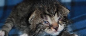
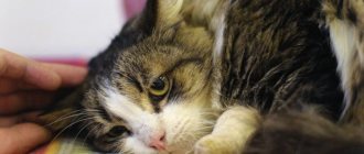
I had to face a situation where it was necessary to provide emergency assistance to a cat with an eye injury from a cat’s claw, and it turned out to be possible to make an appointment with a specialist only three days later. An ordinary veterinarian could not examine the eyeball; this requires special equipment. The eye became cloudy and a milky blur appeared.
I understood that I couldn’t wait three days; I urgently needed to provide competent medical care to the pet. While surfing the Internet, I managed to find a very interesting article on this topic. The article was written for specialists and the material is difficult for the uninitiated reader to perceive, but I really needed to understand what to do. Having carefully studied the article, it became clear that an examination is necessary, but for now, regardless of the severity of the injury, it is urgent to carry out “ forced local antibacterial therapy with fluoroquinolones (Tsiprovet 2 drops 6-8 times a day) in combination with aminoglycosides (Tobrex, iris 1 drop 6-8 times a day) and corneoprotectors (solcoseryl, Actovegin 1 drop 4-6 times a day).
«
I bought ciprovet, iris and solcoseryl, drew up an hourly treatment regimen and regularly instilled eye drops, alternating medications.
When we got an appointment with an ophthalmologist, the cat's eyeball was examined. It turned out that the lens was not affected; there was no need to operate the eye. And this is largely due to the fact that the therapy helped to avoid wound infection and significant swelling of the cornea with all sorts of ensuing complications.
The doctor prescribed observation and treatment for us:
4 times a day for another 7 days (the same active ingredient as in the drug Ciprovet, only these are eye drops for people); Indocollir 4 times a day for 7-10 days (eye drops for people);
until complete recovery;
until complete recovery;
until complete recovery.
We were treated for about a month, and as a result, everything went away, the eye was completely restored.
Below is an article that I would like to share with others in case someone needs it.
Authors:
Associate Professor A.G.Shilkin, resident V.V. Oleinik
Traumatic cataracts in dogs and cats are a serious ophthalmic pathology, usually leading to blindness in the affected eye. In severe cases, with combined damage to intraocular structures and the absence of adequate treatment, complications arising from traumatic cataracts can cause the loss of the eye as an organ.
The main factors causing traumatic cataracts are penetrating wounds and contusive injuries of the eyeball.
The purpose of our work was: Determination of rational tactics for microsurgical and conservative treatment of traumatic cataract, depending on the etiology, localization of the process and age of the animal.
Materials and methods:
Over the past 4 years, we have observed 55 animals with traumatic cataracts of various etiologies. In 43 animals, cataracts developed as a result of penetrating wounds to the eyeball. In 12 animals, traumatic cataracts occurred after contusions of the eyeball. The age of the animals ranged from 2 months to 15 years.
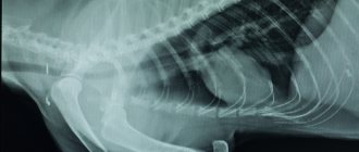
The condition of the anterior segment of the eye was determined by biomicroscopy using a head-mounted binocular microscope. The condition of the posterior segment of the eye was examined using direct and reverse binocular ophthalmoscopy using 20 and 28 D lenses. Intraocular pressure was measured using a standard method with a Maklakov tonometer. Ultrasound scanning of intraocular structures and retrobulbar space was carried out in case of clouding of the internal media of the eye (hyphema, hemophthalmos, etc.) on Honda 2000 and Ausonics devices using segmental sensors 7-7.5 MHz
So, how to provide first aid to a cat in case of various injuries?
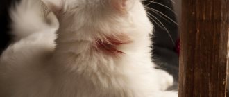
To begin the “treatment”, the pet must first be caught, since after the fight he is extremely excited and behaves aggressively. A better option may be to throw a blanket or soft cloth over the cat to prevent it from scratching your hands and face. After twisting, he will stop resisting, and you can begin providing first aid.
Sometimes a cat just needs first aid after a “showdown” with his relatives
Treatment tactics.
After an ophthalmological examination, all animals were prescribed accelerated local antibacterial therapy.
fluoroquinolones (Tsiprovet 2 drops 6-8 times a day) in combination with aminoglycosides (Tobrex, Iris 1 drop 6-8 times a day) and corneoprotectors (solcoseryl, Actovegin 1 drop 4-6 times a day). If necessary, we prescribe mydriatics or miotics (atropine, pilocarpine, 1 drop 1-3 times a day), as well as local antihypertensive drugs (timoptic, arutimol, 1 drop 2-3 times a day). Systemic therapy included cephalosporin antibiotics and fluoroquinolones (cobactan, tsifran, maxaquin).
In our clinical practice, over 5 years, we observed 7 animals over the age of 1 year, with localized opacities of the lens after penetrating injuries to the cornea and lens. In 5 of them, the injury was caused by a thin sharp object, as well as by cat claws. The damage was localized in the equatorial zone of the lens, and in 2 animals there were needle injuries in the central zone of the cornea and lens. It should be noted that all animals received therapeutic treatment; in none of these animals the injury was accompanied by the development of endocular infection
. The process of localization of turbidity and tissue regeneration took about 2 weeks. We observed these animals for 1 year or more. Throughout the observation period, the eyes remained stable, no increase in opacification or swelling of the lens was noted.
The question of the advisability of urgent surgical removal of cataracts was decided individually in each specific case, depending on the condition of the lens and the degree of intraocular inflammation.
In young animals, when there is swelling of the lens and increased intraocular pressure, surgery is performed on an emergency basis, since swelling of the cataract inevitably leads to the development of glaucoma, boutthalmos and loss of the eye. It is not advisable to wait for such complications, and delayed intervention may reduce the results of surgical treatment.
The indication for simultaneous extraction of traumatic cataracts and implantation of an artificial lens is the absence of an inflammatory reaction and the preservation of the integrity of intraocular structures, as well as the localization of the rupture of the anterior capsule on the periphery. However, the decision to undergo such surgery should be made with great caution.
Technique of operations for penetrating cataracts.
We performed 30 lens removals in animals diagnosed with penetrating traumatic cataract.
The surgical technique was determined depending on the location of the corneal injury and the depth of damage to the endocular structures.
For localized injuries in the peripheral areas of the cornea at the site of perforation, at the first stage, granulation and necrotic areas of the iris were removed with a spatula. Fresh injuries (up to 2 days) were characterized by the absence of cicatricial anterior synechiae and necrotic changes on the outer surface of the iris. The prolapsed iris was peeled off from the cornea along its entire length. A basal iridectomy was performed at the root of the iris using pointed scissors. To avoid bleeding, the jaws of the scissors were directed parallel to the course of the iris vessels. After inserting the scissors to the entire depth of the iris, the branches were carefully moved apart without cutting the iris tissue, while the area of the basal iridectomy was no more than 2 mm2. Removal of the pigment, which was quite pronounced in young animals, was carried out by irrigating the anterior chamber with saline solution. 7-8/0 interrupted sutures were placed on the cornea. The anterior chamber was restored to mild hypertension.
Then, in the opposite segment of the cornea, a cut was made with a sharp-pointed diamond knife corresponding to the expected size of the lens to be removed. The anterior capsule was opened with a cystotome. The absence of a formed lens nucleus (in young animals) is an indication for aspiration of the lens masses through an incision no more than 3 mm long, using a two-channel system. The surgical area of the cornea was closed with interrupted 8/0 sutures.
The presence of adhesions of the iris and cornea at the site of its prolapse in an area of 2-3 mm requires excision and the introduction of air into the anterior chamber in order to create hypertonicity and prevent bleeding. With prolapses of the iris of more than 3-4 mm, after its excision, massive bleeding usually occurs, which can be stopped by tamponade of air into the anterior chamber and abundant irrigation of hemorrhages with solutions of vasoconstrictors. After excision of a sector of the iris, a total hyphema is formed in the anterior chamber, minimizing the effectiveness of the surgical intervention.
The presence of a large corneal wound in the central or peripheral segment suggests a different tactic for removing the lens. Through a traumatic wound of the cornea, we remove retrocorneal moorings, fibrin clots and organized hemorrhages from the anterior chamber. Under high magnification of the microscope, we clarify the localization of the rupture of the anterior lens capsule. In case of stromal edema of the cornea, to facilitate visualization of the edge of the wound, we carefully move apart the edges of the wound with hummingbird-type tweezers, avoiding excess pressure on the cornea and lens. At the site of the detected rupture of the anterior capsule, we make an incision with pointed scissors corresponding to the direction of the corneal rupture. We aspirate the lens masses through the wound opening using a two-channel system. Once the core is formed, we expand the access to the required length to remove the lens. Using a spatula, we rotate the lens upward and move it outside the anterior chamber. This manipulation requires special care. With excessive traction of the lens, rupture of the posterior capsule and prolapse of the vitreous body are possible. We aspirate the remaining lens masses in the anterior chamber using a two-channel system. When the nucleus is formed, the number of masses is usually moderate, but their aspiration is difficult due to clouding of the cornea. Central corneal wounds are sometimes accompanied by rupture of the pupillary diaphragm. In order to form the physiological shape of the pupil, we performed open iridoplasty. After this, the surgical wound of the cornea is sealed with single 7-8/0 sutures. The distance between the sutures is 1 mm, regardless of the area of the cornea. In case of development of foci of septic keratomalacia, the affected corneal tissue is excised, and to close the defect more than 3-4 mm. We use artificial graft transplantation with simultaneous removal of traumatic cataracts. The surgical procedure is described in detail in our article “Simultaneous transplantation of an artificial cornea with cataract extraction through a small incision for penetrating trauma to the eyeball.”
Intraoperative complications.
We operated on 30 animals using both central and peripheral access to the lens through a corneal incision. The main intraoperative complications were:
1. Rupture of the posterior capsule of the lens with prolapse of the vitreous body at the time of removal of the lens nucleus with a central localization of the traumatic defect (2 animals). Both animals required anterior vitrectomy. Subsequently, panophthalmitis developed in one eye, and secondary terminal glaucoma with buvthalmos symptoms occurred in the other eye. Both eyes had to be enucleated at different times.
2. Bleeding from the vessels of the iris, to one degree or another, occurred in most animals at the time of removal of non-viable areas of the iris at the site of its prolapse. Extensive bleeding from the vessels of the iris was observed in 6 animals. In all cases, it was stopped by introducing air into the anterior chamber, applying a hemostatic sponge and abundant irrigation of the wound with solutions of angioconstrictors and hemostatics.
Postoperative period.
The phenomena of postoperative iridocyclitis of varying severity were observed in 16 animals. Iridocyclitis was characterized by the phenomena of aseptic exudate in the anterior chamber. Inflammatory reactions, in the form of pronounced fibrinous inflammation with the formation of moorings stretching from the iris through the anterior chamber to the area of the surgical incision, were noted in 4 animals with an extensive wound in the center of the cornea, necrosis of the iris, an extended rupture of the anterior capsule of the lens, and exit of lens fibers into the anterior chamber , significant time has passed since the injury 3 - 4 days. In 2 out of 4 animals, inflammatory reactions were managed with medication, by prescribing additional steroid drugs in the form of parabulbar injections. The consequence of the inflammation was the formation of posterior synechiae in 2 animals and a fibrinous pupillary membrane, which required dissection when removing the corneal sutures.
Septic panophthalmitis developed in 2 animals that were admitted 4 and 5 days after receiving penetrating wounds caused by cat claws. It should be noted that the preoperative condition was very severe, and the operations were carried out at the insistence of the owners, as an attempt to preserve the eye as an organ, without restoring visual functions. After surgery, on the 5th day, both animals, despite systemic and local antibiotic therapy, developed septic panophthalmitis, which required enucleation.
Another serious postoperative complication was the development of secondary hypertension in 5 animals, as a result of the formation of circular anterior synechiae in the preoperative period. Dissection of synechiae during surgery led to a positive result in 3 animals. And in 2 animals, the severed iris tissue formed adhesions with the posterior capsule of the lens, resulting in bombardment of the iris and pupillary block. Hypertension developed quite quickly. Drug treatment aimed at reducing intraocular pressure did not bring the desired result; boutthalmos developed; after 14 days, these 2 animals underwent enucleation.
In the remaining animals, the postoperative period proceeded according to the severity and extent of the injury received. Suture adaptation was complete. The depth of the anterior chamber in conditions of aphakia exceeded the physiological one by 2-3 mm. Corneal edema regressed starting from the 7th day and disappeared completely by 18-21 days. In animals with total preoperative corneal edema, scarring of the surgical wound was more severe and lasting. The inflammatory reaction lasted until day 10 and was characterized by some opalescence of the anterior chamber fluid and Tyndall's syndrome. There was fibrinous exudate in the anterior chamber, which resolved within up to 14 days. In 10% of animals, the presence of posterior synechiae of a local segmental type was noted. The posterior capsule remained intact in 70% of animals.
In case of eye damage
In this case, veterinarians recommend using Diamond eye drops. They are used when it is necessary to remove dried exudate in the corners of the eyes, after injury to the eyes by foreign bodies, with excessive lacrimation, in case of inflammation of the mucous membrane, and with “red eye” syndrome. They are dripped 1-2 drops up to three times a day until complete recovery.
To prevent your pet from scratching its eyes, further injuring them, we recommend putting a protective collar on it.
Protective collar
In addition, it is very useful to wash your cat’s eyes with chamomile infusion (not tea). And behind the lower eyelid you can put tetracycline eye ointment.
So, if you notice that after a fight your mustachioed fighter’s eyes are watery and this brings him discomfort, use our tips.
Contusion cataract.
Most often (80% of cases) contusion cataract is recorded in dogs of brachiocephalic breeds. The main factors causing traumatic contusive cataracts are bruises to the head and the eyeball itself, as well as non-penetrating bites from other animals.
Clinically, with an eye contusion, as a rule, eyelid hematoma, subconjunctival hemorrhages, hyphema of varying severity, secondary iridocyclitis, and hemophthalmos of varying degrees are observed. In severe cases, scleral rupture with prolapse of the choroid is diagnosed echologically. Contusional rupture of the sclera is most often localized in the equatorial zone, behind the aponeuroses of the eye muscles, where the sclera is thinnest. Scleral rupture is not biomicroscopically determined. It may be indirectly indicated by total hyphema, in combination with severe hypotony of the eye.
The condition of the lens in mild to moderate contusion injuries of the eye is characterized by the absence of a violation of the integrity of the anterior capsule. Traumatic cataract develops in a period of several hours to 10 days, depending on the nature of the contusion. With severe contusions, 30% of animals experience subluxation or luxation of the lens into the vitreous body.
Treatment tactics
For contusion cataracts, we perform surgical hyphema removal only in fresh cases (no more than a day from the moment of injury). We do not perform urgent removal of cloudy lenses.
The main treatment is aimed at relieving inflammation and vascular disorders in the intraocular membranes. To relieve inflammatory reactions, we prescribe instillations of antimicrobial drugs - Tsiprovet, Tobrex, Iris, Colbiocin, 1-2 drops. 4-6 times a day; corticosteroids – prenacid, dexamethasone 1 drop 3-4 times a day; non-steroidal anti-inflammatory drugs – naklof, indocollir 1 drop 2-4 times a day. In order to relieve the pain component and prevent posterior senechias, we prescribe mydriatics - atropine or tropicamide, under the control of intraocular pressure. Among vascular drugs we use a 1% solution of emoxypine. General treatment includes therapy with systemic hemostatic and angioprotective drugs – aminocaproic acid, etamsylate.
In case of choroidal detachment, we do not perform surgical treatment. 3 years ago, we performed 3 posterior scleral trepanation operations in dogs with TCA of 6-12 mm. confirmed by ultrasound. A positive effect was not observed in any case. On the contrary, there was a sharp exacerbation of uveitis and edema of the choroid. In 2 out of 3 dogs, after the operation, total hemophthalmos and subatrophy of the eyeball occurred.
Treatment results.
After treatment, in 6 out of 12 animals the signs of inflammation were completely relieved, the hyphema was resorbed, and intraocular pressure was within the physiological norm. In 2 animals, fibrin deposits were determined echographically in the vitreous body.
The surgical intervention was performed 2 months after the clinical signs of the inflammatory process had subsided.
Cataract extraction with IOL implantation was performed using the standard method of extracapsular cataract extraction through a corneal incision. In most animals, the surgical intervention took place without complications. In one case, due to hidden subluxation of the lens, a small hernia of the vitreous body formed in the upper segment. After anterior vitrectomy, the lens was implanted on the remains of the posterior capsule; the supporting elements of the haptic part eliminated problems with IOL fixation by 90%.
Of the postoperative complications, it should be noted that in 1 animal, reactive iridocyclitis and ASD of 2 mm were treated with medication.
In the remaining animals, the postoperative period was calm. Among the features, it should be noted that there was a slightly more pronounced fibrinous reaction in the anterior chamber, which required additional parabulbar steroid therapy.
In 4 animals observed after severe contusion of the eyeball with ruptures of the sclera and loss of the inner membranes of the eye, the post-wound process was quite difficult. Sustained hypotension, recurrent hyphema, and secondary hemorrhagic uveitis were observed. Traumatic cataract was characterized by complete clouding of the lens. One dog had grade 4 luxation of the lens into the vitreous, and 2 dogs had grade 2-3 lens subluxation. Despite the therapeutic measures taken, in 2 out of 4 animals the outcome of the contusion was subatrophy of the eyeball, which formed within 2 months. These animals underwent evisceration or enucleation.
In 2 out of 6 dogs, the post-traumatic period proceeded more favorably. Intraocular pressure was maintained within the physiological norm, and subatrophy was not observed. During the B-scan, hemophthalmos and tractional retinal detachment were visualized. In the anterior chamber, hemorrhagic uveitis and hyphema were periodic. A pupillary membrane was diagnosed on the anterior capsule of the lens.
The animals received local treatment with steroids and mydriatics for a long time. Surgical treatment was not performed on these animals due to inflammatory reactions in the anterior chamber and the presence of retinal detachment.
Help with serious bleeding
In case of bleeding from the neck or torso, it is necessary to apply a thick layer of gauze or napkins, or absorbent cotton wool and bandage tightly. In order to speed up the stop of bleeding, proceed as follows: moisten the dressing in a decoction of chamomile, or oak bark, in a solution of alum or in iodoform. Collagen sponge is considered the most effective hemostatic material.
Sometimes an animal gets seriously injured in a fight. Then you can’t do without the help of a veterinarian
Basic information
Traumatic effects can be caused by a blunt or sharp object. “Blunt” wounds are much safer, since they do not involve penetration of a foreign body into the deep layers of the eyeball. The danger of this type of damage is different. They can lead to displacement of the eyeball relative to the orbit (proptosis), displacement of the lens, bleeding, retinal detachment, and fractures of the orbital bones. In the latter case, there is a very high risk of rupture of the walls of the eyeball with bone fragments.
Accordingly, penetrating type injuries occur when the injury is caused by some sharp foreign body or object. Similar incidents often occur as a result of cat “battles” in March, when thorns, thorns from plants, grass stubble, parts of children’s toys get into the eyes (often with the “invaluable” help of the children themselves).
Of course, a cat of any gender and age can get an eye injury. But in practice, they are received either by very young kittens, or by seasoned cats that are injured during fights with competitors. Eye injuries are common in animals living near major roads. And this is not just a matter of collisions with vehicles (as a rule, in such cases the cat simply dies). It’s just that small crushed stone and other debris often fly out from under the wheels of cars, which, if they get into a cat’s eye, cause great damage to it.
The more “domestic” your cat is, the more likely it is that it will get injured if it is outside. In addition, for obvious reasons, neutered cats and female cats are much less likely to get into fights, and therefore their eyes are at significantly less risk.
Note that eye damage is not always mechanical in nature. In some cases, such pathologies may well arise due to the fault of overly zealous owners who arbitrarily prescribe potent drugs to their pets. Thus, thoughtless and unnecessary treatment with antibiotics (especially tetracyclines) may well lead to both blindness and deafness.
The animal needs to be calmed down
Remember that the best sedative is kind, comforting words spoken by the owner in a gentle and calm tone. Panic and screaming, on the contrary, will make the cat even more anxious. In addition, you can give your pet a little sedative, for example, “Cat Bayun”. When the animal has calmed down, you can wash the fresh scratches with a solution of hydrogen peroxide and wipe the wounds with a gauze pad that needs to be soaked in brilliant green. Algorithm for treating wounds.
In case of any, even the most minor injury, the wound still needs to be treated with antimicrobial drugs. We propose to do this using the following algorithm. After the bleeding has stopped, carefully trim the hair that surrounds the damaged area. If there are foreign objects in it (sand, grass, pieces of glass), they must be removed with tweezers. Next, we treat the wound with three percent peroxide or a weak solution of potassium permanganate in order to eliminate bacterial flora and remaining dirt. We recommend disinfecting the skin around the wound with a solution of brilliant green, alcohol or iodine.
This is followed by treatment of the damage with “Sanatol” or “Streptocide”. If you see a deep defect, then you first need to stop the bleeding, treat everything according to the above method and then apply a sterile bandage to the wound, the edges of which are fixed with a regular adhesive plaster.
Symptoms
Most cats diagnosed with hyphema have some degree of vision problems and discomfort. In cats, due to hemorrhages, visual acuity decreases and vision deteriorates.
Symptoms of hyphema:
- frequent blinking, squinting of eyes;
- tearfulness;
- inadequate reaction to pupil light;
- changes in behavior (depression, apathy, drowsiness, inactivity);
- pupil dilation;
- redness of the mucous membrane;
- swelling of the eyelids;
- decreased vision (monocular blindness if one eye is affected).
Important! Some cats experience acute pain and severe discomfort when opening or closing their eyes.
Actions after injury
So, what should you do if your cat scratches your eye? The best option, of course, is to go to the clinic to see an ophthalmologist. Although minor scratches usually heal on their own within a few days, there are times when the doctor prescribes medications or contact lenses to the patient. The latter are needed to reduce discomfort from injury and speed up healing.
If the cat scratched more seriously, comprehensive treatment will be required. It includes :
- Eye drops or ointment to prevent infections
- Antibiotics to block the development of the inflammatory process
- If necessary, painkillers
Today, ophthalmologists prescribe new generation medications. Medicines that combine an antibiotic and a pain reliever help much faster.
Please note that all of the above applies to pets that are regularly examined by a veterinarian. If the injury was caused by a stray cat, a visit to a specialist is required, regardless of whether the incident occurred during the day or evening. Delay may result in the development of a serious infection.
In conclusion, let us remind you that, despite the affectionate nature of our pets, cats by their nature remain predatory animals. A loving owner knows his pet well and is able to determine his mood, while avoiding cat scratches.
At first I just blinked, then my eye became inflamed and drool with bubbles came out from behind my mouth, please tell me how to treat it. DO NOT APPLY TO THE HOSPITAL, DIFFICULT SITUATION.
Woman.ru experts
Find out the opinion of an expert on your topic
Nekrasova Natalia
Psychologist. Specialist from the site b17.ru
Ekaterina Denisova
Psychologist, Medical psychologist. Specialist from the site b17.ru
Antakova Lyubov Nikolaevna
Psychologist, Consultant. Specialist from the site b17.ru
Victoria Kiseleva
Psychologist, Gestalt therapist. Specialist from the site b17.ru
Nikulina Marina
Psychologist. Specialist from the site b17.ru
Fortunatova Oksana Vasilievna
Psychologist, Psychosomatologist. Specialist from the site b17.ru
Svetlana Borisova
Psychologist. Specialist from the site b17.ru
Elena Vladimirovna Akchalova
Psychologist, Clinical psychologist. Specialist from the site b17.ru
Boyko Inessa Borisovna
Psychologist, Online consultant. Specialist from the site b17.ru
Causes
There are a number of predisposing factors that can cause a dog to suffer eye injuries.
Among them:
- Chasing cats through thickets of grass and bushes. There are branches sticking out strongly, which pose a huge danger to the organs of vision;
- Explosions of firecrackers. This is especially true during the New Year holidays. In addition to the fact that many dogs are frightened by loud, sharp noises, fireworks can get into the eye. This often leads to the pet's blindness;
- While hunting or serving. While performing service work, dogs often have to deal with sand, dry leaves, grass and branches. This carries increased risk and the possibility of injury increases. This especially applies to animal ages. Wounds and scratches regularly appear on them;
- Activity, youth and neurosis. Pets that are too nervous or easily excitable often injure their own eyes by fighting with other animals or playing with them. These factors should be taken seriously. If you have the slightest suspicion of injury, you should immediately contact your veterinarian.
Damage to the eyelids
They are divided into end-to-end and non-through. A piece of the eyelid may come off. The most effective treatment for injuries to this part of the eye can only be achieved by contacting a veterinarian on the first day after receiving the injury. If you cannot show your pet to a specialist within 2 hours, then you must immediately wash the wound. To do this you will need a solution of furatsilin 1:5000. It is safe for the animal and will not cause any negative effects if it accidentally gets into the eye.
In case of heavy bleeding, it is recommended to apply a napkin to the damaged area. It must be sterile. Carefully bandage the injury site.