Diagnosis and treatment
In most cases, the presence of a tumor can be determined during an initial visual examination. A mouth examination is mandatory at every visit to the veterinarian. If the tumor is located in visually inaccessible places, examination methods such as ultrasound or x-ray are used.
If any neoplasm is detected, a biopsy is required. To do this, a small amount of tissue is collected for analysis. This is necessary to determine the nature of the tumor - benign or malignant.
A disease such as oral cancer in cats requires surgery and removal of the tumor. The greatest difficulty is presented by malignant formations. They tend to grow into all surrounding tissues and spread quite quickly. After removal of one tumor, relapses often occur. If the malignant tumor has managed to affect a large area of tissue in the cat's mouth, then complete or partial removal of the lower jaw may be required.
The success of treatment largely depends on timely seeking medical help and the stage of the disease in the animal. After complete recovery, you should regularly examine the cat several times a year and undergo an examination by a veterinarian in order to timely monitor a possible recurrence of the tumor. Unfortunately, at the moment, oncology treatment does not always give a 100% successful result, neither in humans nor in dogs and cats.
Source
Characteristic signs of neoplasms in a cat’s mouth
A tumor in a cat’s mouth is accompanied by characteristic signs of the development of a benign or malignant disease. The most common aspects include the following:
- foul odor from the animal's mouth;
- saliva with bloody streaks is released;
- there is open bleeding in the oral cavity;
- drooling profusely;
- tooth enamel is destroyed and teeth begin to crumble;
- the muzzle takes on an asymmetrical appearance;
- the release of exudate from the nasal passages and eyes is recorded;
- the cat sneezes often;
- the pet tries to actively scratch its face against pieces of furniture and the floor;
- lymph nodes enlarge;
- the pet refuses to take hard food or bite toys during play;
- There is a decrease in appetite and body weight.
If one or more of the above signs are detected, you should take your pet to the veterinarian and undergo a detailed examination. Once the diagnosis is made, systematic treatment is required. It is very important to determine the nature of the tumor. In most cases, cats develop benign lumps, but it is still recommended to do an oncology test every time.
Squamous cell carcinoma in cats
The development of squamous cell carcinoma in cats begins with the epithelial cellular structures of the skin or oral cavity. Skin cancer is called solar dermatitis, although it is not inflammatory in nature. A malignant neoplasm in the oral cavity develops on the mucous membranes of the upper and lower jaws of the animal.
The course of squamous cell carcinoma varies, but treatment generally involves surgical excision of the tumor. For small localized pathological processes, the affected area is cut out through surgery.
If the formations are extensive, occupying large areas of the mucous membrane or epidermis, it is recommended to use radiation therapy and treatment by introducing into the body chemicals, essentially poisons, that destroy mutated cells from the inside.
Main signs of a tumor
Suspicions about the presence of a tumor in the cat’s oral cavity may arise from the following symptoms:
- bad breath;
- bloody spots in saliva;
- bleeding from the mouth;
- destruction of tooth enamel;
- excessive salivation;
- violation of the symmetry of the muzzle;
- frequent sneezing;
- nasal discharge;
- the desire to actively scratch in the mouth area;
- enlarged lymph nodes;
- avoidance of chew toys;
- weight loss;
- lack of appetite.
If one or more signs from the list are present, you should consult a veterinarian and conduct a detailed examination of the cat. But don’t panic if you find tumors in your mouth. Many of the neoplasms may be benign in nature.
General description and mechanism of the disease
Squamous cell carcinoma in cats is a commonly diagnosed disease. As a rule, the localization of a malignant neoplasm is located in open areas of the skin, where ultraviolet rays penetrate without problems.
Cancer in an animal can occur against the background of chronic inflammatory processes, long-term non-healing wound surfaces and ulcerations.
In most cases, veterinary medicine specialists diagnose tumors that arise in epithelial cells. These are single neoplasms in the form of erythematous papules, nodes or plaques. Malignant tissue proliferation is accompanied by increased peeling of the skin and bloody ulcerations. The skin around the cancer tumor will not change.
Scientists have identified a number of factors that can provoke mutation of healthy cells in the body. The main ones are:
There are two types of squamous cell skin cancer – endophytic and exophytic. The ulcerative-infiltrative or endophytic form of a malignant neoplasm is characterized by the appearance of a small spot on the skin, which quickly develops over 2-3 months into a dense, nodular, sedentary formation.
The form of squamous cell carcinoma is papillary or exophytic, growing rapidly immediately from the onset of tumor formation. It is a dark brown bulge that rises above the skin. The tumor itself is not mobile and can grow into the deepest parts of the epidermis and surrounding structures.
There are also such types of squamous cell cancer as keratoacanthoma, lesions of the animal’s oral cavity against the background of leukoplakia, verrucous form of the tumor and cancer of the skin of the lips.
Classification
Cysts, lipomas, papillomas, and lymphangiomas usually form under the tongue. The most common types of formations that form on the oral mucosa include:
- Sialadenitis.
- Salivary gland cyst.
- Lipoma.
- Papilloma.
- Hemangioma.
- Lymphangioma.
Each type of neoplasm is characterized by certain symptoms that appear as the lump grows. They also have different causes.
Sialadenitis
Appears against the background of inflammation of the salivary glands. The causes are usually viruses, infections and pathogenic microorganisms. They penetrate into the oral cavity not only with food, but also from inside the body.
In medicine, the disease is called salivary stone disease, in which a white lump forms. A stone formed in the cavity of the formation clogs the duct and causes stagnation.
Symptoms of the disease include:
- Painful sensations in the area of the salivary glands.
- Decreased salivation and constant dry mouth.
- Difficulty swallowing.
- Enlarged gland.
You may also feel pressure and tension in the area of the duct. Treatment for salivary stone disease occurs surgically.
Cyst
A cyst under the tongue is rare. The disease is characterized by the formation of a tumor in the maxillofacial area. The cavity of the neoplasm is filled with secretion. There are several types of cysts:
- Dermoid. This is a small cone of white or gray color. At the initial stage, there are no symptoms other than mild discomfort. Over time, difficulty swallowing and chewing food appears. This type is a congenital disease and is removed surgically.
- Mucous. The cone does not reach 1 cm in diameter and has a bluish tint. It disappears upon spontaneous rupture.
- Ranula. It looks like a mucous cyst and is considered a type of cyst. It has the ability to grow up to five centimeters. After the break it appears again. The tumor can be completely removed surgically.
All about the ball on the frenulum under the tongue
A ranula is a white lump with liquid inside. The reason for its appearance is the inability of the secretion to pass through the duct due to its blockage or inflammatory process.
Treatment is prescribed depending on the type of cyst. Some of them may go away on their own, while others require surgical removal. Only a doctor can diagnose the type of tumor.
Lipoma
It is formed from adipose tissue located under the mucous membrane of the oral cavity. The tumor has a benign course. Rarely diagnosed. The cone itself is always located towards the capsule and is separated by bridges.
Removal of the tumor is carried out in cases where it has reached a large size and becomes a cause of discomfort.
Papilloma
Formed from the epithelium of the mucous membrane. The cone is round or oval in shape and has a pale pink color. Papilloma can be single or multiple. In some cases it develops into a malignant formation. In medical practice, cases of spontaneous resorption are known.
Hemangioma
The lump begins to form from blood vessels as a result of embryogenesis pathology. Tissue changes are already noticeable in newborns. Has two types:
- Capillary. In appearance it resembles a small pink spot. It differs in shape, size and shade. Does not rise above the surface of the mucous membrane.
- Cavernous. When pressed, it decreases, but once again takes its previous shape. The tumor is convex and has a blue or purple tint.
Hemangioma is a congenital disease characterized by benignity and rapid growth. It may be located under the mucosa or have a part formed on the surface of the membrane.
Lymphangioma
Looks like a wart with blisters. Characterized by frequent inflammation after mechanical damage during eating. Appears more often in children of the first year of life.
In adults, according to experts, lymphangioma is formed against the background of bad habits or inflammation in harmful conditions. May have a malignant course.
When diagnosing, it is important to establish what type of neoplasm the formed lump belongs to. In certain cases, special research methods are required to establish the nature of the flow. Only after an accurate diagnosis is made, treatment is prescribed.
How it manifests itself
The symptoms of skin cancer directly depend on the stage of development of the pathological process and its type. Like any malignant neoplasm, not only in the cat’s body, skin cancer does not have a clear clinical picture in the first stages of development.
As it progresses, the following characteristic signs appear:
It is especially difficult to notice the development of a tumor on the oral mucosa in a cat. Ulcers form in the cat's mouth, constantly bleeding and, as a rule, infected. The addition of pathogenic bacterial microflora aggravates the process and causes complications.
The animal develops a foul odor from the mouth, and the process of chewing food is disrupted. It is painful for the cat to eat food, so there is a lack of body weight.
The owner of the animal may notice damage to the cat's tongue, but not pay due attention to it, referring to the usual damage to the mucous membrane. A particular difficulty in diagnosing cancer based on clinical signs lies precisely in the fact that in the vast majority of cases, oral ulcers are perceived as an infection rather than a malignant tumor.
Unfortunately, if the diagnosis is incorrect, the animal receives the wrong treatment, which at best does not help, and at worst leads to serious complications, accelerating the destructive processes carried out by the tumor.
Oral diseases: list and characteristics
Cats suffer most from gum disease that develops as a result of feeding poor quality food. Symptoms: hyperemia, swelling, ulcers, similar to signs of scurvy.
- Inflammation of the gums.
A strong odor from the mouth indicates tartar, an accumulation of food particles between the teeth. During the inflammatory process, there is a danger of tooth decay and the development of periodontitis.
Upon examination, reddened, swollen gums are visible, painful with areas of bleeding. If left untreated, the gums gradually recede from the tooth, forming pockets in which food accumulates. Putrefactive inflammation, caries, and periodontitis develop. The cat refuses to eat, becomes weaker, has unkempt fur, and saliva may flow from its mouth.
- Inflammation of the tongue.
Glossitis indicates immunodeficiency conditions, feline leukemia, acute respiratory disease, viral immunodeficiency syndrome. Sometimes the inflammatory process appears when exposed to external factors, licking highly irritating substances. Symptoms: profuse salivation, sometimes foamy, anorexia, pain. Externally, the cat looks unkempt. After healing and disappearance of the inflammatory process, the surface of the tongue is smoothed, it becomes varnished, smooth without a hard brush. Erosion and ulcers often form.
- Stomatitis.
Oral disease is manifested by severe salivation and inflammation of the mucous membrane. The cat refuses to eat, inspection of its mouth is difficult, it rubs its face with its paws and shakes its head.
Redness and swelling are noticeable on the mucous membrane, gums bleed, and there is a strong odor from the mouth. The cat seems to be “ruffling up”, looking unkempt and disheveled. In young cats, candidiasis (thrush) is diagnosed; the disease becomes more pronounced when treated with antibiotics, steroids, with low immunity, or after a serious illness. Symptoms of thrush: a whitish film on the tongue, gums; if left untreated, ulcers form on the mucous membrane.
- Pharyngitis.
This is a rare disease of the oral cavity, occurring as a secondary pathology against the background of a viral infection, diseases of the mouth and pharynx. With pharyngitis, the temperature rises, pain, cough, and nausea appear. No appetite.
- Tonsillitis.
Inflammation of the tonsils is rarely diagnosed in cats. Hyperthermia (above 39.4C), lethargy, and anorexia are observed. The underlying cause of the disease is a bacterial infection.
In case of recurrent tonsillitis, removal of the tonsils is indicated, since greatly enlarged tonsils interfere with the normal passage of air into the lungs, cause attacks of suffocation, and interfere with food intake.
- Neoplasms of the salivary glands.
Cysts and tumors form under the influence of external factors. As a rule, the salivary gland is damaged in cat fights, when a foreign object enters. The accumulating fluid ruptures the duct and a cyst is formed - a mucocele.
The submandibular gland is most often affected; when palpated under the tongue, a smooth, large cyst can be detected. Neoplasms disturb the animal, making it difficult to breathe and swallow. More often, complete removal of the damaged salivary gland is required; punctures and rinses do not give the desired effect.
In older cats, tumors are (usually) malignant and appear as hard, slow-growing lumps on the side of the face or neck.
- Foreign bodies.
Needles, bones, fragments, chips, thorns, threads get into the oral cavity. The specific structure of the tongue with papillae curved inwards does not allow a foreign object to fall out. It is easy for a breeder to understand that something has got into the cat’s mouth; she shakes her head, meows, and tries to reach with her paws what is bothering her. Severe drooling, restlessness, refusal to eat.
Particularly dangerous are the bones from fish, which dig into the soft tissues of the mouth and pharynx and “sit” there without causing signs of concern. After a few days, the bone rots, a strong odor from the mouth appears, intoxication, weakness, and apathy develop. If the outcome is successful, the bone comes out with a ruptured abscess and pus.
- Jacobs ulcer.
It is found in the middle of the upper lip, sometimes on the lower or oral mucosa. It looks like a yellow or reddish shiny spot that turns into a weeping erosion without pain or itching. Diffuse growth: the ulcerated surface increases, teeth and gums are exposed. Jacobs ulcer has a tendency to develop into cancer or fibrosarcoma. The cause of the pathology is not clear; a connection is suggested with dental infections and the feline leukemia virus.
Features of treatment
Before determining further treatment tactics, the veterinarian must conduct a thorough diagnosis. Methods for diagnosing squamous cell carcinoma are aimed primarily at identifying the tumor process in the first stages. Treatment methods and further prognosis depend on this.
If, in the owner’s opinion, suspicious spots appear on the cat’s skin, or ulcerative lesions that do not heal for a long time, it is recommended to immediately consult a specialist. In a hospital inpatient setting, the veterinarian performs a general clinical examination and medical history.
To accurately diagnose squamous cell carcinoma on the skin, several methods are used, but the main ones are dermatoscopy and biopsy. Dermatoscopy is a type of research that makes it possible to study tumors under various magnifications, as well as assess their malignant or benign nature. A biopsy of the affected areas allows you to examine the tumor tissue as closely as possible.
In addition to the main diagnostic methods, additional studies are widely used to assess the degree of destruction in the body in squamous cell carcinoma and the presence of metastases. For these purposes, X-ray examination, computed resonance or magnetic resonance imaging, ultrasound diagnostics and scintography are used.
It is worth noting that squamous cell carcinoma of the skin in cats rarely metastasizes to all organs. As a rule, in the last stages of the pathological process, nearby lymph nodes are affected. Therefore, treatment of this type of oncology involves a local type, which includes surgery to remove the affected area, radiation and cryotherapy.
For cancer of the ears and nose, veterinary experts recommend removing soft tissue and the ears themselves. The animal will have a cosmetic defect, but will have a chance to recover or prolong its life with the disease.
Chemotherapy for skin cancer in pets is rarely used because it does not produce good results. It is more advisable to inject chemicals that kill cancer cells directly into the affected area, especially if these are hard-to-reach areas such as lips or eyelids.
Chemotherapy is also used in those animals diagnosed with squamous cell carcinoma that cannot undergo surgery (severely weakened patients or elderly cats).
Prevention of squamous cell cancer of the skin and oral mucosa consists of several rules, following which the owner will reduce the chances of a malignant neoplasm. It is important to prevent direct sunlight from coming into contact with the animal’s skin, and if this is not possible, provide shade.
Do not allow your animal to come into contact with household chemicals and other toxic substances. The cat's diet should be balanced and the food should be of high quality. It has been proven that atopic dermatitis and food allergies also negatively affect the condition of the skin, like sunburn, provoking mutations in the epithelial cells of the skin.
The appearance of the first characteristic symptoms of solar dermatitis requires immediate contact with a veterinary clinic for further diagnosis and appropriate treatment. Caring for your pet can significantly increase the chances of a successful outcome of the disease, especially if it is diagnosed in the early stages.
Source
Causes of a lump under the tongue
A tumor can occur for various reasons. Usually this is mechanical damage to the mucous membrane and inflammation during chewing of hard foods. In this case, the lump will disappear on its own after some time.
What could be a growth on the gum?
Other reasons for the formation of tumors under the tongue include:
- Wearing low-quality dentures. Sharp edges can injure the surface of the mucous membrane.
- Infections. They get into microcracks formed as a result of injury and become the cause of inflammation. If left untreated, the process affects the deep layers of the mucosa, and a tumor begins to form.
- Salivary stone disease. The lump occurs in the area where the stone is formed.
- Genetic predisposition.
- The first stage of syphilis. A ball forms under the tongue, which causes pain when pressed or during conversation. After a while he disappears.
A tumor under the tongue can form against the background of the development of oncological pathologies. That is why it is important to promptly contact a specialist who will determine the cause and nature of the disease.
Squamous cell carcinoma in a cat
Squamous cell carcinoma (squamous cell carcinoma) is a malignant epithelial tumor that develops from squamous epithelium, which is involved in the formation of the skin, lining the oral cavity, tongue, esophagus, nail bed, paw pads and other organs/tissues.
Abbreviations : SCC - squamous cell carcinoma; SCC—squamous cell carcinoma; FIAB—fine needle aspiration biopsy; PDT - photodynamic therapy;
Squamous cell carcinoma in cats accounts for 15% of all skin tumors and up to 70% of all malignant oral tumors. Older cats aged 10-12 years are at risk.
Predisposing factors for the development of squamous cell carcinoma, as well as its behavior, treatment and prognosis, depend on the location of the tumor and will be discussed separately.
The main reasons for the formation of bumps in the mouths of cats
A tumor in a cat’s mouth can be caused by the following negative factors:
- Fibroma. This is a benign growth that is not difficult to detect since the lump grows right on the gum line. In this case, the color of the tumor may be identical to the gum tissue, or a little lighter. When palpating the lump, the cat does not show any symptoms of pain or discomfort. Fibroma grows quite large and can cover several teeth at once;
- Epulis. The disease is also benign in nature. It grows exclusively on the gums. It is small in size, capable of covering only one tooth. It appears on both baby teeth and permanent teeth, therefore it affects the cat’s body at any age;
- Squamous cell carcinoma. This is a malignant neoplasm. It is diagnosed quite often, so furry owners should definitely show their pet to a doctor and get tested for a biopsy. At the initial stage, the disease affects the tissues of the gums and tongue, and then spreads to the entire oral cavity. Carcinoma can penetrate any tissue, so in severe cases there is a tumor all over the pet’s face, and the head becomes simply huge.
Oral squamous cell carcinoma
Malignant tumors of the oral cavity account for 3-10% of all neoplasms in cats and squamous cell carcinoma occurs most often in this location (Bergkvist GT. et al.. 2011; Gendler A. et al.. 2010).
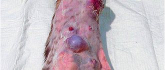
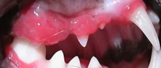
Animals may exhibit a variety of symptoms: pain, hypersalivation (increased salivation), oral bleeding, anorexia (refusal to eat), tooth loss, dysphagia (difficulty swallowing), inability to eat or groom, weight loss, and halitosis (smell). from the oral cavity).
When examining the oral cavity, you can detect an inflamed or ulcerated area of varying size and location (photo 1-3). Differential diagnoses are: eosinophilic granuloma, lymphoplasmacytic stomatitis, periodontopathy and other tumors (photo 4).
Attention! Some pathologies may be clinically very similar to squamous cell carcinoma. The final diagnosis can only be made after a biopsy!
Biological behavior
In cats, squamous cell carcinoma is a locally aggressive tumor that can occur in any part of the mouth. Unfortunately, the tumor is often noticed when it has already reached a significant size and is difficult to treat, which is due to the difficulty of examining the oral cavity in most domestic cats.
According to statistics, with conservative treatment of the tumor, the median life expectancy from the moment of biopsy is on average 33 days (Hayes AM. et al., 2007). Even with radical surgery, radiation and/or chemotherapy, the 12-month survival rate is approximately 10% (Moore AS. et al., 2001).
Distant metastases are not common; in one study of 49 cats with advanced disease, 31% had mandibular lymph node metastasis and 10% had evidence of thoracic metastasis (Soltero-Rivera MM. et al., 2014) .
Etiology (causes of occurrence)
In humans, papillomavirus is the cause of approximately 25% of cases of oral squamous cell carcinoma, and in the remaining 75% of cases, RCC develops due to exposure to carcinogens such as tobacco and alcohol (Munday JS. et al., 2013).
According to a number of studies in cats, the risk of developing oral SCC increased when eating canned food and passive inhalation of tobacco smoke for 5 years or more (Bertone ER. et al., 2003; Snyder LA. et a., 2004; McNiel EA et al., 2007).
Diagnosis and stage of the disease
Due to the presence of secondary inflammation, infection, and tumor necrosis, aspiration biopsy (TIB) is rarely used in animals with squamous cell carcinoma. An incisional biopsy is usually performed to make an accurate diagnosis, avoiding areas of inflammation or necrosis. The bleeding that occurs during this procedure is stopped using coagulation or manual compression of the tissue.
During a clinical examination of the animal, regional lymph nodes must be palpated to exclude their metastatic lesions. If necessary, a lymph node biopsy is performed.
X-rays or computed tomography of the oral cavity are performed to determine the boundaries of the tumor when planning surgery or radiation therapy. An X-ray examination of the chest organs is performed to exclude distant metastases and determine the stage of the disease.
Stages of the disease
| TNM classification of oral tumors in dogs and cats | |
| Stage | Description |
| T: size of primary tumor | |
| Tis | Tumor in situ |
| T1 – up to 2 cm in diameter (a – without bone invasion; b – with bone invasion) | |
| T2 – 2-4 cm in diameter (a or b) | |
| T3 – more than 4 cm in diameter (a or b) | |
| N: state of regional lymph nodes | |
| N0 | No signs of metastasis |
| N1 – Enlarged ipsilateral lymph nodes (a – reactive hyperplasia; b – tumor metastases) | |
| N2 – Enlarged contralateral lymph nodes (a or b) | |
| N3 – Fixed and immobile lymph nodes with metastases | |
| M: Distant metastases | |
| M0 | No evidence of distant metastases |
| M1 | There are distant metastases |
| Disease stages based on TNM classification | |||
| Stage | Primary tumor | Regional lymph nodes | Distant metastases |
| I | T1 | N0, N1a, N2a | M0 |
| II | T2 | N0, N1a, N2a | M0 |
| III | T3 Any T | N0, N1a, N2a N1b | M0 M0 |
| IV | Any T Any T | N2b, N3 Any N | M0 M1 |
Treatment Options
The approach to treating oral SCC in cats depends on the location and size of the tumor. Surgical resection can be performed in animals with tumors localized in the lower or upper jaw, as well as small lesions of the tongue.
Partial glossectomy is well tolerated by cats, but in practice it is rarely advisable to perform it due to the frequent location of the tumor in the area of the root of the tongue (photo 3). Total or subtotal glossectomy involves the removal of more than 75% of the tissues of the tongue, which is not recommended for cats because it is a mutilating operation that leads to the inability to swallow and consume food independently (Liptak JM et. al., 2012).
One study (Northrup NC. et al., 2006) evaluated the results of surgical treatment of oral neoplasms using unilateral or rostral bilateral mandibulectomy in 42 cats (see below for a schematic depiction of mandibulectomy). In half of the cases (21 animals), cats were diagnosed with squamous cell carcinoma: 12 animals underwent unilateral mandibulectomy, 5 had rostral bilateral mandibulectomy, and 4 had more than 50% of the length of the lower jaw removed. In this study, 72% of cats had temporary postoperative dysphagia, and 12% of cats were unable to adapt to eating after surgery. Despite this, the majority of owners (82%) were satisfied with the results of the treatment. The median life expectancy (even with incomplete resection margins) was 217 days. The location of the tumor is an important prognostic factor , for example, animals with rostrally located tumors had a life expectancy of 911 days, which is associated with the possibility of more radical tumor removal and rapid adaptation after surgery.
Radiation therapy alone has limited benefit in cats with oral SCC, regardless of tumor location or protocol used (Bregazzi VS et. al., 2001). For example, an irradiation protocol with a daily dose of 4.8 Gy and a total dose of 48 Gy in cats with the T1-3N0M0 stage of the disease allowed us to obtain a median life expectancy of 174 days (in animals with the T1N0Mo stage the relapse-free period was 590 days) (Poirier VJ et. al ., 2013). Results with similar median life expectancy of 86, 106 and 120 days were obtained in other similar studies (Fidel JL. et. al., 2007; McDonald C. et. al., 2012; Yoshikawa H. et. al., 2013 ).
Tonsillar squamous cell carcinoma is rare in cats and is considered an extremely aggressive tumor in dogs. However, in one study, it responded well to radiation therapy in cats and the median survival was approximately 2 years (compared to 5 months for SCC in other oral sites (Fidel J, et. al., 2011).
A number of studies have examined the effectiveness of combined chemoradiation therapy in cats with oral SCC. The results of these studies showed the relatively low effectiveness of this approach.
| Results from studies of chemoradiotherapy in cats with oral SCC | ||||
| Study | Number of animals | Radiotherapy protocol | Chemotherapy protocol | results |
| Ogilvie et al. | 11 | 44-65 Gy, 10-15 fractions | Mitoxantrone in various doses | Complete remission in 73% Median relapse-free period: 170 days |
| Jones et al. | 8 | 6 fractions of 6 GR | Gemcitabine at a dose of 25 mg/m2, 2 times a week | Complete remission in 25% Partial remission in 50% Median life expectancy 111 days |
| Fidel et al. | 31 | 14 fractions of 3.5 Gy for 9 days | Carboplatin at a dose of 90-100 mg/m2 on day 1 and on days 4-5 | Median life expectancy 163 days Cats with SCC of the tonsils and cheeks had a median lifespan of 724 days |
| Marconato et al. | 6 | 2 fractions per day for 5 days until a total dose of 48 Gy is achieved | Bleomycin, piroxicam and thalidomide | Complete remission of 50% within 759, 458 and 362 days. Two animals died from other causes. One had metastases on day 144 |
The use of mitoxantrone as monotherapy in 32 cats led to complete remission of the disease in one animal and in three cats to partial remission of the tumor within 60 days. Similar results were obtained using doxorubicin and cyclophosphamide protocols (Ogilvie et al., 1993).
It is known that oral squamous cell carcinoma cells express receptors for COX-1 and COX-2 to varying degrees, which suggests the possible treatment of this tumor with anti-inflammatory drugs. However, further research on this topic is required.
Prognostic factors
In cats with oral squamous cell carcinoma, the influence of various factors on disease prognosis was studied: histological grade, mitotic index, proliferation marker Ki67, presence of epidermal growth factor receptor (EGFR) and degree of tumor vascularization. However, the results of these studies are very variable and ambiguous, which requires further study of these prognostic factors (Bergkvist GT. et al., 2011; Yoshikawa H. et al., 2012, 2013; Looper JS. et a., 2006).
Diagnostics
In the early stages, the disease is difficult to detect, since the lump is small and does not show symptoms. A preliminary diagnosis is established based on the patient's complaints and visible signs. The doctor carefully examines the tumor to determine its characteristics.
For confirmation, a histological examination is prescribed. To do this, cells from the tissue of the cone are collected with a special instrument, and then the sample is sent to the laboratory.
In some cases, consultation with other specialists, such as an oncologist, may be necessary.
Squamous cell skin cancer
Cutaneous squamous cell carcinoma can be divided into two forms:
Pigmented areas of the skin and hair are natural barriers from the harmful effects of sunlight, so SCC of the skin occurs more often in cats with light coat color in such areas of the body that lack sun protection, such as the nasal planum, ears and eyelids. The disease is extremely rare in long-haired cat breeds with dark coat colors.
A single animal may have multiple lesions at varying stages, from carcinoma in situ to low-grade squamous cell carcinoma, as all these areas of skin are exposed to ultraviolet rays over a long period of time. Clinically, the lesions are similar to long-term non-healing ulcers and erosions. It is not uncommon for owners to think that these lesions are just an ordinary wound that for some reason does not heal and increases in size.
Etiology (causes of occurrence)
The main cause of squamous cell skin cancer is chronic exposure to ultraviolet rays.
There is an assumption that papillomavirus contributes to the development of squamous cell skin cancer in humans (Munday JS. et al., 2010). Similar studies were conducted in cats with squamous cell skin cancer, but no clear evidence of the effect of the virus on the development of skin cancer was obtained (Munday JS. et al., 2011; Munday JS. et al., 2013; Fgberink H. et al., 2013).
In humans, there is also evidence that papillomaviruses induce the development of oral squamous cell carcinoma. Increased expression of the p16 protein in people with oral squamous cell carcinoma indicates a papillomavirus etiology of tumor development (Munday JS. et al., 2011). Studies in cats have shown the presence of papillomavirus DNA in a small number of animals with oral SCC, but the significance of this finding is unknown (O'Neill SH. et. al., 2011; Munday JS. et. al., 2009).
Diagnosis and biological behavior of the tumor
To make a diagnosis, an incisional, excisional or punch biopsy is performed. Fine needle aspiration biopsy is rarely helpful due to the presence of secondary inflammation or infection. In some cases, it is necessary to carry out several biopsies from different places or lesions to make a diagnosis, since some areas may be in the precancerous stage of dysplasia.
Squamous cell skin cancer has a low tendency to metastasize. Regional lymph nodes and lungs are usually affected. In practice, tumor size and location are important prognostic factors.
To determine the stage of the disease, X-rays of the chest organs are performed in several projections and TIAB of regional lymph nodes. The presence of distant metastases greatly influences the treatment method and prognosis of the disease.
Squamous cell carcinoma in situ does not invade the basement membrane, so distant metastases are unlikely. However, over time, cancer in situ can develop into a more malignant form of cancer with invasive growth and the possibility of metastasis.
| Clinical TNM classification of skin tumors in dogs and cats | |
| Stage | Description |
| T: primary tumor | |
| Tis | Cancer in situ |
| T0 | There is no primary tumor |
| T1 | The tumor is less than 2 centimeters in diameter, exophytic or superficial. |
| T2 | Tumor 2-5 cm in diameter or minimally invasive, regardless of size |
| T3 | Tumor greater than 5 cm in diameter or with invasion of subcutaneous tissue, regardless of size |
| T4 | Tumor with invasion into underlying structures: fascia, muscles, cartilage tissue and others |
| N: state of regional lymph nodes | |
| N0 | There are no signs of metastasis to regional lymph nodes |
| N1 | Mobile ipsilateral lymph nodes (a – absence of tumor cells or b – there are signs of tumor lesions on biopsy) |
| N2 | Mobile contralateral or bilateral lymph nodes (a – absence of tumor cells or b – there are signs of tumor lesions on biopsy) |
| N3 | Fixed, immobile lymph nodes |
| M: distant metastases | |
| M0 | No distant metastases |
| M1 | There are distant metastases |
Treatment Options
When surgically treating carcinoma in situ, it is usually enough to make a small indentation from the borders of the tumor to completely remove it. If the lesions are multiple in nature or located in places difficult to access for surgical treatment, then imiquimod (Vartotsid, Keravot) can be used - this is an immunomodulator with antiviral activity. Imiquimod is used as a 5% cream to treat actinic keratoses, basal cell carcinoma and genital warts in humans.
In one study, imiquimod was used to treat Bowen's disease or carcinoma in situ in 12 cats. Treatment was carried out every other day or three times a week (the same effect was observed). All animals showed a significant response to treatment, but 9 animals developed new lesions over time and also responded well to treatment with imiquimod. Of the side effects, three cats had peeling and erythema of the skin, and one had neutropenia and increased alkaline phosphatase activity, which disappeared after discontinuation of the drug (Gill VL. et. al., 2008).
Imiquimod was used to treat sun-induced squamous cell carcinoma of the auricle in one cat, similar to the treatment of solar keratoses in humans (Peters-Kennedy K. et. al., 2008; Samaro A. et. al., 2013). However, there are many treatment options for this form of SCC in cats that have some effectiveness: surgical resection, photodynamic therapy, cryodestruction, diathermy, plesiotherapy and radiation. The choice of treatment depends on the location of the tumor, the stage of the disease and the capabilities of the doctor, clinic and owners.
If the tumor is located in the area of the auricle, then surgery is usually the method of choice. To achieve adequate resection margins, it is recommended to make an indent of 1 cm from the tumor borders. For better wound healing, it is necessary to stitch the skin edges over the ear cartilage.
When the tumor is localized in the eyelid area, it is necessary to make an indentation of 4-5 mm from the visible edges of the tumor (Ayres SA. et. al., 2012). Removing a tumor in this location often requires plastic reconstruction of the eyelid defect, which is sometimes not the easiest task. In one study, when following the recommended resection margins in cats with SCC in the eyelid area, the median relapse-free period was 319 days (range 95 to 1510 days) (Schmidt K. et. al., 2005). In another study, 5 out of 6 animals had a disease-free period of more than 12 months (Hunt Gd. 2006).
The choice of treatment for tumors in the area of the nasal planum depends on the stage of disease of the primary tumor. Invasive tumors at stage T3 or T4 may respond well to treatment with radiation therapy. The most effective way to completely cure cancer in this area is to remove the tumor tissue with a minimum distance of 5 mm from the boundaries of the tumor, which means complete removal of the nasal planum. This operation leads to a cosmetic defect, but does not significantly impair the animal’s respiratory function or quality of life. More superficial tumors in stage Tis or T1-3 can be successfully treated with photodynamic therapy, cryosurgery, strontium-90 plesiotherapy and radiation (Bexfield NH., et. al. 2008; Buchholz J. et al., 2007; Lana SE. et al., 1997, Clarke RE. 1991; Jarrett RH. et. al., 2013; Goodfellow M. et al., 2006; Hammond GM. et al., 2007; Theon AP. et al., 1995; Melzer K et al., 2006).
Photodynamic therapy (PDT) uses a photosensitizer drug that is absorbed by tumor cells. The active substance is activated by a wave of light of a certain length, which leads to cell death with minimal impact on surrounding tissue. In one study, topical application of photosensitizing cream or 5-aminolevulinic acid resulted in 85% remission in 55 cats, relapse was observed in 51% of cases, and the disease-free period was 157 days (Bexfield NH., et. al. 2008). In another study of 61 cats with cutaneous SCC, PDT with an intravenous photosensitizer resulted in remission in 49% of animals (61% had 1-year disease control), and in another study of 18 cats there was 100% remission and 1-year disease control in 75% of animals. (Buchholz J. et al., 2007). The problem with photodynamic therapy is the limited penetration of the rays and this treatment is more suitable for superficial tumors (in all studies, the best result was obtained for superficial tumors).
Various results have been obtained with the use of cryosurgery, which is due to the difficulty of assessing the exact limits of the effect. The best results were obtained for superficial tumors (stages T1-T2). The disease-free period was 254 days in a study of 11 cats with SCC in the area of the nasal planum and auricles (Lana SE. et al., 1997). In another study of 102 cats, the median remission was 26.7 months, and tumors located in the area of the nasal planum and eyelids responded best to treatment (Clarke RE. 1991).
In one study from New Zealand, 34 cats with pathologies affecting less than 50% of the nasal planum were treated with curettage and diathermy for three cycles. 16 animals had actinic changes, 9 had carcinoma in situ and the rest had full-blown squamous cell carcinoma. In 94% of animals, the relapse-free period was more than a year (Jarrett RH. et. al., 2013).
Strontium-90 plesitherapy is a successful and cosmetic therapy for stage Tis, T1 and T2 squamous cell carcinoma, as beta rays penetrate only a few millimeters into the tissue. In one study, 13 of 15 cats had a median lifespan of 692 days, and two animals that did not respond to treatment responded after the second course (Goodfellow M. et al., 2006). In another study, 88% of animals responded to treatment with a median survival time of 1071 days (Hammond GM. et al., 2007).
Radiation therapy (orthovoltage, megavoltage, electron, and proton therapy) has also been studied in cats with nasal planum SCC with similar success. Cats with stage T1 RCC had an 85% chance of surviving more than 1 year, compared with animals with stage T3 disease, of which 45% of animals survived more than 1 year (Theon AP. et al., 1995). In another study, 16 of 17 cats with stage T1-2 disease had a median disease-free period of 414 days. In this study, electrons were used (10 fractions of 4.8 Gy over 5 days, total dose 48 Gy) (Melzer K. et al., 2006).
There are publications on the use of chemotherapy in cats with RCC, either alone or in combination with radiation therapy. In one study, animals with stage T2-T4 disease were treated with intratumoral carboplatin and orthovoltage therapy. All animals responded to treatment, and the relapse-free period was 52-562 days (4 of 6 animals were in remission at the time of writing (de Vos JP. et al., 2004). In another study, 7 out of 9 animals treated with electrochemotherapy and intratumoral bleomycin had remission for up to 3 years (Spugnini EP. et al., 2009).
Immunohistochemical studies have revealed the expression of receptors for COX-2 in cutaneous squamous cell carcinoma cells, but its role is not fully understood (Bardagi M. et al., 2012).
Prognostic factors
Prognostic markers Ki67, EGFR, p16 were studied in cats with squamous cell skin cancer to assess the prognosis of the disease. In one study, a high Ki67 index was associated with a more transient response to radiation therapy (Melzer K. et al., 2006). In another study, animals whose tumors were positive for EGFR expression had a worse prognosis with a reduced disease-free period and life expectancy (Sabattini S. et al., 2010). Positive expression of p16 is associated with increased lifespan in cats with nasal planum SCC (Munday JS. et al., 2013).
Squamous cell carcinoma elsewhere
Squamous cell carcinoma can occur anywhere where squamous epithelium is present. There are publications about SCC in the area of the renal pelvis, corneal limbus, uterine stump of a sterilized cat, lungs and esophagus (Gomez Selgas A. et al., 2014; Scurrell EJ. et al., 2013; Hayashi A. et al., 2013; Berube D. et al., 2009).
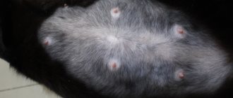
Another relatively common site of squamous cell carcinoma is the phalanges (Plates 10 and 11), which develop in older cats and often involve several toes or a large portion of the foot/hand. One study reported a median duration of only 73 days but included only 7 animals (Wobeser BK. et al., 2007).
Source
Prevention
In order to prevent the development of any oral disease, you should follow the rules of hygiene. It is also important to choose the right toothbrush, since bristles that are too hard can injure the mucous membrane.
If an infectious disease develops, you should undergo the entire course of treatment, since the inflammatory process can spread throughout the body, reducing immunity and causing the formation of a lump.
A tumor under the tongue may indicate the development of serious diseases, such as oncology, or appear as a result of improper oral hygiene. When the first symptoms occur, you should contact your dentist, who will determine the cause and extent of the disease.