The oral cavity in cats is a rather vulnerable place, which is very often subject to various diseases and inflammatory processes. Our four-legged friends have caries, periodontal disease, tartar, plaque, and gingivitis. Pathological processes lead to swelling and redness of the gums. A tumor in a cat’s mouth is not diagnosed as often as the above diseases. But the phenomenon is very dangerous and requires immediate contact with a specialist who, based on tests and hardware studies, will make an accurate diagnosis and prescribe adequate treatment. The owner should remember that growing tumors in the oral cavity in cats significantly worsen the life of the pet, preventing them from eating and being active. Along with the tumor, ailments are manifested by a number of other accompanying symptoms. Severe processes can lead to negative consequences and even death.
What it is?
This unusual term refers to a tumor of fibrous tissue of a benign nature . Despite its benign origin, this type of neoplasm often behaves quite aggressively. In particular, one type of epulis causes complete or partial destruction of not only the fibrous tissue by which the teeth are attached to their alveoli, but even the bone. Moreover, the degree of destruction is such that in severe cases it is necessary to completely (!) remove the upper or lower jaw. You understand that an animal with such an injury definitely becomes disabled, since it no longer has a chance for a normal life.
Important! There is still no reliable information about the exact causes of the disease, and therefore even prevention of this disease is impossible. The genetic and viral theories have the most supporters.
Interestingly, it is not uncommon for practicing veterinarians to encounter gum pathologies during medical examinations of animals. But there are quite a lot of aspects to their treatment, it all depends on the specific case and the condition of the sick pet. So, in some cases it is necessary to trim the gums, in others it becomes necessary to “extract” a tooth, sometimes all teeth have to be removed ... Moreover, you need to decide how much theoretically healthy tissue needs to be left along the edge of the gums. Finally, after all these issues have been resolved, the question of the degree of operability of the tumor and the need to use radiation/chemical therapy arises. Thus, epulis is a rather “complex” disease that requires a balanced and differentiated approach to treatment.
Diseases of the teeth and oral cavity
If a cat's lower jaw is swollen, the likely cause is oral disease. In older cats we are talking about dental diseases. With age, teeth begin to loosen, lose hardness, and begin to crumble. Any microcracks in the enamel lead to the development of a pathological process, which results in complete decay of the tooth. In cats, the roots of the teeth are located deep in the gum tissue, so tooth decay can lead to the formation of a seal on the lower jaw. Associated symptoms:
- refusal to eat;
- jaw turned to one side;
- lethargy and weakness;
- no pain when pressed.
Typically, animals experiencing dental problems refuse to eat. At the same time, the cat experiences severe hunger and can in every possible way attract attention to its own bowl, but as soon as it is filled, the pet will turn away and leave. This is because chewing food causes pain.
The structure of the tumor will tell you about the possible causes. If a hard neoplasm is noticeable upon palpation, but the cat breaks out and does not allow itself to be touched, a cyst of the lower jaw is a possible cause. This neoplasm can be caused by dental diseases and is an overgrowth of bone tissue in the lower jaw.
If a soft and heterogeneous structure is noted upon palpation, the possible cause is an abscess, the development of which is due to the presence of pathogenic microorganisms in the oral cavity. This happens when pathogenic agents penetrate deeply into the root of the tooth. The abscess must be opened surgically, but there are cases when it breaks out on its own.
You might be interested in: Why do my cat's ears fester?
Diseases of the oral cavity and teeth in cats are indicated by foul breath, red gums and impaired jaw movement during eating. This pathology can only be treated surgically - it is necessary to remove rotten teeth, open an abscess or cyst. If the abscess has opened on its own, antiseptic treatment should be carried out several times a day to avoid secondary infection of the wound cavity. During surgical removal, the doctor must prescribe antibiotics to prevent infection of healthy oral tissues.
Differentiation of epulis types, diagnosis
Today there are three types of the disease. In some ways they are similar, but their other characteristics can differ significantly:
- Fibrous epulis. The most common, “classic” type of the disease. It is characterized by fibrous growths along the edge of the gums.
- "Ossifying" type. It is characterized by a gradual thickening of the tumor, impregnation with mineral salts.
- The most severe type of epulis is acanthomatous. It is precisely characterized by the phenomena that we wrote about below. The pathology causes destruction of bone and fibrous tissue, as a result of which the dog can either become toothless (and this is in the best case, by the way), or remain disabled if the upper/lower jaw is removed.
Treatment methods
The presence of a tumor can always be detected by visual examination. At each visit to the veterinary clinic, the doctor must conduct a diagnosis of the oral cavity. In some cases, a tumor grows under the cat's tongue, so it is very important to elevate the organ and identify the growing tumors.
In modern clinics, doctors always take a biopsy test. The procedure involves removing a small amount of tissue from the swollen area. Only after receiving the data, the doctor determines the nature of the lump, benign or malignant.
Treatment of the disease in most cases is carried out by surgery. Only removal can quickly save the animal from the cone. If the tumor covers most of the oral cavity, the doctor performs a partial removal of the jaw (usually the lower jaw). The positive result of surgical intervention directly depends on the speed of access to the clinic. The sooner help is provided, the easier it is to prevent dangerous consequences. After the procedure, it is very important to regularly diagnose the cat’s health. Not often, but recurrent cases do occur when the tumor grows again in the oral cavity. When removing a malignant lump, it is not possible to guarantee a 100% positive result. There is always a risk that metastases have already affected a large area of tissue and the tumor will begin to grow again.
To exclude severe processes, it is recommended to bring your pet for examination and health diagnostics every six months.
Therapeutic techniques
So, how is epulis treated in cats? As a rule, there is only one way out - surgery . The affected areas of tissue are excised, and “faulty” teeth are removed. Naturally, everything is done exclusively under general anesthesia. If there is a possibility of developing a secondary infection (and in an exhausted and weakened cat it is very high), additional antibiotic therapy is resorted to. In addition, anti-inflammatory corticosteroids are sometimes prescribed, but this should only be done after weighing the pros and cons.
The main reasons for the formation of bumps in the mouths of cats
A tumor in a cat’s mouth can be caused by the following negative factors:
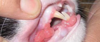
- Fibroma. This is a benign growth that is not difficult to detect since the lump grows right on the gum line. In this case, the color of the tumor may be identical to the gum tissue, or a little lighter. When palpating the lump, the cat does not show any symptoms of pain or discomfort. Fibroma grows quite large and can cover several teeth at once;
- Epulis. The disease is also benign in nature. It grows exclusively on the gums. It is small in size, capable of covering only one tooth. It appears on both baby teeth and permanent teeth, therefore it affects the cat’s body at any age;
- Squamous cell carcinoma. This is a malignant neoplasm. It is diagnosed quite often, so furry owners should definitely show their pet to a doctor and get tested for a biopsy. At the initial stage, the disease affects the tissues of the gums and tongue, and then spreads to the entire oral cavity. Carcinoma can penetrate any tissue, so in severe cases there is a tumor all over the pet’s face, and the head becomes simply huge.
Establishing diagnosis
If you find any of the listed signs in your pet, take it to a veterinarian who will provide qualified treatment. Before this, he will determine the exact cause of the development of a tumor on the cat’s gum. Set of diagnostic measures:
- External examination of the oral cavity. The presence of a neoplasm is established. This is a mandatory examination.
- Ultrasound examination or radiography. Detection of tumors in places inaccessible to visual detection. An additional purpose is the detection of metastases. The chest and skull are examined.
- Biopsy is the removal of biological material for microscopic examination. It is carried out to determine the nature of the neoplasm: benign or malignant.
The success of treatment largely depends on the correct diagnosis.
Forms of the disease
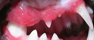
A severe form of the disease is the plasmacytic-lymphocytic form . It is caused by various viruses - rhinotracheitis, panleukopenia, hyperreaction of the immune system. This form has complications. The gums begin to bleed and swell. Without treatment, complications develop.
The dental form of the disease is accompanied by sore gums, bleeding and swelling. Diseased tissues become bluish in color. The submandibular lymph nodes begin to swell. Unlike the plasmacytic form, the disease is caused not by a virus, but by accumulated plaque. The dental form is treated faster and rarely causes complications.
Juvenile gingivitis is a form of the disease characteristic of cats under the age of 1.5 years, also called “juvenile” gingivitis. Inflammation of the gums and pharynx occurs immediately after the eruption of permanent teeth. If left untreated, periodontitis may develop.
Chronic gingivostomatitis (caudal stomatitis) in cats
Gingivostomatitis is an inflammation of the gum tissue and oral mucosa. In the chronic course, ulcers are formed, the affected areas are most often the gums, the mucous membrane of the cheek and palate, the larynx, the mucous membranes of the posterior surface of the oral cavity, the area of the palatoglossal arch and tongue.
The etiology of caudal stomatitis has not been established.
It is believed that the development of gingivostomatitis may be influenced by:
- Calcivirus (common).
- Immunodeficiency and leukemia (less common).
- Bartonella henselae (less common).
- The intensity of the inflammatory response to bacterial plaque (most often).
Clinical manifestations of the disease are unpleasant odor from the mouth (halitosis), swallowing disorder (dysphagia), refusal to eat, lack of appetite, increased salivation (hypersalivation).
To make a diagnosis it is necessary:
1. Rule out viral infections.
2. Visual examination of the animal.
3. X-ray according to indications.
4. Tissue biopsy according to indications.
Indications for which radiography or tissue biopsy are necessary are suspicion of a neoplasm.
The goal of treatment is to eliminate the causes of inflammation; more often, conservative (drug) treatment does not produce any results, therefore, in animals with caudal stomatitis, it is recommended to remove all premolars and molars; canines and incisors can be left if the inflammation of the gums has not spread. If the inflammation has spread, it is recommended to remove the entire arcade of teeth (upper and lower); radiography after complete extraction is necessary for control; if the removal is not done well, stomatitis will remain against the background of root fragments. As a rule, this gives positive dynamics in the treatment of this pathology.
If the owners refuse radical measures, you can try sanitation of the oral cavity (ultrasonic cleaning, polishing) in combination with drug treatment, there is a positive trend, but not significant, often such treatment is temporary, while taking or stopping taking antibacterial drugs there are risks of relapse big enough.
Cat salivary glands:
- The parotid gland is the largest, paired, located at the base of each ear behind the temporal bone.
- The submandibular gland is a steam gland, located under the root of the tongue.
- The sublingual gland, the ducts of which exit under the tongue of the animal.
- Molar gland - in the front part of the lower jaw with ducts emerging in the area of the front teeth.
- The infraorbital gland, or zygomatic gland, is located in the bones of the upper jaw, very close to the lower orbital circle.
Control and prevention measures
But! If the cyst is small, soft, does not interfere with the body's functioning in any way, and is filled with fluid, the veterinarian may well decide to leave it alone. And this is quite justified: an old or very young animal may not survive the operation. In addition, such a cyst (in many cases) can actually resolve on its own. Even if it ruptures, there is nothing to worry about: the liquid will flow out, and the resulting wound will heal quickly. It is much worse when the doctor suspects the infectious or parasitic nature of this formation.
Among the promising methods used in the treatment of cysts, electrocautery and the procedure of freezing the tumor with liquid nitrogen and subsequent removal of the affected tissue have proven themselves to be effective. These methods help minimize the trauma that is inevitable during surgery. Of course, when removing a cyst on the gum, you can do without anesthesia, but when treating an internal organ, it is necessary.
If your pet constantly suffers from cysts, it would not hurt to conduct a full examination, including blood biochemistry. It is possible that these measures will help identify the specific reason why tumors are constantly appearing in the cat’s body. Be sure to consult with a veterinarian or an experienced breeder and prepare a complete diet for your pet, including all the necessary vitamins and minerals. Practice shows that with normal feeding, cysts form in cats much less frequently.
Finally, let us remind you once again: if some strange swelling has formed on your pet’s stomach or in another place, be sure to show your cat to the veterinarian, since cancer cannot be ruled out, and a simple cyst may well cause many problems.
Inflammatory processes developing in the oral cavity of a domestic cat are called stomatitis. Pathology can have many varieties depending on the factors that provoked the development of the disease. One of the serious types of stomatitis is papillomatous, provoked by a viral agent.
As the papilloma virus penetrates the cat’s body, characteristic changes develop, including on the oral mucosa. This type of stomatitis is easily diagnosed - specific growths are found on the mucous membrane of a sick animal, especially in the gum area. The new growth looks like a cauliflower inflorescence.
Animals with strong immune defenses independently get rid of the manifestations of a viral infection after 2-3 months. If the animal is weakened and has a number of chronic diseases of internal organs, then surgical intervention cannot be avoided.
If you find growths on the gums in your pet’s mouth, it is recommended to contact a veterinary hospital for advice and help. In the clinic, specialists will develop an individual treatment regimen, which includes not only the removal of growths, but also the prescription of medications that suppress the activity of the virus and increase the functioning of the immune system.
Symptoms
Some diseases may not appear. For example, epulis, when small in size, does not cause much discomfort or pain in the pet. Signs of the appearance of growths and compactions:
- bad odor, often putrid, from the mouth;
- salivation (intense secretion of saliva) mixed with blood;
- tooth decay with subsequent bleeding;
- distortion of the muzzle due to an increase in tumor volume;
- itching in the mouth area, so the cat is constantly scratching;
- sneezing caused by irritation of the mucous membrane;
- refusal to feed, inactivity, lethargy;
- emaciation.
With the subsequent development of the process, the animal stops eating, the coat becomes coarser and dull, and signs of dehydration appear, since drinking water causes pain. Symptoms of the severe stage are blackening of the stool due to swallowing a large amount of blood from the affected gums; chamfering of teeth.
Other reasons
With a strong immune system, pathogens do not cause significant harm to the cat. The animal's saliva contains thiocyanate ions, which destroy bacteria, and lysozyme, an enzyme that lyses food particles and prevents microorganisms from feeding.
Lysozyme destroys pathogenic cells or slows down their reproduction by facilitating the access of thiocyanate ions.
When the body’s defenses are weakened, the oral cavity and pharynx are at the forefront; the gums (gingivitis) and tonsils are especially affected.
The formation of tartar begins with plaque; when bacteria attach, the plaque thickens and gradually increases.
Swelling of the lower jaw and lips in cats can be caused by household injuries. An older cat may accidentally scratch his lip when chewing hard food. As a result, it will swell and upon external examination it will appear that it is a swollen jaw. Burns to which animals that like to chew wires or taste food on the stove are also susceptible.
Malignant melanoma
It is a rapidly growing tumor, usually accompanied by ulceration and/or necrosis. Malignant melanoma can be pigmented or non-pigmented (amelanotic melanoma). Amelanotic melanoma is often difficult to diagnose and has a very aggressive course.
It is considered the most common oral malignancy in dogs and accounts for 30–40% of all oral malignancies in this species. In most cases, it occurs in older dogs due to some degree of oral pigmentation. Malignant melanoma is rare in cats, but its biological behavior in this species is the same as in dogs.