5263Administration
Cats are very active, move a lot and are constantly near people, vigilantly monitoring the activities of their owners. All this also requires a lot of energy and strength. And the cat’s heart copes with the function of saturating the body with oxygen and energy.
What is the function of a cat's heart? What is its structure? What is the most common disease in a cat?
© shutterstock
Heart of cats and dogs
HEART POSITION
The longitudinal axis of the heart in a dog forms an open cranial angle of 40° with the sternum, in a cat it forms an angle of 25–30°, with the apex of the heart directed towards the diaphragm. The base of the heart in a dog is oriented craniodorsally and lies approximately at the level of the fourth rib.
The right (“cranial”) ventricular edge runs along the sternum, from which it is a short distance away, the left (“caudal”) ventricular edge follows the cranial edge of the VII rib. Both edges meet at the apex of the heart, which faces slightly to the left and reaches approximately the VII costal cartilage.
Thus, the heart is located 4/7 to the left and 3/7 to the right of the midline and occupies the space of the III-VI intercostal spaces in this plane, and in the frontal plane the space between the sternum and the middle of the chest cavity.
The heart is close to the chest wall only with its left surface in the area of the cardiac notch of the left lung.
The topography of the lateral chest wall is important in such methods of clinical examination of the heart as palpation, percussion, auscultation, and radiography. The chest in its cranial part is covered by the scapula and part of the humerus, as well as the muscles associated with them.
The line of the triceps muscle, linea musculi tricipitis, is formed by the caudal border of the triceps brachii muscle and stretches from the thoracic angle of the scapula to the ulnar tuberosity. This line is clearly visible through the skin.
By pulling the thoracic limb forward, you can free the lateral chest wall in the area of the IV-VII ribs for examination.
Rice. 3. Position of the heart in a standing dog fixed in formalin (after Schummer, 1984)
A vertebra cervicalis VII; In vertebra thoracica I; With vertebra thoracica VI; D costa I; E costa VI; F scapula; Ghumerus; Hsternum; I radius; To ulna
a auricula cordis dextra, b conus arteriosus; with atrium sinistrum ef auricula cordis sinistra; d sulcus coronarius; e sulcus interventricularis paraconalis; f ventriculus dexter, g his margo ventricularis dexter; h ventriculus sinister, i his margo ventricularis sinister; k apex cordis; l diaphragm contour
1 truncus pulmonalis; 2 arcus aortae; 3 aorta thoracica with aa. intercostales dorsales; 4 v. cava cranialis; 5 v. cava caudalis; 6 a. pulmonalis sinistra, 6′ vv. pulmonales; 7 ligamentum arteriosum (Botalli); 8 a. subclavia sinistra; 9 truncus brachiocephalicus; 10 a.m.
subclavia dextra; 11a. carotis communis dextra; 12 a.m. carotis communis sinistra; 13 a.m. vertebralis; 14 v. jugularis externa sinistra 15 a. et v. axillaris sinistra, 15′ a. et v. thoracica interna; the dashed line corresponds to the caudal edge of m. Triceps brachii (linea mi.
tricipitis or anconaea)
Knowledge of the topography of the heart allows the use of various methods of intravital heart diagnostics.
The apical impulse of the heart, which occurs as a result of contraction of the heart muscle and leads to a shaking of the lateral chest wall, in a dog is heard clearly on the left in the lower third in the IV-VI intercostal space, especially well in the V intercostal space, and slightly worse on the right in the IV-V intercostal space .
Using percussion, as a rule, it is possible to detect an area of absolute cardiac dullness in the part of the heart not covered by the lung on both sides in the IV-VI intercostal spaces. The dorsal border of this area is formed by the symphyses of the IV-V ribs.
Auscultation of heart sounds or pathologically altered heart murmurs in a dog should be carried out in the 5th intercostal space for the left ventricle and in the 4th intercostal space for the right ventricle.
For intracardiac injection, only the right ventricle is suitable and the needle should be inserted from the right side into the 5th intercostal space as close to the sternum as possible.
In a cat, the caudal edge of the triceps brachii muscle runs parallel to the IV intercostal space, the heart is located closest to the chest walls on the left side between the IV and VI ribs, on the right side - under the V rib.
In these places, the apical impulse of the heart is clearly perceived and heart sounds are clearly audible. The 5th intercostal space is also suitable for intracardiac injection in cats.
In a standing cat, this point is located directly above the elbow tubercle.
Size and weight of the heart in carnivores
Table 1. Heart mass in dogs of various breeds (according to Ballmer, 1937)
| Breed | Absolute mass | Average mass | Relative mass |
| Saint Bernard | 200—500 | 301, 0 | 0, 64% |
| Great Dane | 130—470 | 293, 1 | 0, 71% |
| Cop | 100—350 | 233, 8 | 0, 78% |
| Setter | 100—200 | 158, 6 | 0, 73% |
| German Shepherd | 100—300 | 199, 6 | 0, 75% |
| Airedale Terrier | 100—300 | 185, 0 | 0, 76% |
| Schnauzer | 40—150 | 95, 6 | 0, 71% |
| Spaniel | 30—120 | 92, 8 | 0, 76% |
| Spitz | 15—100 | 58, 4 | 0, 76% |
| Dachshund | 40—100 | 75, 2 | 0, 73% |
| Miniature Pinscher | 10—80 | 48, 0 | 0, 70% |
| Fox terrier | 24—120 | 67, 7 | 0, 73% |
Table 2. Mass and size of the cat’s heart (according to Sichert, 1935)
| Floor | Body weight, in g | Heart mass | Diameter | District hearts, in mm | ||
| absolute, in g | relative, in % | sagittal, in mm | transverse, in mm | |||
| male | 3360 | 18, 4 | 0, 55 | 33, 0 | 24, 3 | 90, 3 |
| female | 2480 | 12, 7 | 0, 51 | 25, 5 | 19, 3 | 77, 6 |
Structure of the heart wall
The heart wall consists of three layers: the epicardium, epicardium, myocardium, myocardium, and endocardium.
The epicardium corresponds to the visceral plate of the serous pericardium. It covers the surface of the heart with a smooth, shiny, transparent membrane.
The outer layer of the epicardium consists of mesothelium, the cells of which, depending on the degree of stretching of the heart, have a flat or cubic shape.
Below them is the lamina propria, lamina propria, in which bundles of collagen fibers are arranged to follow the changes in the shape of the heart.
This is followed by a subepicardial layer of collagen and elastic fibers associated with the connective tissue skeleton of the cardiac muscle. Large vessels and nerves pass through this layer. In the coronary sulcus and in areas of the interventricular grooves close to the base of the heart, there are deposits of adipose tissue, the amount of which depends on the age and fatness of the animal.
The red muscle layer, the myocardium, consists of membrane-covered longitudinal muscle cells of a special structure with a nucleus located in the center. Individual cells are connected to each other through processes extending at sharp angles and form a kind of network.
Lines of adhesion, or intercalary plates, disci intercalares, unite muscle cells into more extensive systems of fibers covered with sarcolemma, sarcolemma.
Those, in turn, form bundles united into a three-dimensional mesh, the cells of which are filled with delicate connective tissue - endomysium, endomisium.
This internal connective tissue of the myocardium in some places continues into the collagen fibers of the tendons of the papillary muscles, connecting the muscle fibers of the papillary muscles with the tendon strings of the leaflet valves. The number of blood capillaries in the myocardium approximately corresponds to the number of muscle fibers.
The endocardium is a membrane of connective tissue and elastic fibers and a layer of endothelium that lines the internal cavities of the heart and valves. The endocardium is movably connected to the base, which allows it to adapt to changes in the stretching of the walls during cardiac activity.
Source: https://privetstudent.com/referaty/biologiya-referaty/page,2,1074-serdce-koshek-i-sobak.html
Normal organ sizes
The size and weight of the heart are not constant and depend on many factors:
- The age of the pet plays a big role (in old dogs the organ often becomes flabby and enlarges).
- Floor. In males, the mass of the organ is up to 20% greater than in females.
- Animal fatness.
- Physical activity. Service dogs and hunting dogs regularly used for their intended purpose have a noticeably larger heart.
It is also known that in the embryonic state this organ (relative to body weight) is simply huge. This is easily explained by the fact that at the stage of fetal maturation, the puppy’s heart pumps blood not only throughout its own body, but also through the placental network.
Congenital and acquired heart diseases in cats
Pets are susceptible to various ailments from birth and throughout their lives. Among internal non-contagious pathologies, heart disease in cats occupies one of the leading places.
According to veterinary statistics, every tenth pet suffers from heart disease to one degree or another.
A feature of myocardial abnormalities is the hidden nature of the disease, difficulty in diagnosis and long-term treatment.
Types of pathology
The heart is a unique type of muscle tissue that can contract throughout the life of the body. Due to these contractions, blood supply occurs to all organs and tissues of the animal. The health of the heart muscle, myocardium, determines not only the cat’s life itself, but also its quality. Disturbances in the functioning of the heart in animals are both congenital and acquired.
About what to do if a cat’s kidneys are enlarged. You will learn about the reasons that cause their increase, diseases, the manifestation of which is a change in the size of the kidneys, diagnosis and treatment. And here is more information about what to do if a cat’s kidneys fail.
Congenital
Congenital myocardial defects are diagnosed by veterinarians in approximately 1% of newborn babies. In this case, pathological underdevelopment of the heart valves, especially the mitral valve, impaired development of the arterial ducts, as well as pathologies of the conduction of nerve impulses through the organ are most often observed.
Anatomy of a healthy cat heart
Breeds such as the Maine Coon, Abyssinian cat, as well as popular British and Scottish breeds have a genetic predisposition to congenital heart pathologies.
Veterinarians diagnose the following congenital defects of the heart muscle.
| Congenital heart defects in cats | Short description |
| Atrioventricular atrial septal defect | The most common congenital pathology among newborn kittens. With this type of defect, the fusion of the septum between the atria is disrupted. As a rule, the anomaly is accompanied by insufficient development of the mitral valve. The prognosis is unfavorable. |
| Patent ductus botallis | Normally, the Botallian duct, which connects the aorta and pulmonary artery, closes a few days after the animal is born. With a genetic defect, the closure of the ductus arteriosus does not occur, which leads to blood entering the pulmonary vessels. As a result, the vasculature and the left side of the myocardium are overloaded. If the right side of the heart is not involved in the process, then the prognosis is positive. |
| Subaortic stenosis (narrowing of the aorta) | A rare congenital pathology among representatives of the feline family. Heart disease in cats is associated with the development of pathological tissue below the aortic valve, which complicates the work of the heart and increases the load on the left ventricle. Unlike other congenital pathologies, subaortic stenosis does not lead to early death of the body, but significantly worsens the quality of life of the pet. |
| Tricuspid valve dysplasia | As a result of the pathology, complete closure of the atrioventricular valve is impossible, which leads to reverse flow of blood into the atrium. The right side of the organ increases in size, congestion and heart failure develop. Pathology is rarely diagnosed in cats. |
Animals with congenital anomalies of the organ, as a rule, die in the first year of life due to the fact that the structure of the cat’s heart is changed. This is due to severe overload of the myocardium, the development of heart failure and concomitant vascular diseases. Defects such as aortic stenosis and tetralogy of Fallot are serious diseases with a poor prognosis.
If congenital anomalies are detected in the offspring, the parents are excluded from the breeding program.
Appeared with age
Myocardial diseases often develop gradually and appear in adulthood or closer to old age. Experts attribute this to the fact that cats are not very active animals and do not experience serious physical exertion. With age, couch potatoes may develop the following pathological conditions of the heart muscle:
- Pathologies of inflammatory non-infectious nature. Often, such diseases are a consequence of the use of medications (antimicrobial agents, painkillers and anti-inflammatory drugs). Cats most often suffer from myocardosis. At the same time, dystrophic processes develop in the heart muscle. There are many causes of the disease: unbalanced feeding, concomitant non-communicable diseases, helminthic infestations.
- Cardiac pathologies of an infectious nature. Transferred viral and bacterial infections lead to the fact that pathogenic microorganisms enter the heart muscle through the bloodstream due to the weakened immunity of the animal. The most common diagnoses in cats are myocarditis and endocarditis. The disease is observed in both acute and chronic forms.
The cause of the pathology is septic processes, pancreatitis, panleukopenia, purulent processes in the uterus, as well as acute intoxication.
- Hypertrophic cardiomyopathy . This disease is accompanied by pathological thickening of the myocardium, which leads to the development of severe heart failure. The pathology is widespread among representatives of the cat family, especially among Maine Coons, Scottish Folds, Norwegian Forest cats and representatives of the British breed.
The disease develops gradually. Thickening of the myocardium leads to stagnation and enlargement of the left side of the organ. At later stages of development of the pathology, pulmonary edema, the appearance of fluid in the pleural area, and death may occur. A timely diagnosis can extend the life of a sick animal for many years.
Secondary cardiomyopathy develops against the background of diseases of other organs, most often due to hyperthyroidism. High levels of thyroid hormones lead to constant tachycardia, as a result of which the heart wall thickens and the organ works under overload.
A complication of the disease in the form of thromboembolism poses a threat to the pet. Blockage of large vessels usually leads to death.
As for cardiac arrhythmias, veterinary specialists do not classify these disorders (except congenital) as a separate group of diseases. Most often, arrhythmia occurs due to a malfunction of the endocrine system and itself leads to the development of cardiac pathology.
For information about hypertrophic cardiomyopathy in cats, watch this video:
Symptoms and signs that should alert you
It is quite difficult to suspect heart problems in a domestic cat. This is due both to the lack of activity and physical activity, and to the blurred clinical picture of the onset of the pathology. However, owners should be aware of the symptoms of heart disease in cats:
- Decreased motor activity, lethargy, apathy, drowsiness . The cat, feeling weak, moves less.
- Rapid breathing . Normally, a cat makes 20 - 30 breathing movements per minute. If the number of inhalations and exhalations is more than 40, you should show the animal to a veterinarian.
- Heavy breathing, shortness of breath . The animal experiences difficulty breathing both during rest and during increased physical activity. The owner often observes the pet breathing with its mouth open and its tongue hanging out. In this case, breathing movements are carried out not by the chest, but by the stomach. Often, a cat takes a pose of oxygen starvation: crouching on the ground with its belly, the pet stretches its head and neck forward. Elbows placed at the sides improve oxygen supply to the chest.
- Decreased appetite . During meals, the animal holds its breath, which, against the background of oxygen starvation due to heart pathology, leads to poor appetite in the pet.
- Pale or blueness (cyanosis) of the mucous membranes.
- The owner can observe how strongly the cat's heart beats as the disease progresses by placing his palm on the chest area. Tachycardia accompanies a number of cardiac pathologies, including myocarditis and endocarditis.
- In the later stages of the disease, the owner may observe a loss of consciousness in the pet . Fainting is caused by impaired blood supply to the brain.
- An unfavorable symptom is paralysis of the hind limbs . Thromboembolism (blockage of large blood vessels) is often a complication of hypertrophic cardiomyopathy.
Unfortunately, characteristic clinical symptoms in cats become apparent already with the development of heart disease, when the pathological process has affected not only the myocardium, but also other organs and systems. Often, a serious pathology goes unnoticed by the owner, and only sudden cardiac arrest in a cat indicates that the animal was sick.
Signs similar to heart pathology
Uncharacteristic clinical manifestations complicate the timely detection of heart pathology in a pet. In addition, similar symptoms may be a sign of various systemic diseases.
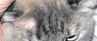
Enlargement of the left lobe of the thyroid gland
Tachycardia, for example, develops in diseases of the thyroid gland and often accompanies chronic intoxication. Vague symptoms such as poor appetite, apathy, and weight loss are characteristic of many diseases.
Worm infestation and lung diseases can masquerade as cardiac pathology. A symptom such as ascites (accumulation of fluid in the abdominal cavity) is characteristic not only of heart disease, but also of cancer. Only a veterinarian can make a correct diagnosis, based on special research methods.
Diagnosis of the condition
It is possible to establish cardiac pathology and make a diagnosis only in a specialized clinic. Veterinary specialists, after collecting anamnesis (information from the owner), use the following techniques and methods for diagnosis:
- Auscultation using a phonendoscope . Allows you to listen to the work of the heart muscle, determine the presence of tachycardia, and identify heart murmurs.
- Tonometry . Allows you to identify deviations in blood pressure.
- Electrocardiography (ECG) allows you to recognize circulatory rhythm disturbances.
- Radiography . The most informative method, thanks to which a veterinary specialist determines congestion in the heart muscle and the presence of pulmonary edema. Pictures are taken in two projections.
- The main method for diagnosing heart disease in veterinary practice is ultrasound. Using echocardiography, you can determine not only the size of the organ, but also the thickness of the muscle walls, size, shape, condition of the atria, and the diameter of large heart vessels (aorta and pulmonary arteries). Ultrasound detects blood clots, determines the rhythm of contractions, and evaluates blood flow.
It is heart screening in cats using ultrasound that makes it possible to identify pathology without the presence of clinical signs, at an asymptomatic stage. The owner should resort to this procedure if the pet is more than 7 years old, as well as before each surgical intervention that requires the introduction of narcotic sleep.
What does the doctor recommend?
Treatment of cardiac pathologies in domestic cats does not involve surgical intervention. Treatment of heart disease in cats is based on conservative methods using medications.
First of all, the sick animal is prescribed rest. Stress plays an important role in the development of heart disease. A sick animal should be protected from unnecessary worries and a calm, friendly environment should be created.
Good nutrition plays an important role in maintaining normal heart function. Balance of nutrients, enrichment of the diet with vitamins and biologically active substances helps to improve the condition of the pet.
Drug treatment is prescribed depending on the type of pathology detected. Heart medications can be used in courses for life.
The main goal of cardiac therapy is to facilitate the work of the myocardium, supply the heart muscle with oxygen, nutrients, and improve blood flow. If necessary, medications are used to normalize blood pressure.
For pronounced arrhythmias, therapy uses medications that correct the heart rhythm.
We recommend reading about how cats die. You will learn about the signs that the inevitable is approaching, the actions of the owner, the reasons why pets leave home, and whether you should go to the veterinarian. And here is more information about how many times a day to feed a cat.
Prevention of disease development
To reduce the risk of heart disease in cats, veterinarians recommend following these tips:
- Maintaining the animal's muscle activity. The cat must play and move. Physical inactivity leads to the risk of developing myocardial diseases.
- Controlling your pet's weight. Obesity is one of the factors leading to heart failure.
- Screening studies before surgery using anesthesia, as well as once a year to diagnose the disease in the preclinical stage.
Heart pathologies are often the cause of ill health and sudden death of an animal. Diagnosis is made difficult by the peculiarities of the course of the disease and the lack of expression of clinical signs. Screening cardiac examinations are the only method for early detection of heart disease.
Source: https://zootvet.ru/bolezni-serdtsa-u-koshek/
Prevention
Cats suffering from heart failure need preventative care to keep them active. We need to try to “stir up” animals that lead a “sofa” lifestyle. Obese cats are at risk for heart disease. You need to ensure your pet is eating properly. An annual checkup with a veterinarian will protect him from heart disease.
Sphynx, British, Persian, Scottish cats, and Maine Coons are also susceptible to heart disease. This does not mean that all cats of these breeds, sooner or later, have heart problems. This statement means that representatives of these breeds develop heart disease at an earlier age.
Heart failure in neutered cats is quite common because these animals are very lazy. They lead a sedentary lifestyle and are obese.
It is necessary to pay increased attention to neutered cats, as they are more susceptible to heart disease.
A diagnosis of heart failure in cats is not a death sentence. The main thing is to notice the first symptoms of the disease in time, carry out regular examination and treatment. Monitor your pet's diet. With proper care and care, a cat can delight its owner with affection and beauty for a long time.
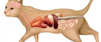
Internal structure of a cat
The location and functioning of the internal organs, as well as the internal structure of the cat, are in many ways similar to the structure of other mammals. However, there are also differences that are unique to this species of animal.
The main organ of the circulatory system is the heart - a hollow muscular organ located inside the chest, namely behind the median sternum. It is worth noting that the weight of a cat's heart depends on body weight, namely 0.6 percent of body weight. The heart consists of two atria and two ventricles.
A cat has 2 circles of blood circulation . Blood circulation occurs through arteries leading from the heart to capillaries, penetrating all internal organs and tissues.
Metabolism takes place there, then blood, which is saturated with carbon dioxide and waste products of the body, flows through the veins leading to the heart. Veins form the 2nd, pulmonary circulation .
Venous blood enters the right ventricle of the heart, then through the pulmonary arteries into the lungs.
A cat's respiratory system is designed to function well in a variety of environmental conditions. The task of the organs is to ensure gas exchange and oxygen delivery to the tissues of the body. They also play the role of excretory organs, through which excess moisture and harmful gases are removed, and also participate in heat exchange, removing excess heat from tissues.
A cat's respiratory system consists of:
- nose,
- larynx,
- nasopharynx,
- trachea,
- bronchi,
- lungs.
The lungs are the main organ of the respiratory system.
A paired organ that consists of 2 lobes that occupy most of the chest.
They consist of alveoli - that is, pulmonary vesicles that are tightly intertwined with a network of capillaries, serving as conductors during gas exchange.
Something else interesting: Cat skeleton
In cats, the respiratory organs are covered with a mucous membrane that serves as their protection.
When breathing, air enters the larynx through the nose, and from there into the bronchi and lungs.
If the body or environment temperature is elevated, or the animal is in an excited state, its breathing rate increases. There are times when a cat breathes with an open mouth.
The cat's digestive system consists of:
- oral cavity,
- throats,
- esophagus,
- stomach,
- small and large intestines,
- pancreas,
- gallbladder,
- duodenum.
The cat's chewed food from the oral cavity enters the esophagus, which is a muscular tube that can increase in diameter if necessary to push food into the stomach.
The esophagus is covered on the inside with mucous membrane.
Saliva allows food to be partially broken down while still in the mouth. Next, the digestion process continues in the stomach, which is located in the front of the peritoneum.
The cat has a single-chamber stomach, covered from the inside with a mucous membrane, which produces gastric juice necessary for the subsequent processing of food.
2 cone-shaped openings open from the stomach, the 1st of which connects the stomach with the esophagus, the 2nd with the duodenum.
Food from the stomach enters the small intestine, where the final processing of food occurs. The small intestine is a long thin tube twisted into several loops. Its length is 4 times the length of the cat's body.
Food inside the intestines is affected by a pancreatic enzyme.
From the small intestine, unprocessed solid food remains enter the large intestine, where they are enveloped in mucus, which is secreted by the walls of the large intestine.
It consists of 3 elements:
- cecum (appendix),
- colon,
- the rectum, which removes compressed feces from the body.
A cat has anal glands on the sides of the anus, which secrete a secretion with a pungent odor. In addition to the excretory function, the rectum maintains bacteriological balance in the animal’s body.
Something else interesting: Biological characteristics of cats
The organs of the urinary system are needed to remove excess fluid from the animal’s body. Consists of:
- kidneys,
- bladder,
- urinary tract - ureters.
Urine is formed in the kidneys, or more precisely, in the renal pelvis, from which it flows through the ureters into the bladder, where the closing muscle prevents spontaneous urination.
urethra is different: stenoses are special narrowings that serve to facilitate the faster passage of sediment present in the urine.
The reproductive system in cats consists of testicles and vas deferens, which open into the urethra. Through it, sperm enters the reproductive organ.
Testicles are the sex glands of cats. They are located in the scrotum, which is a fold of skin at the base of the penis. They produce male reproductive cells, namely sperm.
Internal genitalia of a cat:
- Ovaries. They produce eggs.
- the fallopian tubes;
- uterus.
External genitalia of a cat:
In cats, the endocrine glands are of great importance for life:
- Adrenal glands;
- Hypothalamus;
- Thyroid.
Source: https://ourkoshki.ru/stroenie-koshki/vnutrene-stroenie-koshki.html
Prevention
The most important prevention is changing the animal’s lifestyle to a more active one.
If a cat does not eat properly and, as a logical consequence, suffers from obesity, these are the main enemies for her heart. Therefore, an attentive owner should also ensure that the cat has plenty of healthy (!) food.
If a cat does not eat properly and, as a logical consequence, suffers from obesity, these are the main enemies for her heart.
Also, for prevention, after acquiring (or if the owner found it or took it for free, but after this acquisition), the pet must be examined at a veterinary clinic; in case of any risk, the cat must be checked at least once a year.
Cats with a “sofa” lifestyle are more likely to develop diseases such as heart failure.
Treatment consists of drug therapy, which will make the heart work easier, eliminate pain, improve blood circulation, and also nourish the myocardium, since, unfortunately, heart surgery is not performed in cats.
Structure and functions of the cat's heart
Cats are very active, move a lot and are constantly near people, vigilantly monitoring the activities of their owners. All this also requires a lot of energy and strength. And the cat’s heart copes with the function of saturating the body with oxygen and energy.
What is the function of a cat's heart? What is its structure? What is the most common disease in a cat?
© shutterstock
Function and principle of action of the heart
The heart is an internal pump, a muscle that pumps blood, supplying the cat’s body with oxygen and nutrients and removing carbon dioxide from the organs. In this case, one side of the heart, the right, pumps blood only to the lungs. The left one pumps blood to the rest of the cat’s body organs.
Interesting information about cat blood. It turns out that the formula and composition of a cat’s blood does not correspond to any other animal.
Blood clots very quickly and its reaction to some chemicals is unique (this point is still being studied). But that's not all. Scientists have found that the composition of the blood of each cat is different, just like that of humans.
There is a certain system that divides blood into groups. This is all that the researchers have found out so far.
The essence of the heart's action is to force blood to move through the cat's body. The heart pumps blood into the lungs. There it is enriched with oxygen and returned to the muscle.
Then it is sent through the vessels throughout the body, and some of it seeps into the tissues and collects in the lymphatic system. In this system, the blood is purified by passing through the glands and receives a certain number of lymphocytes. And all this movement is driven by the heart pump. It’s worth getting acquainted with the structure of this pump and finding out what the structure of a cat’s heart is.
© shutterstock
Anatomy of the heart
The structure of the cat's heart muscle looks the same as in birds, and in humans, and in any mammal, it consists of 4 parts, chambers:
- The upper chambers are called the left and right atria.
- Next comes the middle layer. This muscle is called the myocardium.
- The lower hollow chambers are the ventricles.
Blood moves through the body due to the contraction of the ventricles.
What is the size of a cat's heart? It is believed that while the anatomy of the structure and arrangement of organs is similar to other animals, the cat’s heart is small. Depending on the breed and age, we can say that it occupies only 2 intercostal spaces - this is a very small size. And the heart doesn’t look special or big from the outside; it weighs from 12 to 15 grams.
If the cat’s body is healthy, it has a sufficient amount of hormones and everything is normal with the autonomic system, then this affects the main organ, and the cat will not have heart problems.
The rhythm, the force with which the heart muscle contracts and the frequency of contractions, the work of the vascular system, its contraction and expansion, all depend on whether the cat has certain problems or hormonal heart disease.
How do you know if there is a disease in the heart itself? Several factors may indicate this. For example, a cat’s weight exceeds the norm. A symptom may also be that the cat is tired, or that after intense games her heart almost jumps out of her chest. This, if not a sign of the disease itself, is a reason to start worrying and start checking.
Cat heart disease
What do pet owners need to know about what heart problems there are in cats and how to recognize them? Most often, furry owners encounter a problem such as cardiomyopathy; the causes of the disease are different, and therefore there are several varieties :
- Hypertrophic;
- Dilation;
- Restrictive;
- Intermediate.
© shutterstock
Types of heart failure
According to the characteristics of the process, this disease can be divided into acute and chronic. Typically, cats are characterized by chronic insufficiency.
Acute heart failure
Occurs in people with a sudden disruption of the blood supply to the myocardium (infarction). Cats do not have the main factor that leads to blockage of coronary vessels - atherosclerosis. This feature of the cat family lies in their diet, which does not clog its blood vessels with cholesterol plaques. Heart attacks occur due to a sharp imbalance in the functioning of the nervous and endocrine systems during severe stressful situations. The stress response can be so severe that it causes the heart to stop completely.
The most stressful (and deadly) veterinary procedure is the operation of pulling out claws, which leads to disability of the pet due to the removal of the first phalanges of the fingers.
Chronic heart failure
It often affects our pets, regardless of breed and age.
Good to know
- Can cats hide pain?
- Serval cats
- Urinary retention in cats
- Causes of pain syndrome in cats
- Feline Orofacial Pain Syndrome
- Catnip for cats
- How to care for an old cat?
- How to properly dry a cat?
- How to Choose a Kitten for your Home
- Pneumonia of Cats
- Causes of Sneezing in Cats
- Anatomy and physiology of the liver in cats
- The structure and functions of the thyroid gland in cats
- Measuring blood pressure in cats
- Current role of dexmedetomidine in clinical veterinary and medical anesthesia and intensive care
- Thyroid function testing in cats
- Gabapentin in cats and dogs
- Information content of hematological and echocardiographic preoperative screening indicators
- Historical background on hyperthyroidism in cats
- Hyperthyroidism in older cats
- Cardiac hypertrophy in cats
- Analysis of anamnestic data during preoperative examination of animals
- Study of anamnestic data in cats before general anesthesia
- Method for measuring blood pressure in cats
- ICD in cats (diagnosis and symptoms)
- Hyperthyroidism is suspected, but serum T4 concentration is normal
- Acute gastroenterocolitis in dogs (etiology, pathogenesis, diagnosis and treatment)
- Reducing mortality during anesthesia is an important task of veterinary anesthesiology
- Thrombosis in cats (general information, etiology, pathogenesis, clinical picture, diagnosis and treatment)
- Urolithiasis of small animals (etiology, pathogenesis, diagnosis and treatment)
- Congestive cardiomyopathy in cats (What is it? How to protect your pets)
- Gastrointestinal tract in dogs (anatomy and physiology)
Alarming symptoms
Normally, the anatomy of a cat's heart is not very different from a human's, except for the size of the organ, and it consists of 4 chambers. The pet's internal organ is located above the ulnar tubercle. Owners are not always able to recognize heart pathologies on time, since many diseases do not show any clinical picture for a long time.
With myocardial disease, the animal is forced to stick out its tongue while breathing.
Most heart pathologies are characterized by the following symptoms:
- decreased motor activity;
- lethargic and apathetic state;
- constant desire to sleep;
- rapid breathing, in which the cat takes more than 40 breaths per minute;
- shortness of breath not provoked by physical activity;
- protruding tongue when breathing;
- problems with appetite;
- pale and blue visible mucous membranes;
- tachycardia, in which the heart beats rapidly.
Muscular system
Cats have an unusually developed muscular system. This is proven by their amazing jumps over fairly long distances and fast running. Also, a set of muscles helps the cat maintain its aristocratic bearing.
- heart muscle;
- smooth muscles, which control internal organs and work involuntarily;
- striated muscles that the cat controls itself.
Special fibers are part of all muscles. Cat muscles contain 3 types of cells:
- are strongly contracted, but work for a short time - thanks to them, the cat is able to jump long distances; the strength of these cells is not able to act for long;
- with a strong contraction they work for a long time - a cat has few such cells, which explains its inability to run long distances;
- they contract quietly and work for a long time - this type of muscle cells is used in a cat when hunting, when it sits in ambush for a long time, and also sneaks quietly and gently.
To take a step, the cat pushes off with its hind paws, and the front paws participate in the braking process. Thanks to the elasticity of the back muscles, the cat can easily curl into a ball and assume other bizarre poses.
Causes
Epilepsy attacks in cats do not have a breed or gender predisposition. But statistically, more cases are registered in exotic animals, where a modification of the skull is observed, and cats suffer from this pathology more often than females. There is no explanation for this - just statistics. The disease can be transmitted genetically, but not necessarily directly from one generation to another, maybe through a generation or two.
The most important causes of epilepsy are disturbances in the conduction activity of nerve impulses in the brain. They are acquired during life, or cats are born with them. Various additional factors act as provocations of attacks. These factors are cerebral (structural pathologies of the brain) and extracerebral (toxic effects on the brain without primary changes in its structures).
- Structural changes in the brain: vascular pathologies, hemorrhages, inflammatory processes with edema, trauma, neoplasms, etc.
- External factors: poisons and toxins, hypoxia (oxygen starvation), jumps or drops in blood enzyme levels, etc.