When talking about common feline diseases, one cannot fail to mention cancer. Yes, unfortunately, animals, like people, have a fairly high risk of developing cancer. A tumor of the mammary gland in cats is quite common, and in four out of five cases the disease takes a malignant course. This serious illness can be completely cured only with early diagnosis. The owner should closely monitor the health of his pet and, if a small lump or lump appears in the mammary glands, be sure to contact a veterinary clinic for advice.
What is this?
A mammary gland tumor (MBT) in cats is a neoplasm that can be benign or malignant. Unfortunately, in 9 out of 10 cases it is malignant, and most often it is a fairly aggressive carcinoma.
The risk group includes adult cats - from 10-12 years old. Oncology is less common in young animals. However, it is impossible to determine the nature of the tumor during routine examination and palpation. To make an accurate diagnosis, you will need to undergo mandatory studies and tests.
Tumor diagnosis
In order to establish the shape and type of neoplasm, a veterinary specialist, in addition to a general clinical examination and palpation, will prescribe the following diagnostic methods:
- general and biochemical blood test (to assess the general condition and identify concomitant pathologies);
- ultrasound and x-ray examination of the chest (allows us to identify not only the location of the main tumor, but also the presence of metastases);
- biopsy or fine needle aspiration of damaged tissues, as well as lymph nodes, followed by cytological analysis.
The type of tumor can also be determined by histological examination of the affected tissue. This examination helps determine treatment options and prognosis for the animal.
We recommend reading about when you can sterilize a cat. From it you will learn about the best age for sterilization, periods when it is not recommended to sterilize a cat, operations during pregnancy and after childbirth. And here you will learn about how to care for a cat after sterilization.
Causes of tumor appearance
The causes of mammary tumors in cats are not fully understood. However, very often the disease is diagnosed in pets taking hormonal medications aimed at suppressing sexual arousal.
As a result of disruption of the functioning of the endocrine glands, severe hormonal disruption occurs. It entails improper functioning of internal organs, including the organs of the reproductive system. In 99% of cases, animals that have not had their ovaries removed are susceptible to cancer.
Note! Another common reason is a genetic predisposition.
It is formed when a mutated gene is transmitted, leading to uncontrolled cell division.
Preventive measures
To prevent neoplasms and early diagnosis of diseases, you should:
- Sterilize your cat before her first heat;
- do not use hormonal drugs without the advice of a doctor;
- see a veterinarian annually;
- monitor the cat’s weight, do not overfeed;
- provide active exercise in the fresh air.
A cat can lead a full life even with a diagnosis of a tumor and live long enough if the owners treat it in a timely manner.
Please rate the article. We tried our best:)
Main signs and stages
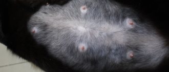
Symptoms of mammary tumors in cats do not appear in the early stages. The exception is neoplasms that have the form of small nodules and are located in the abdominal area. Unfortunately, most owners do not notice these changes.
Signs of a malignant tumor:
- Stage I
The size of the tumor ranges from 0.3-0.5 to 1-1.5 cm. When palpating the abdominal area, you can feel a slight “graininess”. At this stage, the pet is not bothered by pain. His activity remains and his appetite is normal.
- Stage II
New growths rapidly increase in size - up to 2-3 cm. New, not yet enlarged, lesions can be palpated on nearby mammary glands. However, the pet's condition is normal and he does not experience any discomfort.
- Stage III
The size of the neoplasms reaches from 3 to 5 cm. The behavior and mood of the pet changes. More often he is depressed and refuses food. When palpating the abdominal area, it reacts painfully. An ammonia smell appears.
- IV stage
Ulceration of the tumor usually occurs. It grows noticeably and begins to penetrate nearby tissues. An unpleasant odor appears. The pet's condition worsens, weight loss and difficulty breathing are observed.
If you notice one or more signs of cancer in your pet, contact an experienced veterinarian immediately. Do not try to make a diagnosis or prescribe treatment at home, otherwise you will not be able to avoid irreversible consequences.
Stages of cancer in cats
Cancer develops very quickly. The problem is that in the first stages the disease does not manifest itself in any way and does not interfere with the animal’s life. There are several stages of development of a malignant tumor:
- At the first stage, the size of the seals does not exceed one centimeter in diameter. The tumor can be detected by palpation, but the cat itself is not yet bothered by its presence;
- The second stage is characterized by an increase in the tumor up to 3 centimeters. Metastases do not appear at this stage;
- The tumor reaches 5 centimeters in diameter. At this stage, the formation of metastases is possible, which most often affects the lymph nodes located next to the mammary glands;
- The tumor is constantly growing, and metastases reach the liver, lungs and other organs.
Diagnosis of AMF
If you notice any changes in your cat's mammary glands, do not hesitate - make an appointment at the veterinary clinic. Only a specialist can make an accurate diagnosis for your pet.
The first stage of diagnosis is an external examination. Particular attention is paid to the area where the mammary glands are located. The doctor probes each lesion and gland so as not to miss changes. If necessary, a biopsy (examination of cell samples) is prescribed.
Additionally, blood samples are taken for biochemical and clinical analysis. This will allow you to find out how the internal organs function and whether there are accompanying pathological processes in the body.
The next stage of tumor diagnosis is an X-ray examination of the lungs, which allows one to exclude or confirm the presence of metastases in them. If the cancer spreads to the lungs, the cat may have difficulty breathing and develop shortness of breath.
Symptoms
Adenoma of the third century
– a pink tumor forms on the eye in the area of the external lacrimal canal, which stands out strongly against the background of the eye and eyelid. The formation irritates the surface of the eye and the disease is accompanied by the complication of conjunctivitis.
Sebaceous gland adenoma
– has a benign course and rarely causes discomfort in cats. Most often located on the head, tumors can rarely be found on the body or paws. The appearance of cauliflower is characteristic - in the form of a warty formation, hard, raised above the surface. They can be pink or yellow and even brown, they have no hair. They appear greasy, with a shiny, oily surface. There are often many adenoma nodules located on a cat.
Breast adenoma –
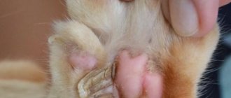
Dense, knotty joints can be felt on the cat’s chest. They are not fused to the skin and have a round shape (unlike cancerous formations). There may be several of them in the mammary gland, and their sizes increase over time. Often the gland is deformed and enlarged. The main danger is possible necrotic-purulent transformation or malignancy.
Pituitary adenoma –
the tumor is a hormone-producing tumor and changes in the body are associated with overproduction of sex hormones, glucocorticosteroids and mineralcorticoids.
Depending on the part of the affected gland, several types of diseases can occur:
- Cushing's disease is associated with increased production of cortisol, which is produced by the adrenal glands. Since the pituitary gland has a direct influence on them, when it is damaged, secretion may increase. One of the first signs is increased thirst and urination. Then comes the “symptom of fragile skin.” The skin is easily damaged, becomes dry and thin, and over time, its pigmentation and sagging folds are noticeable. Wounds and abrasions quickly become inflamed, leading to purulent complications. Changes in the fur are observed - it is disheveled, and focal alopecia may appear. The disease is also characterized by muscle wasting, lethargy and weakness.
- Acromegaly – due to overproduction of growth hormone, there is a change in the size of the paws, lower jaw and skull in cats. They grow slowly, often becoming noticeable after a few years. The disease causes non-insulin-dependent diabetes, so its first symptoms are polyuria, increased thirst, and obesity. The disease is also accompanied by an increase in internal organs and endocrine glands.
- Panhypopituitarism is a disease that is accompanied by a decrease in hormone production due to compression of the pituitary gland structures by the adenoma and loss of any functions. The disease is accompanied by hypothyroidism, hypocortisolism and hypogonadism. The production of only one hormone may be reduced; these are called “isolated”. Such a decrease leads to atrophy of the thyroid and gonads, as well as the adrenal glands. Cats' character changes, they become withdrawn and often hide. As the tumor grows, headaches occur and pressure on the optic nerve increases, which can cause the cat to go blind.
Often the tumor can be asymptomatic and diagnosed during routine examination. In almost all cases, adenoma is accompanied by diabetes mellitus, which can make it difficult to diagnose and find out the true cause of the disease.
Adrenal adenoma
– with this neoplasm, the diseases observed are the same as with the pituitary due to the close connection of the endocrinological system – the pituitary-adrenocorticoid system. Insulin-resistant diabetes mellitus occurs first. Cushing's disease is also possible.
When mineralcorticoid production is affected, a decrease in potassium levels and arterial hypertension are observed.
Overproduction of sex hormones leads to feminization and infertility.
Treatment of breast tumor
What to do if your cat has a mammary tumor? Immediately after diagnosis, the specialist will develop an individual treatment plan, taking into account the pet’s age, the characteristics of its body, the nature and stage of tumor development.
The main goal of treatment is to destroy tumor cells and stop their division. This will not only prevent the further development of oncology, but will also completely eliminate its recurrence in the future. Among the main treatment methods:
- surgical intervention,
- radiation therapy,
- cryodestruction,
- chemotherapy,
- hyperthermia, etc.
The choice of treatment method depends on the location of the tumors and their sensitivity to medical intervention. If necessary, combination therapy is prescribed, combining several types of possible treatment for the pet.
Characteristic symptoms
The main symptoms appear when the disease has already entered the advanced stage. At this stage, the animal’s general well-being worsens and its appearance changes. The tumor may appear as single or multiple nodes. The inguinal and axillary lymph nodes are inflamed. The lesion may involve several lobes of the mammary gland. Sometimes its true size can only be assessed after shaving the fur over a fairly large area of the body. The main clinical signs at this stage are:
- the neoplasm is significant in size;
- there is quite severe inflammation of the surrounding tissues;
- the cat is in quite a lot of pain;
- body temperature may rise;
- the animal loses weight sharply, there is no appetite;
- Bleeding and discharge of pus from the opened tumor are possible.
If a cat's mammary gland is swollen and painful, this is not always associated with cancer. Very often, some non-tumor conditions of the mammary glands have similar signs. Basically, these are hyperplasias (tissue growths) of various etiologies and some other conditions:
- hyperplasia of the gland ducts;
- breast cysts;
- lobular hyperplasia;
- fibroadenomatous hyperplasia;
- false pregnancy;
- true pregnancy;
- consequences of the administration of progesterone hormone drugs.
Surgical removal
The most common way to treat a mammary tumor in a cat, whether benign or malignant, is surgery. Sometimes it is enough to remove a local lump (neoplasm), but usually it is necessary to remove the entire mammary gland and the lymph nodes draining it.
If a malignant tumor affects two or more mammary glands, then they are removed together - the entire row on the left or right side. In particularly severe cases, a bilateral mastectomy may be prescribed, i.e. removal of all the mammary glands of the cat and the lymph nodes draining them.
Sterilizing an animal before puberty significantly reduces the likelihood of tumor formation. In some cases, it is performed already during the treatment of oncology. Sterilization does not affect the growth of a malignant tumor, but can lead to regression of dysplasia.
In the final stages of cancer, all mammary glands and lymph nodes have to be removed. However, even in this case, the pet’s complete recovery cannot be guaranteed. Additionally, systemic chemotherapy may be prescribed before/after surgery.
Treatment
Since an adenoma is a benign neoplasm, in the absence of discomfort and pathological growth it is simply observed. This management of the disease is especially typical for sebaceous gland formations, but only after cytological examination and confirmation of benignity.
Laser or cryological removal of an adenoma is also possible if it causes discomfort, a significant cosmetic defect or trauma.
Breast adenoma is removed in the same way as cancer. For complete removal, a study is carried out and the quality is determined. This technique is needed to exclude malignancy in the future. The only difference is the absence of chemotherapy. It is also advisable to carry out sterilization with complete removal of the uterus and ovaries to stabilize hormonal levels.
Lacrimal gland adenoma is also treated surgically under general anesthesia. The blockade is also done locally, by instilling lidocaine into the eye and injecting a mixture of novocaine and adrenaline into the formation itself. After 10 minutes, the tumor is cut off without suturing. To prevent complications after surgery, instillation of chloramphenicol solution is recommended.
In case of pituitary adenoma, its complete removal is recommended. This approach often leads to the elimination of diabetes and normalization of hormonal levels. This type of operation is quite complex and is performed using the latest microsurgical equipment.
Alternatively, if tumor excision is not possible, adrenal gland removal is performed with lifelong administration of glucocorticosteroids and mineralcorticoids as replacement therapy. To stabilize the condition and prevent sepsis, blood clots and poor wound healing, adrenal hormone synthesis inhibitors - metyrapone, ketoconazole or trilostane - are prescribed.
Adrenal adenoma can only be surgically removed, with a complete ectomy of the gland. If surgery is not possible, methothane or Vetoril is used to suppress excess secretion of adrenal hormones.
What should the owner do?
If your cat is diagnosed with a mammary tumor, you will need to prevent rubbing, scratching, or licking the affected mammary glands until surgery. This will reduce:
- inflammatory processes,
- itching,
- ulcers,
- risk of infection.
Note! Any damage to your pet's skin must be kept clean.
After surgical removal of a benign or malignant lesion, ensure that the surgical suture remains dry and clean. If inflammation or suture separation is detected, immediately contact your veterinarian.
Possible causes of breast swelling and swollen nipples in a cat
New growths of the mammary gland that affect a cat are malignant and benign in nature. In addition, they differ in the characteristics of their growth. Benign tumors under the skin develop slowly, but eventually reach impressive sizes and do not metastasize. A malignant compaction in the area of the mammary glands causes metastasis, the development of necrotic processes, as well as intoxication of the body. Cats with this disease do not live long. Benign pathologies of the organ are mammary hyperplasia. It manifests itself in the following forms:
- Focal;
- Fibroepithelial, cystic.
Moreover, both types develop due to high levels of progesterone in the cat’s blood, which is typical for unsterilized females.
Timely treatment of benign pathologies has a favorable prognosis if it was an unopened cyst or a complicated formation. In this case, there is a significant risk of wound infection. In total, benign tumors are detected in 15% of all pathologies in this organ.
Veterinarians give an extremely cautious prognosis for such a tumor due to the fact that when it reaches a size of more than 3 cm, it is extremely unfavorable. This is due to the frequent development of metastases in other organs, as well as the brain. This disease is also characterized by a high chance of relapse, even if it has already been operated on.
Inflammation can be caused by the penetration of pathogenic microorganisms into the gland; a tumor often indicates the development of mastitis and mastopathy. Hormones have a great influence on the condition of the mammary glands. Hormonal imbalances also often lead to swollen nipples and the appearance of a variety of discharge. Most often, cats develop cancer. The risk of cancer increases in animals aged 10–12 years.
If the glands are swollen, this does not always indicate the presence of pathology. Natural causes of this condition are pregnancy and breastfeeding. Nipple enlargement occurs in the fourth week of pregnancy. This sign is especially well expressed in nulliparous females.
If the female is not pregnant, then most likely the tumor is caused by pathological processes. They can occur as a result of hormonal imbalances, the use of hormone-based drugs, poor ecology, and poor nutrition. The most common breast diseases are:
- neoplasms;
- mastitis (serous, purulent);
- mastopathy;
- false pregnancy.
Postoperative care and restrictions
Most cats are discharged from the hospital after surgical removal of the tumor after 2-5 days, depending on their condition. You must make an appointment with an oncologist 12-16 days after the operation so that the specialist can re-examine the patient and remove the sutures.
Restrictions during the rehabilitation period (10-14 days):
- collar or blanket,
- limitation of mobility,
- proper nutrition,
- caring for the suturing area.
If the neoplasm is malignant, you will need to visit a veterinary clinic every 3 months.
Prevention of mastopathy
In order not to endanger the pet, the owner needs to take care of its health in advance:
- provide a balanced diet and dosed drinking to increase immunity weakened by childbirth and form proper lactation;
- carefully inspect and feel the milk bags of a nursing cat; if it is very swollen or painful, express;
- Trim kittens' claws to reduce the risk of nipple injury;
- timely deworming, getting rid of fleas, antiseptic treatment of scratches and injuries;
- stop using medications containing hormones and hormone-like substances;
- exclude contact between your pet and infected animals;
- Use products made from natural materials as a lounger.
If mastopathy is suspected, veterinarians recommend unscheduled mating. If the owner is not committed to caring for the offspring, the best solution is to sterilize the cat before the first heat. We must not forget about regular preventive examinations with a veterinarian. A simple procedure will save the owner’s nerves and the health of the four-legged household.
What you need to know about benign tumors?
Benign tumors of the mammary glands are diagnosed in cats in 1% of cases, precancerous conditions - in 20% of cases. They are usually formed from the epithelium that produces milk; less often they affect other glandular tissues.
If your cat is diagnosed with a benign tumor, do not refuse treatment. Over time, they can become malignant (for example, under the influence of a certain group of diseases). The most effective treatment is surgical removal of tissue in the early stages.
Diagnostics
The only way to determine the presence of a tumor with one hundred percent accuracy is a biopsy. The procedure is carried out very carefully, since tissue injuries can lead to the development of metastases. Along with the examination of the mammary glands, an examination of the lymph nodes is carried out.
Other diagnostic methods are also used for examination: CT, MRI, ultrasound. A blood test is performed to determine the stage of cancer.
Once the diagnosis is confirmed, the animal is prescribed chemotherapy. Biochemistry testing allows you to determine the type of treatment that is appropriate for a particular animal.
Cancer prevention
To reduce the likelihood of developing cancer in your cat, it is necessary to carry out sterilization in a timely manner. The recommended age for a pet is before the animal reaches puberty. At the same time, sterilization does not 100% exclude the appearance of neoplasms.
As soon as the animal reaches the most dangerous age (from 8 years), it is recommended to undergo a thorough examination at a veterinary clinic - at least once every six months. This will allow you to identify the tumor in the initial stages and completely cure your pet.
Proper care, attentive attitude and professional veterinary care are the best prevention of cancer for every cat!
Types of tumors
Among all cases of tumors in cats, more than 80% are malignant. This type of disease tends to develop very quickly, spreading metastases into the animal’s body almost at lightning speed. Often, compaction occurs in several places at once. The rapid growth of the tumor causes a deterioration in the pet’s condition, which can result in death.
Benign tumors are characterized by slow growth. They are localized in a specific tissue without affecting neighboring organs. In this case, no necrotic processes are observed, which is why the type of disease is called “benign.”
The onset of the disease in both cases is characterized by the same symptoms: a small compaction appears in the cavity of the mammary gland, which does not bother the animal at the initial stage.


