Polycystic ovary syndrome is a common reproductive system disease that occurs in cats of different ages and breeds. Most often it is caused by hormonal imbalance or unprofessional sterilization. Without timely and proper treatment, polycystic ovary syndrome is dangerous not only to the health, but also to the life of your pet.
Here you can learn all the most important things about the common causes, symptoms and treatments for this disease.
See this article:
Polycystic ovary syndrome - what is it? Why are multiple cysts dangerous? Causes of the disease Main symptoms What to do if your cat has polycystic ovary syndrome? Disease prevention: useful tips
Types of cysts in cats and places of their occurrence
In veterinary medicine, the following classification of cysts in domestic animals is accepted, based on the nature of the contents of the blisters.
Atheromas are widespread in the cat population.
Based on location, veterinary specialists distinguish the following types of neoplasms:
- Epidermal tumors. This form of cyst-like cavities is characteristic of the outer layers of the animal's skin. Cysts are soft swellings with clear boundaries that do not cause concern to the pet. Most often, epidermal neoplasms are observed on the back and side surface of the body. Often the cyst breaks out on its own, releasing a paste-like content of gray-yellow color.
- Hair cyst. It is usually formed when the sebaceous glands of the skin become clogged and inflamed and is diagnosed in animals after 5 years of age. Such a cavity is most often filled with keratin contents.
- Dermoid cystic tumor. It occurs most often in young individuals and is localized in the auricle. It is innate in nature. The cavity of the dermoid formation is filled with a sebum-like secretion. The cyst has a spherical shape, is soft to the touch, and does not cause pain to the animal.
- Internal cysts. Neoplasms can affect many organs: ovaries, kidneys, uterus, liver. Internal tumors are not as harmless as external ones. Polycystic kidney disease is dangerous due to the development of secondary bacterial infection and the development of sepsis.
- Mucocele . A separate type of tumor that occurs when the salivary ducts are blocked. The disease is divided by localization into sublingual, pharyngeal and zygomatic mucocele. This type of neoplasm often degenerates into an oncological tumor.
- Breast cyst . The most common formation in domestic cats. The main reason for the formation of this type of pathological cavities is hormonal imbalances in the female’s body.
The variety of cystic tumors often frightens owners. Most cysts are benign. However, some formations can degenerate into malignant tumors and threaten the health and life of cats.
Life forecast
Predicting future life depends on the aggressiveness of the pathology, the state of the immune system, stage, and age of the pet. A good prognosis is given to cats in which polycystic disease was detected in the early stages of development and undergoing treatment. Modern screening techniques for young individuals allow them to live with changes in the kidneys for a long time.
Checking kittens
An unfavorable development of events threatens cats with symptoms of chronic kidney failure and large areas of damage to paired organs. The deterioration in survival statistics is associated with:
- with pain syndrome that arose at an early age;
- arterial hypertension that cannot be controlled with drugs;
- secondary infectious process in the kidneys;
- regular appearance of blood particles in the urine;
- other chronic diseases.
How long does the process last: depending on the aggressiveness of the course, polycystic disease can last from 3-4 months to several years. The literature describes cases where a sick pet (with proper nutrition and symptomatic therapy) lived for at least 14 years.
Animals at risk
The reasons for the formation of cystic cavities are not fully understood. It is believed that some types of cysts are caused by a hereditary predisposition, while others appear due to a violation of the hormonal status of the body. The conditions of keeping and feeding the pet are also important.
Veterinary specialists and experienced breeders believe that the following pets are at risk:
- Unsterilized cats. Estrus, pregnancy, childbirth, and feeding offspring are often accompanied by disruptions in the female’s hormonal system.
It is the disruption of hormonal status that is the main cause of the development of polycystic ovary syndrome and cystic tumors of the mammary gland.
- The use of hormonal drugs to control estrus and sexual behavior is a serious blow to the cat’s endocrine system. Veterinary experts consider the use of tablets, drops, and injections based on sex hormones to be the main cause of the development of ovarian and mammary cysts in domestic cats.
- Injuries and mechanical damage. If the integrity of the skin is damaged, a keratin-like substance can penetrate into the layers of the skin. Epidermal cysts often develop through this mechanism.
- Breeds such as the Persian, Himalayan, British Shorthair and Exotic Shorthair are at risk for developing polycystic kidney disease.
- Keeping pets in poor hygienic conditions and the presence of thorny indoor plants provokes the development of hair and epidermal tumors.
- Violation of feeding rules. Eating bones is a risk factor for the development of mucocele in the domestic cat.
Clinical symptoms of a cyst in a cat
Identifying an external neoplasm in an animal is not difficult for an attentive owner. Typically, the disease has the following symptoms:
- A spherical or spherical compaction is visible above the surface of the skin.
- On palpation, the tumor is often soft and elastic. In the case of a keratinized formation, the cavity is dense when palpated.
- The animal does not experience pain on palpation.
- The general condition of the animal is usually satisfactory. The cat does not lose its appetite and physical activity.
- When opening the cavity, a complication in the form of wound infection is possible. In this case, the symptoms will depend on the nature of the inflammatory process.
Mucocele has characteristic features. The owner observes excessive salivation, poor appetite, and loss of body weight in the cat. With the development of the pathological process, saliva contains blood impurities.
It is not difficult to detect mammary cysts in a domestic cat. At the first stages of development, the pathological formation does not cause concern to the pet. However, breast tumors are prone to rupture and infection.
Damage to internal organs is much more difficult for the owner to determine. So, polycystic kidney disease has no obvious symptoms. Clinical signs of the disease are similar to those of renal failure. The animal becomes lethargic and urination is delayed.
Polycystic kidney disease in a cat
Symptoms
Cysts are not malignant neoplasms. This is a benign process that is not characterized by the property of pathological growth. Symptoms of the pathological process depend on the location of the neoplasm, its size and type. Cysts are characterized by clear, regular shapes, and inside the bladder there can be both the secretion of the sebaceous glands and serous fluid, up to bloody exudate or pus. Cystic neoplasms can become inflamed, provoking the development of suppuration processes.
Cysts of the epidermal type are rarely diagnosed in cats in veterinary medicine. They are characterized by soft structures, reaching about 5 cm in diameter, without pain. Epidermal cysts are characterized by spontaneous rupture with specific fluid leaking out. In the presence of pathogenic bacterial microflora, infection occurs, followed by a purulent-necrotic process.
The second type of cyst, dermoid, is diagnosed in the head area of a pet. They are denser, there is no fluctuation. Dermoid cystic neoplasms are characterized by long-term development. Exposure to strong traumatic factors leads to the formation of fistulas with further release of blood and purulent exudate.
Symptoms of an ovarian cyst in a cat, which should alert the owner and prompt a visit to a veterinarian, are:
- problems during periods of sexual heat - more than 4 times a year or vice versa, less than 2 times;
- heat period lasting longer than 14 days;
- the cat's inability to become pregnant;
- problems during the period of gestation of kittens and during the birth process - weak labor, miscarriages.
Benign neoplasms in the ovaries can be single or multiple. They are provoked by disturbances in hormonal balance and further systemic disruptions in the functioning of the reproductive system. As a rule, the vast majority of clinical cases in veterinary medicine do not have pronounced symptoms, although a strong increase in cysts leads to compression of internal organs and provokes pain. When injured, cysts can rupture, leading to peritonitis.
Characteristic features of cysts formed on renal structures are their size. They are small - from 0.1 to 1 cm in diameter. They can increase in size, gradually replacing healthy kidney tissue, causing the development of failure. Chronic renal failure in the absence of timely treatment leads to the death of the pet.
Benign neoplasms can be located in the pancreas, uterus or liver structures. Signs of cysts on internal organs include cat lethargy, urinary problems, and loss of appetite.
With a salivary gland cyst, the signs are increased secretion of salivary fluid, swelling of tissue structures. Depending on which gland was affected by the cyst, localized symptoms may occur. The lesion may be in the area of the pharyngeal, sublingual or zygomatic gland. The danger lies in the fact that this particular type of cyst from benign neoplasms can turn into malignant.
Cysts located in the area of the sublingual gland are characterized by the following symptoms: impaired chewing, impaired appetite, and the appearance of blood in the saliva. Cystic neoplasms in the area of the pharyngeal gland, in addition to problems with swallowing food, are characterized by impaired breathing. Cysts of the zygomatic gland are manifested by swelling under the eyes. With the development of pathology, damage to vision and hearing occurs.
Diagnosis of cyst formation in an animal
In a specialized institution, your pet will not only be diagnosed, but also the type of cystic formation will be determined. Modern veterinary medicine has the latest techniques. The sick animal will first undergo a biopsy. Using a puncture, the veterinarian will puncture the pathological bladder and take its contents for examination.
The procedure is important for the differential diagnosis of a cyst from an abscess, inflammatory infiltrates, and various types of malignant formations.
Histological examination of biological material allows for a correct diagnosis.
If the development of cyst-like cavities in the internal organs is suspected, the veterinarian uses the following methods for diagnosis:
- Ultrasound examination of the kidneys, ovaries and other internal organs. Diagnostics allows the animal to painlessly determine the presence of pathological blisters in the ovaries, kidneys, uterus, liver, estimate their number, and determine their exact location.
Ultrasound of a cat: polycystic kidney disease
- An X-ray examination of the abdominal organs allows one to identify pathological structures in the kidneys and uterus. An X-ray of the cat's head will allow us to determine the exact location of the mucocele.
For a differential diagnosis, methods such as general and biochemical blood tests, determination of hormone levels, and urinalysis can be used.
For information on diagnosing polycystic kidney disease, watch this video:
Treatment of cysts in cats depending on location
If a formation of any size is detected, the most important thing is to consult a doctor. Only he can know exactly how to act in a given situation, which examination will more accurately show the problem, and also prescribe the most effective treatment.
Should I always delete
Surgical treatment does not always have indications. If the animal is elderly or weakened by disease, the operation can be dangerous.
Contraindications to tumor removal are often concomitant diseases or conditions in which anesthesia is undesirable.
If surgical intervention is not possible, fine-needle puncture is performed. The procedure makes life easier for the pet with large tumors and reduces the risk of spontaneous opening and infection.
Surgery as an option to get rid of a cyst
The most effective way to cure a sick animal is surgical removal of pathological cavities. Excision of the tumor occurs within healthy tissue. The capsule must be excised.
Treatment
Once a pet has been given a disappointing diagnosis of a tumor, removing the cat’s mammary tumor is often the only way to save his life. If the pathology is benign, then after removal surgery there will be an almost complete recovery. But it is important to do this before inflammation or ulceration begins.
In cases where surgical treatment is not possible for certain reasons, conservative methods are used. Thus, for benign pathologies, the essence of such therapy will be to stop the growth and development of the pathology, as well as to prevent infection of the abnormal tissues. In addition, it is necessary to increase the animal’s immunity level.
For this purpose, various antitumor and antibacterial agents are used. This could be Dokosorubicin. Also, if there is cancer in a pet, Cyclophosphamide is used, which has a cytostatic effect. Non-steroidal anti-inflammatory drugs are also prescribed, which include Meroxicam, and painkillers and antibiotics are used.
When adenocarcinomas are detected, they are surgically removed. In this case, resection of pathological tissues is carried out, along with all tissues of the organ, to prevent metastasis. The procedure consists of several stages, due to the fact that a complete mastectomy when performed in one stage is extremely difficult for animals to tolerate. Next, a course of chemotherapy is prescribed. Due to their strong toxicity, blood and urine tests of the cat should be regularly carried out all the time they are taken. All this time the pet is in a hospital setting.
Prognosis for an animal with a cyst and after removal
Veterinary specialists do not classify cystic formations as life-threatening pathologies for pets. However, this is true for cavities that are not complicated by infection. Spontaneous opening of tumors is often accompanied by the development of a secondary bacterial infection. This phenomenon leads to a deterioration in the general condition of the animal, an increase in temperature, and the development of pain. The risk of developing sepsis increases.
After removal of the cyst, veterinarians usually give a favorable prognosis if the tissue does not degenerate into oncological formations.
Preventing the appearance of cysts in cats
Preventive measures depend primarily on the type of cystic pathology:
- If a pet is diagnosed with polycystic kidney disease, the owner must decide to remove the cat from breeding, since the pathology is hereditary.
- It is necessary to protect the animal from injury and mechanical damage to the skin.
- Timely sterilization of the cat before the start of the first heat allows to minimize the formation of pathological cavities in the ovaries and uterus.
- Do not use hormonal drugs that affect sexual heat in reproductive animals.
- Keeping your pet clean.
- Exclusion of bones from the diet, follow the diet.
- Carrying out regular deworming and vaccination.
The formation of cysts in furry pets has a different etiology. The variety of forms and types of neoplasms requires a qualified approach in diagnosing pathology. The most effective treatment method is surgical removal of the cyst. The prognosis for uncomplicated forms is usually favorable.
How dangerous is hypertrophic cardiomyopathy in cats and how is it treated? Types of cysts in cats, methods of diagnosis and treatment.
The difference in the forms and manifestations of cysts in cats is associated with a variety of reasons leading to the development of pathology.
It is worth noting that salivary gland cysts in cats are often prone to recurrence, therefore, even after successful treatment.
Polycystic ovary syndrome - what is it?
This is a dishormonal disease accompanied by multiple formation of cysts. On average, their diameter ranges from 0.5 to 5 cm. However, large cysts with a diameter exceeding 10 cm can also occur.
In cats there are:
Functional cysts . They occur against the background of minor hormonal disorders, in particular they can be caused by long-term use of hormonal drugs. However, functional cysts resolve on their own and do not require special treatment.
Organic cysts . Their main feature is their rapid increase in size. Organic formations can grow to large sizes, and therefore often lead to negative consequences for the entire body. Requires veterinary treatment.
Skin cysts
Svetlana Belova, Estonian University of Life Sciences, www.vetderm.eu Photos of the author are used in the article
A cyst is a pathological cavity (in our case in the skin) with a wall lined with epithelium. The contents of the cyst depend on the secretory activity of this epithelium. Cysts can be acquired (for example, blockage of the excretory duct of the skin gland) or congenital.
In the skin, there are cysts of sweat and sebaceous glands, follicular cysts and dermoid cysts/sinuses.
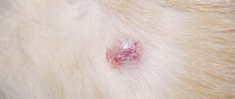
A sweat gland cyst is a small (several millimeters) sized cavity with a bluish tint (photo 1). It is filled with clear liquid (sweat) and protrudes above the skin level. It is relatively rare in both dogs and cats. Single sweat cysts, as a rule, do not cause discomfort and do not require treatment.
Multiple cerumen/ceruminal (modified sweat) gland cysts in the external auditory canal and pinna of cats are a relatively common occurrence and are called ceruminal cystomatosis . While the cysts are small in size (they look like dark gray, almost black smooth elevations a few millimeters in diameter), they do not cause any discomfort (photo 2). However, when they reach a certain size and quantity and block the lumen of the ear canal, they cause external otitis media with secondary infections, soreness and itching (photo 3). Cysts can degenerate into adenomas and adenocarcinomas of the sulfur glands. Treatment consists of surgical excision (preferably laser excision).
Sebaceous gland cysts are small (1–3 mm) elevations of a whitish or yellowish hue (photo 4). They are relatively common, especially in dogs. They do not cause discomfort and do not require treatment.
Follicular cysts (FC) are most often formed by the epithelium of the mouth of the hair follicle and are a cavity filled with dead keratinized epithelium - keratin. FC occurs quite often in dogs (predisposed breeds are German Shepherds, Pekingese, Shih Tzu and Boxers) and rarely in cats. Clinically, FC appears as a rounded raised area of grayish skin ranging from a few mm to 6 cm, often with a hairless and comedonal surface (Figure 5). FC is often mistakenly called a sebaceous gland blockage. They can be single or multiple and have a progressive course. Follicular cysts that have reached significant sizes often have a “pore” on the surface of the skin through which a curdled content (keratin) of a grayish tint is released. When drying, keratin forms a kind of cutaneous horn on the surface of the skin (photo 6). Secondary bacterial infection of the FC is also not uncommon (especially after an attempt to squeeze it out!), in which erythema is noticeable and pain and itching appear (photo 7). Main differential diagnosis: abscesses, tumors. Diagnosis is made based on history, clinical presentation, cytological examination of cyst aspirate (keratin, cholesterol crystals +/- signs of infection) or histology. Treatment consists of surgical excision. Retinoids (isotretinoin 1.5–3 mg/kg/day) may be effective in inhibiting the growth of old cysts in dogs and preventing the formation of new ones.
A dermoid cyst (DC) is a congenital anatomical anomaly in which a fully functioning piece of skin with all appendages (hair follicles, sweat and sebaceous glands) is enclosed in a closed cavity. DC becomes noticeable already in puppyhood, most often on the midline of the head (photo 8). It looks like one or more soft, round or oval-shaped bumps that can be infected and have a “pore” on the surface, from which a tuft of hair often sticks out. Treatment is surgical excision. A dermoid cyst may be connected by a tubular tubule to the spine (dermoid sinus), which naturally creates a risk of bacterial infection of the nervous system. Rhodesian Ridgebacks are especially predisposed to this anomaly, in which the presence of a crest is predetermined by a mutation of certain genes inherited as an autosomal dominant trait and is associated with the formation of dermoid sinuses (photo 9). The diagnosis is made on the basis of anamnesis, examination, contrast radiography, magnetic resonance imaging. Treatment is surgical excision (photo 10).
What types of tumors are there?
There are cysts of hormonal and non-hormonal nature.
In the first case, the neoplasm occurs when the ducts are blocked on both sides. The duct expands, stagnation of secretion occurs in it, which is covered with a dense fibrous capsule. Hormonal tumors appear when the ovaries produce excessive estrogen, which, in turn, causes breast swelling and dilation of the milk duct. The capsule is filled with transparent or brownish-colored contents and has a thick, liquid or jelly-like consistency. The size of the tumor varies from a few millimeters to 5 cm. Large capsules can cause changes in the shape of the breast. They develop due to hormonal imbalances and can contribute to the development of cancer.
Mostly single-chamber cysts are diagnosed, having one cavity with liquid contents. If septa appear inside such formations, they turn into solitary tumors that have several liquid cavities inside the capsule.
Types of cysts
External or internal factors can provoke the appearance of different types of tumors in the breast.
- Atypical neoplasms are characterized by the presence of intracavitary growths. They have an oval or round shape, surrounded by a fibrous capsule. The contents of the capsule are liquid that fills the dilated duct and clogs it.
- Fibrous cysts are often malignant and contribute to the development of breast cancer. Menopausal women are more susceptible than others to the appearance of malignant neoplasms in the breast. Fibrous tumors are characterized by the presence of cluster-like lumps in the breasts, discharge from the nipples, pain and increased sensitivity in the premenstrual period.
- The solitary cyst has a round shape, it is elastic, and the cavity is filled with fluid. Most often, such a tumor is found in only one breast and has a large cavity. The longer it stays in the chest, the denser its capsule becomes.
- A ductal cyst is a precancerous disease. It is a growth that goes deep into the mammary gland. Characterized by the presence of bloody, greenish or clear discharge from the nipple. There is no pain on palpation of the breast, but small compactions are detected in the area of the milk ducts.
- Multilocular cysts are detected during ultrasound examination. They develop by the fusion and growth of single-chamber capsules. The disease is very dangerous for the female body, as it can develop into a malignant tumor.
Any breast tumors require careful diagnosis and effective treatment. A photo of an ultrasound examination of the mammary glands will allow the doctor to make a more accurate diagnosis and determine the danger that this cyst poses to a woman’s health.
Lumps under the skin in cats
The appearance of strange formations under the skin of a pet is not that uncommon. They can be malignant or benign, become inflamed, cause discomfort to the cat, ooze exudate (serous or purulent) or be absolutely painless and not annoy the pet in any way. But any subcutaneous formations in a pet deserve the owner’s attention.
Under the skin, between the skin and the muscle layer, nodules or capsules with liquid contents may form in your pet. They are “dough-like”, mobile and do not cause trouble for your pet. The lumps may be hard and painful.
Subcutaneous formations consist of degenerated tissues, or are capsules filled with contents (dense or serous). They have normal body temperature or are dense and hot to the touch. At the same time, the animal’s general body temperature may increase, and other signs of general intoxication may appear (weakness, refusal to eat, irritability or lethargy, dyspepsia). Formations prone to rapid growth deserve special attention.
Causes of bumps
Lipoma, also known as fat, is a benign neoplasm that can appear on any part of the body. Prone to growth, can reach large sizes, but does not metastasize. The formation is soft, limited, painless. General symptoms are related to the location, that is, the mechanical pressure of the tumor on surrounding tissues. The formation of lipomas on the limbs can lead to lameness of the animal, and in the area of the carotid artery to death from suffocation. The general condition of the animal is usually normal. Unfortunately, lipomas often occur in association with malignant tumors (fibrosarcomas or sarcomas). More often this happens when the tumor appears on the back of the animal. Lipomas are more common in older animals.
Abscesses in animals can be post-injection, develop as a result of wounds or after insect bites, as a consequence of notoedrosis.
Post-injection abscesses form after an unsuccessful injection, if a bacterial infection was introduced during the injection process or the animal “got a cold” at the injection site. Thin deep punctures often fester. Because anaerobic microflora enters the narrow, deep wound channel and actively multiplies, since it is difficult for oxygen to enter such a wound. Abscess tumors are painful, they lead to dysfunction, and are accompanied by fever and local temperature.
Lymphadenitis or an inflammatory process in the tissues of the lymph nodes, can occur with “feline distemper”, leukemia, tularemia, or fungal infections. This disease is caused by some species of Bartonella, Pasteurella, and Fusobacterium. In this case, swelling appears under the jaw, in the groin and axillary area. Swollen lymph nodes are very painful, even if the swelling is minimal, and they are barely palpable. Lymphadenitis is usually accompanied by general intoxication and a disturbance in the condition of the animal.
Nodules under the skin can form as a result of encapsulation of a foreign body trapped under the skin of an animal or a source of bacterial infection. In this case, the formation is dense and painless. Discomfort in the animal can be caused by pressure on the nodule if it presses on the underlying tissue. The general condition of the cat is not disturbed, the nodule is not prone to growth.
A mite embedded under the skin and feeding is very similar to a small skin formation. You need to carefully examine such a “growth” and remove it. A tick, even one that has eaten, is small, usually no larger than the nail plate of a person on the little finger of his hand. There is no disturbance in the animal's condition if a tick attaches itself to it.
Cat salivary glands:
- The parotid gland is the largest, paired, located at the base of each ear behind the temporal bone.
- The submandibular gland is a steam gland, located under the root of the tongue.
- The sublingual gland, the ducts of which exit under the tongue of the animal.
- Molar gland - in the front part of the lower jaw with ducts emerging in the area of the front teeth.
- The infraorbital gland, or zygomatic gland, is located in the bones of the upper jaw, very close to the lower orbital circle.
Polycystic kidney disease in cats: how long can they live and can it be cured?
Polycystic disease is a pathology in which many cavities (cysts) filled with fluid form in the kidneys. Part of the organ becomes a vesicular formation. The disease affects both kidneys at the same time. Since a significant part of the parenchyma is lost, the function of filtering biological waste is not fully performed. Metabolites accumulate, causing intoxication.
Polycystic disease is a hereditary disease. Man created many breeds of cats according to his own will, endowing them, in addition to the given qualities, with unexpected deformities. Among cats suffering from polycystic kidney disease, the most common are Persian and British breeds.
The pathology does not threaten the pet with instant death, but makes his life a series of painful sufferings. It must be treated immediately after diagnosis. In addition to constant intoxication, the contents of the cyst provide a breeding ground for conditionally pathogenic microflora.
The cavities tend to grow and replace the parenchyma of the excretory organ. Symptoms of polycystic disease become noticeable in cats over 7 years of age, as the enlargement of the cavities occurs slowly.
The cat adapts to life with defective kidneys and for a long time the disease proceeds without revealing itself. The disease cannot be cured. The veterinarian’s task is to stop the development of pathology.
Treatment of any disease is effective if it is detected in the initial phase. This has nothing to do with polycystic disease. Until the cavities occupy a significant part of the kidney area, signs of the disease cannot be detected. If upon palpation you notice pain in the abdominal cavity, the prognosis is poor.
Specific signs of polycystic kidney disease include:
- Loss of appetite.
- Oppression.
- Hematuria.
- Thirst.
- Emaciation.
- Vomit.
- Pollakiuria.
- Nervous phenomena.
Diagnostics
A presumptive diagnosis is established by clinical signs, urine analysis, and in-depth palpation of the abdominal chamber. To confirm the assumption, a biopsy is performed. This study allows us to establish the nature of the cavities and determine the degree of their danger to the life of the cat. Cultures of pathological material reveal the presence or absence of secondary infection of the kidneys and excretory channels.
Ultrasound allows you to see the tumor and determine the ratio of intact and modified tissue in the organ. Modern clinics have mastered the method of gene testing. If both methods indicate polycystic disease, it becomes possible to carry out preventive treatment and prophylactic measures that can slow down the development of cysts. Radiography reveals the disease in its final stage.
Polycystic kidney disease cannot be cured non-surgically. If the pathology is detected in the early phase, and the affected area is small, then there is a chance to get rid of the disease. But such a development of events is possible when the cat owner himself insists on conducting an examination. In most situations, the veterinarian has to deal with the second or even third phase of the disease. The goal of treatment is to ensure satisfactory well-being and prolong life.
If the size of the cystic blisters is large, periodic removal of fluid is carried out with a puncture needle. Drug treatment is aimed at minimizing developing chronic renal failure.
The following methods are used in the treatment of polycystic kidney disease:
- Diet therapy.
- Rehydration detoxification.
- Normalization of blood pressure.
- Gastroprotectors.
- Prevention of anemia.
- Antibiotic therapy.
Diet therapy
They use specialized wet food for cats with kidney disease, with reduced levels of Phosphorus and protein, which provoke the development of symptoms of chronic renal failure. To normalize phosphorus-calcium metabolism, vitamin D preparations are used.
Rehydration detoxification
To remove toxins, parenteral administration of rehydration solutions is necessary.
Normalization of blood pressure
The medical drug Amlodilin is in demand for cats. To maintain the dosage, you will need a tablet knife.
Prevention of anemia
Use of Erythropoietin, as well as drugs for the treatment of anemia.
Gastroprotectors
The main purpose is to relieve attacks of nausea and prevent the formation of stomach ulcers. They use drugs that reduce the production of gastric juice - antacids.
Antibiotic therapy
If bacterial culture reveals the presence of secondary microflora, antibiotic therapy is prescribed. First, universal drugs are used, and when the results of a test for the sensitivity of microflora to antibiotics become available, treatment correction is applied.
When carrying out maintenance therapy at the first stage of the disease, the prognosis is favorable, in the opposite situation it is doubtful or negative. The lifespan of sick cats can be several years or two months.
Prevention
Care should be taken when breeding polycystic-prone Britons and Persians. Pets with this diagnosis are not allowed to be bred and are recommended to be sterilized. For breeds predisposed to the disease, it is necessary to use specialized feed.