Why does atheroma occur?
The appearance of a cyst is caused by blockage of the outlet duct of the sebaceous glands. As a result, the sebum produced by the gland does not come to the surface, but accumulates inside. The duct increases in size, and the body’s defense system, trying to stop this process, forms a cyst of connective tissue in the form of a cavity around the atypical formation.
Factors leading to blockage of the sebaceous glands:
- Injury to the epidermis - skin cells, after being damaged by a blunt object, clog the lumen of the sebaceous gland duct.
- Low level of personal hygiene - dust particles and other contaminants must be regularly removed from the surface of the skin so as not to clog the excretory ducts of the glands.
- Hormonal imbalance, the predominance of male hormones - testosterone, dehydroepiandrosterone - leads to changes in the functioning of the sebaceous glands, increased secretion of sebum and its thickening.
- Postmenopause - during menopause, the concentration of estrogen in the female body sharply decreases, which increases the likelihood of changes in the composition of sebum and the formation of atheroma.
Although atheroma most often occurs in adults, the congenital form of this neoplasm is rarely diagnosed in children. The cyst forms in front of the ear and looks like a small ball up to 2 cm in diameter. The cause of atheroma in children is a small defect of the epidermis in the area of the auricle. In the future, such atheroma does not develop and does not affect the child’s health.
Animals at risk
The reasons for the formation of cystic cavities are not fully understood. It is believed that some types of cysts are caused by a hereditary predisposition, while others appear due to a violation of the hormonal status of the body. The conditions of keeping and feeding the pet are also important.
Veterinary specialists and experienced breeders believe that the following pets are at risk:
- Unsterilized cats. Estrus, pregnancy, childbirth, and feeding offspring are often accompanied by disruptions in the female’s hormonal system.
It is the disruption of hormonal status that is the main cause of the development of polycystic ovary syndrome and cystic tumors of the mammary gland.
- The use of hormonal drugs to control estrus and sexual behavior is a serious blow to the cat’s endocrine system. Veterinary experts consider the use of tablets, drops, and injections based on sex hormones to be the main cause of the development of ovarian and mammary cysts in domestic cats.
- Injuries and mechanical damage. If the integrity of the skin is damaged, a keratin-like substance can penetrate into the layers of the skin. Epidermal cysts often develop through this mechanism.
- Breeds such as the Persian, Himalayan, British Shorthair and Exotic Shorthair are at risk for developing polycystic kidney disease.
- Keeping pets in poor hygienic conditions and the presence of thorny indoor plants provokes the development of hair and epidermal tumors.
- Violation of feeding rules. Eating bones is a risk factor for the development of mucocele in the domestic cat.
Symptoms of atheroma
Since the formation of atheroma is not accompanied by an inflammatory process, there are no symptoms such as weakness, fever, loss of appetite, hyperemia and changes in the surface layer of the skin. If the atheroma does not become inflamed, then no problems other than a cosmetic defect are recorded.
Location of the cyst:
- Back - most often occurs on the skin between the shoulder blades, although another area of this part of the body is not excluded;
- Head – face, back of the head, area around the ears, chin;
- Crotch;
- Coccyx;
- Armpits;
- Popliteal fossae.
In those areas of the human body in which there are no sebaceous glands (palms, feet), atheromas never form.
Differentiation of atheroma from similar pathologies
A sebaceous cyst may be similar in its symptoms to other subcutaneous formations - an enlarged lymph node, fibroma or lipoma. It is important to know their characteristic features in order to distinguish them from each other. Other formations are so rare that their symptoms are not important for diagnosis.
| Sign | Atheroma | Lipoma | Fibroma | Lymph node |
| Mobility of the skin layer over the formation | Inseparable from the skin, as it is formed in the thickness of its layer | They are located under the skin layer, so the skin is mobile | ||
| Appearance | Looks like a round formation rising above the skin | Often invisible without the use of special equipment or magnification | ||
| Density | Consistency is soft | The consistency is thick | ||
| Soreness | Usually painless, with suppuration there is pain on palpation | The formations are painless | Painful | |
The main signs make it possible to differentiate atheroma from similar pathologies.
Diagnosis of the disease
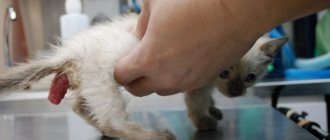
Mucocele in cats is easy to diagnose. On palpation, a tumor can be clearly felt in the area of the affected salivary gland - under the neck or on the front of the cat's head (Fig. 2). Examination of the oral cavity helps determine the presence of disease if swelling is clearly visible under the tongue or in the throat area.
Rice. 2
In order to distinguish mucocele from other diseases, a fine-needle puncture can be performed in the edema tissue to determine the presence of saliva in the cyst. The contents of the cystic sac are quite characteristic. It is usually a mucous (liquid or thickened) exudate, clear or yellowish-brown, often mixed with blood.
When cysts become abscessed, the contents are purulent. For successful subsequent treatment, it is necessary to determine the side of the lesion. When the cyst is localized in the ventral surface of the neck, it is often difficult to immediately understand which ducts of the glands are affected, the right or the left. In such cases, contrast radiography is used.
Inflammation of atheroma
When pathogenic microorganisms enter the cavity of the cyst, its tissue can become inflamed. The cause of inflammation is injury to the epidermis as a result of cuts, punctures of the skin, scraping of the top layer, attempts to squeeze out atheroma on the face, neck, or back of the head.
Signs of inflammation:
- Increase in size of atheroma by several centimeters in the shortest possible time;
- Hyperemia;
- Swelling;
- Pain on palpation of atheroma.
As a result of inflammation and melting of tissues, pus finds its way out, breaking through the skin. After applying a sterile dressing, you need to contact a surgeon to remove the remaining connective tissue of the capsule and sanitation of the wound.
If you do not consult a doctor and self-medicate, the remaining parts of the capsule will cause the re-formation of atheroma.
Polycystic kidney disease in cats: how long can they live and can it be cured?
Polycystic disease is a pathology in which many cavities (cysts) filled with fluid form in the kidneys. Part of the organ becomes a vesicular formation. The disease affects both kidneys at the same time.
Since a significant part of the parenchyma is lost, the function of filtering biological waste is not fully performed. Metabolites accumulate, causing intoxication.
Polycystic disease is a hereditary disease. Man created many breeds of cats according to his own will, endowing them, in addition to the given qualities, with unexpected deformities. Among cats suffering from polycystic kidney disease, the most common are Persian and British breeds.
The pathology does not threaten the pet with instant death, but makes his life a series of painful sufferings. It must be treated immediately after diagnosis. In addition to constant intoxication, the contents of the cyst provide a breeding ground for conditionally pathogenic microflora.
The cavities tend to grow and replace the parenchyma of the excretory organ. Symptoms of polycystic disease become noticeable in cats over 7 years of age, as the enlargement of the cavities occurs slowly. The cat adapts to life with defective kidneys and for a long time the disease proceeds without revealing itself.
The disease cannot be cured. The veterinarian’s task is to stop the development of pathology.
Treatment of any disease is effective if it is detected in the initial phase. This has nothing to do with polycystic disease. Until the cavities occupy a significant part of the kidney area, signs of the disease cannot be detected. If upon palpation you notice pain in the abdominal cavity, the prognosis is poor.
Specific signs of polycystic kidney disease include:
- Loss of appetite.
- Oppression.
- Hematuria.
- Thirst.
- Emaciation.
- Vomit.
- Pollakiuria.
- Nervous phenomena.
[custom_ads_shortcode1]
Surgical treatment of atheroma
The only effective way to treat atheroma is surgical removal, because drug therapy or traditional medicine recipes do not bring any significant results and can cause a relapse.
As soon as signs of inflammation appear around the formation, you should immediately go to the outpatient clinic or the emergency room of a surgical hospital. Uncomplicated atheroma is operated on as planned.
The main goal of surgery is the removal or destruction of the cyst and its contents. Methods of surgical treatment:
| Method | Contents of the method | Advantages |
| Classic method | An incision is made in the superficial layers of the skin, through which the cyst is removed without violating its integrity. A suture is placed on the wound, and then the threads are removed during dressing after healing |
|
| Argon plasma coagulation | Atheroma is destroyed using a beam of plasma at the end of a special scalpel; simultaneously with the destruction of tissue, the blood is stopped |
|
| Laser destruction | The laser knife destroys the cyst and its capsule |
the disadvantage of these methods is their low prevalence due to the high cost of equipment and its absence in public clinics |
| Electrocoagulation | An electric knife removes a cyst using high frequency current | |
| Radio wave knife | Atheroma tissues are burned using high-intensity radio waves |
All surgical methods require local anesthesia. The operation to remove atheroma rarely lasts more than 20 minutes.
Postoperative period
In the first days after removal of the capsule, it is important to carefully monitor the condition of the wound. At first, the surgeon performs daily procedures, changing the drainage device and antiseptic dressings. The total duration of the postoperative period is 10-14 days. It takes place on an outpatient basis. Occasionally, complicated forms of atheroma are treated in a surgical hospital.
After the formation of connective tissue between the edges of the wound, the sutures are removed. This manipulation takes no more than 3-5 minutes and is usually painless.
Dangerous signs of complications after surgery
- Addition of inflammation after removal of an uncomplicated form of the cyst, appearance of pus;
- An increase in temperature indicates tissue infection; normally, hyperthermia disappears within 2-3 days;
- Dehiscence of wound edges – discovered during dressing;
- Leakage of blood through the bandage - increased bleeding is diagnosed in hepatitis, hemophilia, thrombocytopenia, enlarged spleen, as well as in patients using anticoagulants (Aspirin, Cardiomagnyl, Heparin, Thromboass).
The appearance of any of the above signs is a reason to consult a doctor, who will assess the risk of complications and carry out therapeutic measures.
Mammary tumors in cats
If a mammary tumor is detected in a cat, its owner will be very upset due to ignorance. In addition, many do not know the dangers of such education and what needs to be done with it. Pathologies of this type do not appear instantly, but changes are often detected by chance. Therefore, it is worth examining in more detail why a cat develops a tumor on the mammary gland and how it should be treated.
Mammary gland pathologies are often detected in cats. In this case, neoplasms often have a malignant form (in 80% of cases). According to the observations of doctors, there is a relationship between the age of the pet and the onset of the disease.
Therefore, a young cat will be less susceptible to developing this disease. The increase in the number of identified tumor pathologies occurs in direct proportion to the number of years she has lived.
In addition, some breeds of pets have an increased chance of developing such neoplasms. Thus, Siamese and Oriental breeds are more vulnerable. The development of mammary gland tumors in cats most often occurs after reaching 10 years of age.
Exceptions are Oriental breeds, in which the development of pathology occurs even earlier. In addition, the chance of developing AMF is higher in pet breeds with short hair.
[custom_ads_shortcode1]
Answers to frequently asked questions
- Is there a risk of atheroma reappearing?
Yes, even a small number of surviving cells can give rise to the appearance of a new cyst.
- What does a postoperative scar look like? Is it possible to avoid it?
Minimal scars are formed after surgery using radio wave radiation. In second place is the argon plasma method. After these interventions, there may be no scars at all. Traditional surgery leaves noticeable scars.
- How to prevent atheroma?
There are no specific methods of prevention. It is important to control hormonal levels, maintain hygiene, and avoid skin injury.
- What is the difference between atheroma and lipoma?
Lipoma is located in the thickness of the fatty tissue, it is caused by excessive growth of connective tissue and is a benign tumor.
- Can the process become malignant?
No, such cases are excluded, this is not a precancerous disease.
- Can cyst destruction occur on its own?
No, this is impossible, atheroma cannot resolve, it remains unchanged for a long time.
- Is a surgeon required to operate on non-inflammatory atheroma?
Services under the compulsory medical insurance policy may not provide for the removal of non-inflammatory atheroma. The solution is to undergo surgery in a private clinic or wait for inflammation to develop, which does not guarantee the absence of a cosmetic defect after the intervention.
- What are the consequences of self-squeezing atheroma?
The cyst is usually located in areas of intense blood circulation, so there is a high risk of infection spreading to the brain through the blood vessels if bacteria gets into the wound. You cannot squeeze out the cyst - you need to seek qualified medical help.
Author of the article:
Volkov Dmitry Sergeevich |
Ph.D. surgeon, phlebologist Education: Moscow State Medical and Dental University (1996). In 2003, he received a diploma from the educational and scientific medical center for the administration of the President of the Russian Federation. Our authors
Cyst in a cat
Hello! My cat is 10 years old and has not given birth. A cyst was diagnosed.
The doctor said that surgery was needed. I would like to ask how risky this operation is and is it possible to somehow protect the cat from the recurrence of the cyst? And are there any contraindications for the operation?
If so, can this sore lead to more serious consequences if not operated on? Thanks in advance for your answer.
Hello. The operation is indicated; a repeat cyst on the ovaries will not occur, since they are completely removed. Contraindications for surgery are the age of the cat, concomitant diseases, and the state of the cardiovascular system, but if the cat is completely healthy at 10 years old, then the operation can be performed.
It is not the manipulation itself that is dangerous, but the anesthesia. If possible, you should use gas, it is safer.
[custom_ads_shortcode1]