Why does it happen?
Mostly in cats, a diaphragmatic hernia is observed due to traumatic factors. The injuries can be open, resulting in damage to the chest and abdominal cavity, and the diaphragm is also injured. If we talk about closed injuries of a traumatic nature, they are observed during a fall from a height, road traffic accidents, and also in a situation where intra-abdominal pressure increases sharply and greatly.
Congenital diaphragmatic hernia in kittens is also not uncommon. This pathology is usually divided into 4 types:
- Pleuroperitoneal. Rarely diagnosed. In most situations, pets with this diaphragm defect die during childbirth or a few hours after birth.
- Pericardio-pleuroperitoneal. A common type of disease. Mostly found in Persian felines.
- Sliding. It develops due to weakening of the diaphragmatic ligament, which causes the stomach to move upward. No strangulated hernias are observed.
- Paraesophageal. This type of pathology is characterized by fixation of the cardiac region and displacement of the digestive organs into the chest cavity through the esophageal opening, which is excessively dilated. Hernias can be strangulated and cause severe pain.
Peritoneo-pericardial diaphragmatic hernia is a congenital defect, as a result of which a connection between the abdominal cavity and the cardiac membrane is formed in a cat during intrauterine development due to a violation of the integrity of the diaphragm.
What symptoms indicate pathology?
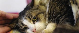
Against the background of this pathology, the animal develops shortness of breath, which is difficult for the owner to notice.
It is important for pet owners to monitor the general condition of the kitten. As the disease progresses, it becomes apathetic and does not make contact with the owner. If the pathology has not been identified and appropriate treatment has not been carried out, breathing problems develop. The animal may choke and experience shortness of breath. Since the body does not receive the required amount of oxygen, changes from the pink tint of the mucous membranes to blue are observed. If the abdominal organs put too much pressure on the blood vessels, there is a risk of pulmonary edema.
With pericardial-diaphragmatic disease, cats may not have any symptoms. This is often due to the fact that abdominal organs slide freely into the pericardium. In such a situation, a hernia is often diagnosed when an x-ray is routinely performed. However, there are pets in whom the pathology provokes loss of appetite, constant vomiting, diarrhea and weight loss. This symptomatology is caused by strangulation of the stomach. Some pets suffer from compression of the heart and lungs.
Is it possible to prevent a hernia in a kitten or an adult animal?
Following these recommendations will help prevent this problem:
- Provide your pet with a balanced diet, which will avoid disruption of the bowel movement process. If possible, the cat's diet, regardless of whether it consists of natural products or prepared food, should be agreed with a veterinarian.
- If constipation occurs, immediately take measures to eliminate it.
- Limit the number of matings of your pet to reasonable limits.
- Protect your four-legged pet from falls from heights. You should not allow your animal to walk on an open balcony or sit on a windowsill near an open window.
- After castration, carefully follow the veterinarian's recommendations regarding postoperative care.
- Conduct preventive veterinary examinations at least once a year.
- Exclude from breeding animals whose immediate relatives had this problem.
Diaphragmatic hernia develops in cats as a pathological condition due to various reasons. But in this case, damage to the “film” of varying intensity occurs, in which organs from the abdominal cavity begin to shift upward, causing pinching of the lungs, heart and large vessels.
- participation in breathing;
- support for organs;
- natural partition;
- providing lymphatic drainage;
- participation in digestion.
A hernia most often appears when the animal is severely injured (road accident, fall from a height) or due to an impact. Along with the hernia, the cat has multiple injuries to the bones and soft tissues, so it is necessary to take it to the RosVet EC, where the operating team will take care of it.
What to do?
Diagnostic measures
Sometimes it is advisable to examine an animal using an ultrasound method in order to accurately diagnose it.
If the owner notices breathing problems in a cat or other undesirable symptoms, it is important not to delay a visit to the veterinary clinic. First of all, the veterinarian interviews the owner and clarifies how long ago the cat’s condition worsened. Then the cat is examined and x-rayed. A routine examination or a technique during which a contrast agent is used is prescribed. If part of the digestive organs is localized in the chest cavity, the preliminary diagnosis of diaphragmatic hernia is confirmed. To detect a diaphragmatic rupture, the cat is prescribed a peritoneogram. Sometimes an ultrasound examination is required.
How is the treatment carried out?
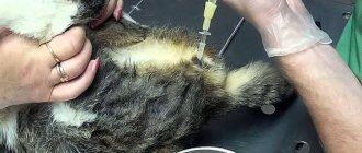
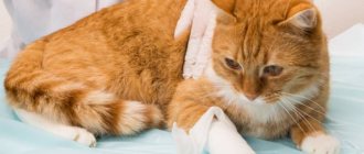
Only surgical intervention can help cope with diaphragmatic hernia in cats. The operation involves opening the chest by a veterinarian. After this, he returns the organs that have moved back to the peritoneum. Then the doctor sews up the diaphragm, but first resorts to plastic surgery. The surgeon places a special mesh on the muscle-tendon plate. In a situation where internal organs are strangulated, their resection is sometimes required.
If after the intervention the animal may develop a bacterial infection, then appropriate medications are prescribed.
12 hours before surgical treatment of a diaphragmatic hernia, the cat is prohibited from giving food. It is not recommended to give water to your pet 2-3 hours before surgery. In most situations, surgery is scheduled in the morning. By evening the animal can return home. The rehabilitation period takes about 14 days on average. After this time, the stitches are often removed. For 2 weeks, the cat's owner will need to give her complete rest and monitor the operated area. Sutures must be treated with medications prescribed by a veterinarian. Sometimes antibacterial treatment is prescribed to avoid infection of the operated area.
Preventive actions
To prevent the occurrence of a diaphragmatic hernia in a cat, veterinarians from the Pride clinic recommend that pet owners be attentive to changes in the cat’s condition. If any problems with digestion or bowel movements are detected, it is important not to delay a visit to the veterinary clinic. Timely diagnosis allows us to identify the possible presence of a congenital hernia and carry out appropriate treatment. If your pet is kept in an apartment on the upper floors, you should not let him sleep on the windowsill or walk along the ledge. Once again, it is not recommended to open the balcony and windows unless necessary. Such precautions are due to the fact that many situations have been recorded when cats, trying to jump onto the windowsill, did not calculate the distance, missed and fell.
Diagnosing a ruptured diaphragm
Auscultation
Auscultation refers to listening to the lungs using a stethoscope. If the diaphragm ruptures, the lung noise on the side of the rupture will be less because the lungs work less well.
Percussion
The veterinarian should tap the cat's chest with his fingers. Usually the lungs are full of air and sound like a drum. If the diaphragm is damaged, the veterinarian will only hear a dull thud.
X-ray
This is the most convenient and accurate diagnostic method. An X-ray will help determine not only the location of the diaphragm rupture, but also other possible organ damage.
What symptoms should alert the owner?
If the organs of the abdominal cavity are shifted through the hole into the chest cavity, then compression of the lungs inevitably occurs, and also the organs themselves, pinched in the diaphragmatic hernia, have their blood circulation disrupted.
The following signs should alert a cat owner:
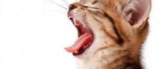
- obvious breathing problems;
- a superficial, shallow sigh;
- the cat deliberately extends its neck and head to improve air passage.
The liver, stomach and intestines “stuck together” cease to function normally, which visually may look like signs of gastrointestinal disorders or liver failure.
Types and characteristics of diaphragmatic hernias
After the examination, the veterinarian determines the cause of the damage; this is what is important to do in order to predict the course of the operation in the future. A defect in the diaphragm appears after an injury; these are the most common cases of cat owners contacting the RosVet VC.
- closed (bumps, falls, road accidents, sharp increase in intra-abdominal pressure);
- open (when, together with the diaphragm, the integrity of the surrounding tissues and bones is disrupted).
Clinical signs of damage are very different and depend on the cause, on which organs and tissues are injured. The respiratory and digestive systems are most often affected.
With a congenital diaphragmatic hernia, there are:
- pleuroperitoneal, which are rare and kittens with similar anomalies die in the first week after birth;
- pericardio-pleuroperitoneal, the most commonly diagnosed injuries in the practice of veterinary surgeons, Persian cats are predisposed to them;
- sliding, develop with weakness of the esophageal-diaphragmatic ligament, while the stomach and esophagus, having lost support, move into the mediastinum. A hernial sac is formed from a fold of the peritoneum and, as a result, the sphincter between the stomach and esophagus does not close well (reflux esophagitis);
- paraesphageal, the fundus of the stomach, omentum and part of the intestine are moved through the dilated esophageal opening into the chest cavity with simultaneous fixation of the cardiac section. The main difference is the possibility of strangulation, pain, nausea and vomiting. In the absence of quick help, the strangulated tissues quickly die, which is irreversible.
Traumatic injuries can cause pneumothorax (air in the chest cavity) or hemothorax (collection of blood). As a result, breathing is impaired until critical situations arise when the cat needs help immediately.
Diagnosis of diaphragmatic hernias in animals
About half of diaphragmatic hernias (35-50%) are accompanied by severe respiratory symptoms: rapid or impaired breathing up to attacks of suffocation, cyanosis of the mucous membranes and tongue. This condition is also characterized by a sunken abdominal wall during inspiration and a decrease in shortness of breath, which is clearly manifested if the animal is lifted by the front of the body. The condition worsens when the dog goes down the stairs.
To confirm this diagnosis, it is necessary to take an x-ray of the chest and abdominal cavities, including with a contrast agent; ECG and ECHO of the heart, ultrasound of the abdominal organs.
The main method for diagnosing diaphragmatic hernias is radiography. On an x-ray you can determine:
Discontinuity of the diaphragmatic circuit
- Contents of the abdominal cavity inside the ribcage
- Displacement of thoracic structures
- Displacement of abdominal organs
- Divergence of the legs of the diaphragm
X-rays can be taken using radiopaque agents or air.
Left - The picture shows a cat with a diaphragmatic hernia.
Right: The same cat after surgery. The difficulty with x-ray examination is that prolapsed organs may spontaneously return to the abdominal cavity.
Ultrasound examination is performed in cases when:
- herniation of the contents of the abdominal cavity penetrates into the chest cavity through a defect in the diaphragm;
- on thoracic radiographs, pleural effusion can hide the diaphragmatic-hepatic silhouette and abdominal hernias;
- a diaphragm rupture occurred, i.e. with loss and interruption of the normal echogenic line (pleuropulmonary interface);
- the contents of the abdominal cavity can be seen through the defect and the chest;
Traumatic ruptures of the diaphragm are often accompanied by pleural effusion. In congenital periteneopericardial diaphragmatic hernia, the appearance of abdominal viscera adjacent to the heart within the pericardial sac and loss of diaphragmatic contour near the midline are considered diagnostic.
Methods for examining diaphragmatic hernias in animals:
- Esophagogastroscopy
- Biopsy of the esophageal mucosa
- Study of acidity in the esophagus
- X-ray of the stomach
Treatment of diaphragmatic hernias in animals
Hiatal hernias are characterized by relaxation and can be sliding or paraesophageal. Mostly, hiatal hernias are treated conservatively (by gastroenterologists). Surgical treatment is resorted to when competent conservative therapy is ineffective, in case of complications and in case of paraesophageal hernias.
Principles of conservative treatment:
- Prevention of reflux of gastric contents into the esophagus
- Reducing the acidity of gastric juice
- Drug protection of inflamed esophageal mucosa
- Treatment of concomitant diseases that provoke the development of a hernia
All traumatic diaphragmatic hernias, due to the risk of strangulation, must be treated surgically, which is carried out immediately after stabilizing the patient.
In this case, preoperative preparation using intensive care methods is very important.