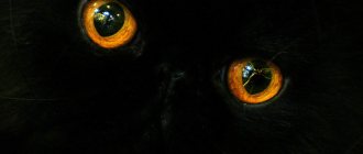
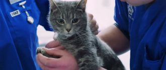
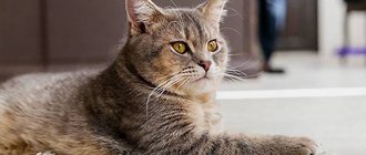
Jaundice is a dangerous condition of the body, characterized primarily by yellowing of the mucous membranes of the mouth, lower eyelid, sclera, and inner surface of the ear. Lemon-yellow skin in a pet should greatly alert a responsible owner, as this symptom indicates the presence of serious health problems. What is this condition and how to deal with it?
Jaundice in cats is not an independent disease; it is a clear symptom of many pathological conditions.
Jaundice is caused by high levels of bilirubin. This useful substance, the so-called bile pigment, is part of hemoglobin, which is contained in erythrocytes - red blood cells that supply tissues and organs with oxygen. They are utilized in the liver, which produces the components it needs from their breakdown products. If the liver cannot cope with this task, bilirubin turns the cat's skin a characteristic color. Naturally, this is an abnormal condition of the body, requiring immediate contact with a veterinary service.
Jaundice in a cat is accompanied by the following symptoms::
- the color of urine and feces changes (urine becomes orange, feces become whitish)
- fever
- severe chills
- weakness
- poor appetite
- bloating
- diarrhea
- vomit
- extreme thirst
- excessive urination
- changes in behavior (aggressiveness or apathy)
Why did the cat turn yellow?
Taking into account a number of reasons
jaundice that causes this pathological manifestation is divided into only three types:
- Hemolytic or suprahepatic jaundice occurs due to increased breakdown of red blood cells. This can happen for several reasons:
- feline dirofilariasis (parasitic helminthic disease), leptospirosis,
- blood parasites (feline hemobartonellosis),
- intoxication with certain drugs and poisons (for example, as part of rodent poison), snake bites, spider bites;
- severe viral infections (leukemia, immunodeficiency virus).
2. Hepatic jaundice is a consequence of liver disease. It could be:
- poisoning with hepatoxins (mercury salts, lead, drugs - paracetamol, diazolin, insectoacaricides);
- infections of both viral and bacterial origin;
- hepatitis (liver inflammation);
- hepatosis (liver failure, metabolic disorders in the liver);
- parasitic liver diseases are leptospirosis, opisthorchiasis and echinococcosis, as well as toxoplasmosis;
- cirrhosis of the liver (degeneration of its tissue into connective tissue with disruption of all functions);
- oncological neoplasms of the liver.
Liver diseases will also be accompanied by vomiting, loss of appetite, and changes in the color of urine and feces.
3. Mechanical (subhepatic) jaundice occurs when bile stagnates in the bile ducts. This is a consequence of diseases such as:
- chronic cholecystitis - inflammation of the gallbladder,
- pancreatitis - inflammation of the pancreas,
- cholelithiasis (formation of stones in the gall bladder),
- pathologies of the sphincter of Oddi (spasm, polyps, etc.).
To what extent does evisceration and enucleation spoil the appearance of an animal?
It all depends on the severity of the pathological process and the traumatic mechanism that preceded the operation. As a rule, the recovery period takes place in at least a month.
Yes, it is necessary, since the animal will definitely “try” to wipe the newly operated eye with its paw. As a result, the sutures will be removed, and pathogenic microorganisms will inevitably enter the fresh wound.
It all depends on the perception of others and on the extent of the owner’s love for his four-legged pet. Of course, one cannot expect the same appearance, but in this case the question arises not about a cosmetic effect, but about saving the life of the animal.
ATTENTION!!! Unfortunately, it is not always possible to save an animal’s eyeball, especially in cases of late contact with an ophthalmologist or in cases of severe eye injury, tumors and complicated cases of eyeball prolapse. The eye is removed only when vision is irretrievably lost and when it causes suffering to the animal, as well as in cases of risk of involvement in the pathological process of organs and tissues adjacent to the orbit or the neighboring eye.
Diagnostic methods
To make the correct diagnosis
For your “yellowed” pet, the veterinarian will definitely interview you in detail about the cat’s condition, nutrition, and behavior. Then he will prescribe laboratory tests such as blood biochemistry, blood sugar, coagulogram, general blood test, tests for viral infections, toxoplasmosis, parasitic infestations, and urinalysis. An ultrasound and x-ray may be necessary. Sometimes a liver biopsy is required. Thanks to these tests, the doctor will reveal where the “culprit” of jaundice is hiding and determine the disease. Having made a prompt diagnosis, a veterinarian will most likely be able to cure jaundice in a cat.
Types, symptoms and treatment
Below is a classification of eye diseases in cats, symptoms characteristic of each disease, as well as an approximate treatment regimen.
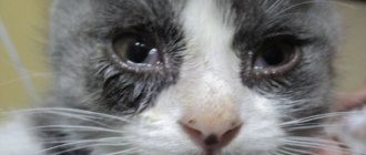
Conjunctivitis
Conjunctivitis in cats is perhaps the most common eye disease.
Inflammation of the mucous membrane of the eyelids is called conjunctivitis. There are many types of it:
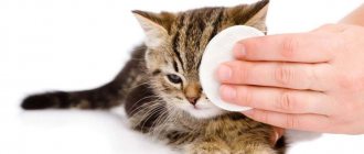
There are many reasons for this inflammation. The most common ones are a foreign body getting into the eye (grains of sand or hair, for example), vitamin deficiency, injuries, infections. In babies (especially kittens), inflammation from the nasal cavity (a seemingly harmless runny nose) quickly spreads to the eyes. Therefore, not only rhinitis is recorded in them, but also inflammation of the nasolacrimal duct and purulent conjunctivitis.
When an animal has conjunctivitis, the mucous membrane of the eyelids turns red, swells, and tears flow. Pus may also appear (but not immediately; first, catarrhal conjunctivitis, which, depending on the cause of its appearance, gradually or extremely quickly “turns” into purulent).
The first step is to determine what caused this inflammation? If it is a foreign body, then you just need to rinse the eye and, to prevent complications, apply tetracycline eye ointment to the lower eyelid a couple of times a day (eye ointment is always 1%!). You can purchase it either at a veterinary pharmacy or at a human pharmacy. This is not a scarce medicine at all and costs a penny. It is best if you always have it in your first aid kit.
Jaundice in cats, necessary therapy
- Therefore, often, while awaiting the results of laboratory tests, the veterinarian will prescribe supportive measures for the cat:
- droppers to combat dehydration, correct the level of electrolytes in the blood if the animal suffers from anorexia, vomiting and diarrhea;
- drugs that lower blood sugar levels (as indicated);
- the use of drugs for anemia, hematopoietic stimulants, donor blood transfusions for severe anemia;
- course of antibiotics, antiemetic drugs.
Once the test results are in and the underlying cause of the jaundice is found, your veterinarian will decide on more targeted treatment. This can be surgical intervention (for example, if stones are found in the gall bladder or it is necessary to remove a tumor), and antiparasitic therapy, and the introduction of antidotes for poisoning with certain toxic substances. As you can see, there are many causes of jaundice in cats, so treatment protocols will vary greatly, as will the prognosis for recovery.
Therapy for jaundice will be aimed not only at eliminating its root cause (for example, infection or tumor), but also at stopping the processes that it caused. This may be pain syndrome (with obstructive jaundice), oxygen deficiency, or intoxication of the body. For these purposes, analgesics, oxygen therapy, droppers with hepatoprotective drugs, etc. will be used.
Another area of treatment is to combat symptoms such as diarrhea, vomiting, anemia, and dehydration.
. This will also require the introduction of intravenous drips, vitamin complexes, electrolytes, drugs against vomiting and increased stomach acidity.
Cat care
Mandatory recommendations when caring for a cat with liver failure are:
- strict adherence to veterinary prescriptions,
- a special gentle diet (consult your doctor to create a treatment menu for your cat),
- control by the veterinarian over the healing process,
- rest, peace, lack of stress for a sick animal.
Owners' fears
Sometimes cat owners, having discovered that they have jaundice, begin to worry about the health of all household members, worrying that the infection does not turn out to be contagious. In most cases, these fears are unfounded; feline hepatitis is not transmitted to humans. Of course, you must monitor the health of your pet, there are diseases that are common to us and our smaller brothers, but in the case of jaundice, you only need to worry about the health of your pet!
If you see symptoms of jaundice in a cat, call our center and you will receive a free consultation to provide first aid to your animal. At your request, the doctor will come to you at any time within 40 minutes if you need emergency help. Many medical procedures and express tests for the presence of certain infections can be done at home without wasting precious time. Remember that in this case, time should work for you, and not for the disease, since the animal often dies without timely help.
Treatment of jaundice requires an integrated approach
, therefore, on the basis of ours, you can examine your pet comprehensively, not excluding ultrasound and x-rays. Severe patients will be admitted to hospital for urgent treatment with modern drugs. After recovery, the treating veterinarian will provide advice so that the cat with jaundice can lead a healthy and active life!
Primary neoplasms of the eyeball in dogs
Anterior uveal melanoma Anterior uveal melanoma (AUM) is the most common primary neoplasm in dogs. Iris melanomas are usually focal, dark, growing lesions on the anterior surface of the iris in young dogs. These neoplasms grow slowly, cause small intraocular lesions, are histologically benign, and rarely metastasize.
PUM most often occurs in dogs over 7 years of age and is usually benign, both histologically and in terms of the nature of the tumor process. The clinical picture is related to the area of origin of the tumor. Most often, the animal will experience changes in the color, shape and texture of the iris and ciliary body. In dogs, this neoplasm will be more discrete than in cats, in which it will most often be diffuse. Typically there is a smooth, raised and pigmented tumor on the iris or ciliary body. Pigmentation of a lesion does not confirm the presence of melanoma, because some melanomas are non-pigmented. Sometimes non-pigmented growths of the ciliary body can push the anterior part of the iris forward and thus resemble a PUMP. Other clinical symptoms of intraocular neoplasm may also be observed. Like other primary eye tumors, this tumor progresses, increasing in size, and without treatment causes secondary uveitis or glaucoma. Uveal cysts may be similar in appearance to PUMs. To differentiate these formations, transillumination is used. Uveal cysts are usually transilluminated, but PUMs are not.
Fine needle aspiration and incisional biopsy are necessary for diagnosis. Fine-needle aspiration of an anterior uveal mass usually has few side effects, especially if the mass is located within the anterior chamber of the eye. An incisional biopsy requires intraocular surgery, which must be performed by an ophthalmologist. Ultrasound of the eye provides more accurate information about the spread and place of origin of the tumor, especially if the intraocular environment is opaque. Enucleation can also be informative, but this operation is performed only in cases of blindness or severe pain. The main treatment is resection, cryosurgery, enucleation and laser photocoagulation. The most successful method is laser photocoagulation, which best leads to remission, reduction of the tumor body, and slowing down the growth of intraocular tumors in dogs. Moreover, laser photocoagulation preserves vision and has minimal side effects. Since pigmented tissues absorb laser energy better, pigmented tumors can be treated more successfully than non-pigmented ones. But laser photocoagulation has also proven effective for treating non-pigmented tumors in dogs. Potential side effects of laser photocoagulation include hyphema, vitreous bleeding, corneal edema, mild anterior uveitis, corneal scarring, and cataract formation (if the lesion is anterior to the lens). With the exception of scarring and cataracts, other side effects will be temporary and usually go away within 2 weeks of treatment. New growths of the ciliary body respond best to this treatment.
Because most intraocular neoplasms in dogs have an extremely low potential for metastasis, we believe that laser photocoagulation is a safe and effective alternative to enucleation. If symptoms of severe secondary uveitis or glaucoma are present, we recommend enucleation and histological examination of the tumor. Before initiating any treatment, the animal should be thoroughly evaluated for systemic metastases, including general examination, complete blood count, chest x-ray, and abdominal ultrasound. Canine PUM metastases can occur in the lungs, liver, kidneys, spleen, adrenal glands and heart. It is also possible for melanoma to grow into the eye from another area.
Epibulbar Melanoma in Dogs Epibulbar melanoma in dogs grows at the junction of the sclera and cornea and typically presents as a smooth, raised, pigmented mass at the limbus of the sclera that may invade the cornea. This tumor has not demonstrated systemic metastasis but is a locally invasive neoplasm. If left to grow, it can invade the eyeball, causing uveitis and secondary glaucoma, in which case enucleation will be required. Common treatment options include full-thickness resection with homologous or synthetic graft coverage, partial resection, cryosurgery, and laser photocoagulation. Because of the high morbidity following full-thickness resection and the difficulty in locating the desired tissue, we consider cryosurgery and laser photocoagulation to be the more optimal treatment. The sooner these surgeries are performed, the safer and more effective the treatment will be.
Choroidal melanoma Choroidal melanoma is more common in older dogs. In contrast to humans, in dogs these tumors have benign histological and prognostic characteristics. They are rare and usually grow slowly, developing into large masses that require enucleation to treat. As they increase, they cause atrophy and retinal detachment. Clinical ophthalmic evaluation reveals well-defined, raised pigmented masses under the retina. When the intraocular environment is cloudy, ultrasound is useful for diagnosis.
Anterior uveal epithelial neoplasms Epithelial tumors of the anterior uveal tract are most often adenomas and adenocarcinomas of the ciliary body, and in dogs are secondary tumors arising from other neoplasms. Most often, these are non-pigmented, pink or white, slow-growing tumors that occupy the posterior chamber of the eye. They usually do not cause obvious eye disease until they become quite large. To distinguish between adenomas and adenocarcinomas, it is necessary to carry out histological differentiation, but this has little effect on the prognosis. Both types of tumors invade local tissues but rarely metastasize. As with PUM in dogs, laser photocoagulation is the best way to maintain vision and effectively treat these tumors, of course, in the absence of severe secondary uveitis or glaucoma.
Medulloepithelioma Medulloepithelioma is rare in dogs and grows from embryonic neuroepithelium.
This neoplasm mainly occurs in young dogs, but if growth is slow, it can be detected several years later. In clinical appearance, this tumor is similar to adenoma and adenocarcinoma of the ciliary body. No local extraocular spread or metastases were noted.
Prevention measures
Cats are incredibly clean and can take care of their eyes on their own. They wash themselves regularly, putting themselves in order, but they are not able to cope with the disease. Owners of four-legged furry dogs should check them regularly. Particular attention is paid to animals that walk outside. To prevent pathology it is necessary:
- Feed your pet high-quality food. Balance your diet and don’t experiment with it.
- Visit the veterinarian, get vaccinations and examination of internal organs.
- Ensure your pet has an active lifestyle, play with him, and go out into the fresh air.
- Protect from contact with sick and unfamiliar animals.
- Regularly cleanse the body of helminths.
- Monitor the length of the claws and trim them in time so that the pet does not injure itself.
Owners of purebred cats prone to excessive tearing should make it a rule to treat their eyes daily. With increased tear production associated with the characteristics of the breed, you may not immediately notice that the animal has health problems. Monitor the nature of the discharge and pay attention to your pet’s behavior.
Simple prevention methods will help protect your cat from various troubles, including eye problems. If you have the slightest doubt about the causes of tearing, contact your veterinary clinic. Remember that home remedies are temporary measures to combat the problem. Only a doctor can rid an animal of the causes of unpleasant discharge.
When a cat develops mucus in the eyes, this is not always a signal of the development of any disease. In some cases, this phenomenon indicates spontaneous cleansing of the animal’s visual organs. However, it is necessary to distinguish when a cat has mucus in the eyes - it is a natural process, and when it is a pathological process. Since in the first case there is no need to do any special manipulations, in the second case you will need to consult a specialist.
Exenteration
This is an operation that is a radical way to eliminate pathological processes in the eye. This method is an enucleation combined with the removal of extraocular muscles, fatty tissue of the orbit, upper, lower and third eyelids, lacrimal gland, as well as all conjunctival tissues.
In some cases, the subsequent placement of an intraocular prosthesis can eliminate the unattractive cosmetic effect after surgery. Such an event is characterized by low trauma. However, a thorough assessment of the safety of the procedure is required. Thus, contraindications to prosthetics include panophthalmitis, uveitis, panuveitis, and suspected intraocular neoplasm.
We invite you to read: Kazakh white-headed breed: description and productivity