Cat blood test
Owners, when bringing a sick cat to a veterinary clinic, are almost constantly faced with the fact that veterinary specialists, in addition to a clinical examination of the sick cat, conduct a blood sample test from the sick animal in order to make an accurate diagnosis of the disease.
Modern effective treatment of a sick animal cannot be based only on examining the sick animal. If not so long ago no one seriously took the possibility of conducting a study to assess the condition of an animal using a clinical and biochemical blood test in the laboratory, today every self-respecting veterinary specialist, and even more so a modern veterinary clinic, considers it the norm to do a blood test on their patients. A blood test is one of the most informative ways to examine pets. By conducting a blood test, a veterinarian has the opportunity not only to confirm or refute the clinical diagnosis, but also to identify hidden pathological processes in the animal’s body (subclinical pathology) that have not yet produced characteristic symptoms.
The main types of research are general clinical and biochemical blood tests. Using the CBC, you can establish the basic composition of the blood. Biochemical research allows a veterinary specialist to more accurately judge the functional state of the animal. Before performing an operation or other intervention, a blood test is a mandatory norm.
Dangerous deviations in indicators in a cat
Deviations from the norm (increased or decreased) indicate that a malfunction has occurred in the body. Monitoring allows you to timely identify the development of the disease and begin treatment.
Bilirubin
Bilirubin is part of bile.
Bilirubin colors the blood, so an increased amount of it leads to discoloration of the skin (jaundice).
An overestimated value indicates the development of liver diseases (hepatosis, hepatitis), as well as blockage of the bile ducts.
Scheme of bilirubin formation in the blood
A decrease in bilirubin levels is observed with anemia and bone marrow lesions.
Total protein
An increase is observed with dehydration due to vomiting and diarrhea. A decrease in protein levels is typical for intestinal diseases, chronic liver diseases (cirrhosis or hepatitis), renal failure, and fasting.
Proteins are involved in all metabolic processes in the body.
Protein synthesis
Creatinine
An increase in blood creatinine levels may indicate the development of hyperthyroidism or kidney failure. A decrease in this value is observed during protein starvation.
Elevated levels of creatinine in both urine and blood indicate that the cat's diet contains a lot of animal protein.
Urea
An increase in urea indicates impaired kidney function and blockage of the urinary tract. Also, an excess of this value is observed when feeding your pet food rich in animal protein.
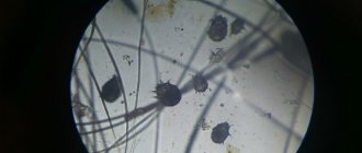
Uric acid crystals under a microscope
A decrease in urea indicates intestinal dysfunction, liver pathologies or protein deficiency in the diet.
Glucose
The reasons for increased blood glucose are as follows:
- Cushing's syndrome;
- diabetes;
- release of adrenaline into the blood due to increased physical activity or severe stress;
- chronic kidney or liver diseases;
- pancreatitis;
- pancreatic tumors.
A decrease in value is observed with an overdose of insulin, prolonged fasting, poisoning with poisons or alcohol.
Blood glucose
Low glucose levels are also characteristic of pancreatic diseases.
Amylase
An increase in the rate is observed in the following diseases: pancreatitis, diabetes mellitus, peritonitis, volvulus, renal failure.
Also, a high level of amylase indicates intoxication of the body due to poisoning.
Amylase
A decrease in the indicator may be the result of taking anticoagulants, poisoning, or necrosis of pancreatic tissue. The analysis determines total amylase and pancreatic amylase. The norm is 500-1200 units/l.
Cholesterol
An increase in cholesterol levels is typical for pancreatitis, diabetes mellitus, hypothyroidism and kidney disease.
Deficiency is observed with the development of malignant neoplasms, impaired absorption of nutrients in the intestines, and unbalanced nutrition.
Cholesterol in the blood
AST and ALT
An increase in these indicators indicates the destruction of liver cells, which was caused by cirrhosis, hepatitis or other diseases. Also, an increase in AST and ALT can be a consequence of injury or heart failure.
A simultaneous decrease in AST and an increase in ALT indicates the development of infectious hepatitis.
Alkaline phosphatase
Increased alkaline phosphatase can occur in pregnant animals and in pets that eat fatty foods.
A high rate is also typical for bacterial infections of the intestines and stomach, and liver diseases.
A decrease in the level of alkaline phosphatase is observed with anemia, vitamin C deficiency, and long-term use of corticosteroids.
Alkaline phosphatase is a whole complex of enzymes that is found almost throughout the body in small quantities.
Phosphorus
An increase in phosphorus is characteristic of leukemia and bone tumors. Also, a high value is observed in renal failure, hypervitaminosis of vitamin D, and disorders of the endocrine system.
Phosphorus deficiency can result from a lack of growth hormone and vitamin D.
Long-term diarrhea also leads to a decrease in the indicator.
Calcium
An increase in calcium is typical for:
- dehydration;
- destruction of bone tissue due to cancer;
- excess vitamin D.
Calcium deficiency is observed with pancreatitis, lack of vitamin D, taking anticonvulsants, and chronic renal failure.
How to properly prepare an animal for blood donation
- Before taking blood for analysis, the cat is not fed for 10-12 hours, and there must be free access to water.
- We limit the animal’s physical activity (refusal of games, etc.).
- Taking any medications is prohibited. If your cat regularly takes any medications as prescribed by your veterinarian, consult about stopping them.
- Before taking blood, therapeutic procedures, ultrasound, x-rays, and massage are unacceptable.
The procedure for taking blood for analysis
Blood is taken from the vein of the animal. To do this, the cat is placed on its side, and if the cat resists, it is secured in a special veterinary bag. Blood is taken from the front paw by shaving off a small area of fur. The injection site is treated with a disinfectant solution, a needle from a disposable syringe is inserted into the vein, blood in the amount of 2 ml is drawn into a test tube with heparin or sodium citrate.
What blood tests are performed in veterinary clinics?
In modern veterinary clinics, two laboratory blood tests are performed:
- General or clinical.
- Biochemical.
General blood test for a cat
A general blood test based on the number and condition of formed blood elements shows the health status of the cat’s body. When performing a general blood test, parasites such as dirofilaria (dirofilariasis), hemobartenella can be detected in a cat’s blood.
What indicators does the clinic’s veterinary specialist receive when conducting a general blood test:
- Hematocrit
- Hemoglobin.
- Average content and concentration of hemoglobin in an erythrocyte.
- Color indicator.
- ESR (erythrocyte sedimentation rate).
- Red blood cells.
- Leukocytes.
- Neutrophils.
- Lymphocytes.
- Eosinophils.
- Monocytes.
- Platelets.
- Basophils.
- Myelocytes.
Biochemical blood test for a cat
Biochemical blood tests allow veterinary specialists to identify subclinical (hidden) cat diseases. A biochemical blood test allows you to determine the functioning of the body’s enzymatic system and provide information about damage to a particular organ in a cat.
A biochemical blood test for a cat includes enzymatic, electrolyte, fat and substrate indicators.
Basic biochemical parameters:
- Glucose.
- Protein and albumin.
- Cholesterol.
- Bilirubin is direct and total.
- Alanine aminotransferase (ALT).
- Aspartate aminotransferase (AST).
- Lactate dehydrogenase.
- Gamma glutamyl transferase.
- Alkaline phosphatase.
- a –Amylase.
- Urea.
- Creatinine.
- Calcium.
- Magnesium.
- Creatine phosphokinase.
- Triglycerides.
- Phosphorus, inorganic.
- Electrolytes (potassium, calcium, sodium, iron, chlorine, phosphorus).
Indicators of blood tests obtained and their characteristics
Each indicator of a blood test shows the functioning of individual organs or entire systems, while the veterinarian takes into account not only each data separately, but also the relationship to each other.
Hematocrit is a conditional indicator showing the ratio of all formed elements of blood to its volume, i.e. determines the thickness of blood. Shows how much blood can carry oxygen.
Hemoglobin is a protein contained in red blood cells that ensures the movement of oxygen and carbon dioxide throughout the animal’s body.
The average concentration of hemoglobin in an erythrocyte shows in percentage terms how much erythrocytes are saturated with hemoglobin.
The color (color) blood index shows how much hemoglobin is contained in red blood cells in relation to the normal value.
ESR is an indicator that determines the presence of an inflammatory process in the body.
Erythrocytes are red blood cells that take part in tissue gas exchange and maintaining acid-base balance. It’s bad when test results go beyond the norm, not only in the direction of decrease, but also of increase.
Leukocytes (white blood cells) - indicate the state of the animal’s immune system. Leukocytes include lymphocytes, neutrophils, monocytes, basophils and eosinophils. For a veterinarian, the relationship between these cells is of diagnostic importance.
- Neutrophils are responsible for destroying bacteria in the blood.
- lymphocytes - speak of a general indicator of immunity.
- monocytes - perform the function of destroying foreign substances in the blood,
- caught in the blood.
- eosinophils - perform the function of fighting allergens.
- basophils - together with other leukocytes, help the body recognize and identify foreign particles that have entered the blood.
Platelets are blood cells responsible for blood clotting. In addition to this function, they are responsible for the integrity of blood vessels. Both high and low levels of them are dangerous for the body.
Myelocytes are located in the bone marrow and normally should not be in the blood.
Blood chemistry
Glucose is an informative indicator indicating the functioning of a complex enzymatic system in the body, including individual organs (liver, pancreas, kidneys). Glucose metabolism in the body involves 8 different hormones and 4 complex enzymatic processes. A disorder is considered to be either high or low blood sugar levels in a cat.
Total protein in the blood reflects the correctness of amino acid metabolism in the body. Shows the total amount of all protein fractions - globulins and albumins. Proteins in an animal’s body take part in almost all life processes of the body. For specialists, both their increased and decreased amounts are important.
Albumin is the most basic blood protein produced by the liver. Albumin in a cat’s body performs a large number of functions (transporting nutrients, maintaining reserve reserves of amino acids for the body, maintaining osmotic blood pressure, etc.).
Cholesterol is a structural component that ensures the strength of cellular structures and takes part in the synthesis of many vital hormones. Veterinary experts judge lipid metabolism in a cat’s body based on cholesterol levels.
Bilirubin is a bile pigment found in the body in two forms - direct and indirect. Indirect bilirubin is formed in the blood as a result of the breakdown of red blood cells, and bound (direct) bilirubin is converted from indirect bilirubin in the liver. Bilirubin shows the functioning of the hepabiliary system (biliary and hepatic). Refers to “color” indicators i.e. when its content in the body is increased, the tissues turn yellow (jaundice).
Alanine aminotransferase (ALT, ALaT) and aspartate aminotransferase (AST, ACaT) are enzymes produced by liver cells, heart cells, red blood cells and skeletal muscles. It is an indicator of the functions of these organs or departments.
Lactate dehydrogenase (LDH) is an enzyme involved in the final step in the breakdown of glucose. Veterinary LDH specialists monitor the functioning of the cardiac and hepatic systems, and also judge the risks of tumor formation.
ɤ-glutamyltransferase (Gamma-GT) – in combination with other liver enzymes, provides insight into the functioning of the hepabilary system, pancreas and thyroid glands.
Alkaline phosphatase is determined to monitor liver function.
ɑ-Amylase – produced by the pancreas and parotid salivary gland. Their work is judged by its level, but always in conjunction with other indicators.
Urea is the result of protein processing, which is excreted by the kidneys. Some remains circulating in the blood. Using this indicator, you can check your kidney function.
Creatinine is a muscle byproduct excreted from the body by the renal system. The level fluctuates depending on the condition of the urinary excretory system.
Potassium, calcium, phosphorus and magnesium are always assessed in complex and in relation to each other.
Calcium is involved in the conduction of nerve impulses, especially through the heart muscle. By its level, you can determine problems in the functioning of the heart, muscle contractility and blood clotting.
Creatine phosphokinase is an enzyme that is found in large quantities in the skeletal muscle group. By its presence in the blood, one can judge the work of the heart muscle, as well as internal muscle injuries.
Triglycerides in the blood characterize the functioning of the cardiovascular system, as well as energy metabolism. Usually analyzed in conjunction with cholesterol levels.
Electrolytes are responsible for membrane electrical properties. Thanks to the electrical potential difference, cells pick up and execute commands from the brain. In pathologies, cells are literally “thrown out” from the nerve impulse conduction system.
Decoding biochemical
A biochemical analysis of a cat's blood examines the condition and functioning of internal organs.
- Glucose, the norm is 3.3–6.3 mmol per l; an elevated level most often indicates diabetes mellitus. Also, the cause of increased glucose can be pathologies of the pancreas and stress. A decrease in values indicates that the cat is malnourished, and in some cases, oncology.
- ALT (normally 19–79 units), when increased, indicates liver dysfunction and pathological processes in its tissues. In addition, an increase in ALT indicates severe poisoning or intoxication of the body.
- AST (normally 9–30 units) increasing, indicates diseases of the liver, heart, and muscle injuries. It is also an indicator of stroke.
- CPK (normally 150–798 units) increases with damage to the heart muscle, stroke, or poisoning.
- Alkaline phosphatase (normally 39–55 units) if increased means pregnancy or regeneration processes in the body. In some cases, it may mean bone cancer or gastrointestinal disease. A reduced value indicates anemia, vitamin C deficiency, and thyroid disease.
- Amylase (normally 580–1600 units) when increased means pathologies of the pancreas, salivary glands, and intestinal obstruction. A decrease in indicators indicates pancreatic insufficiency.
- Bilirubin (normally 3.0–12 mmol/l) increases in cases of liver or bile duct disease. Its decrease often means anemia or bone marrow pathologies.
- Urea (normally 5.4–12.0 mmol/l) with an increased value indicates renal failure and poisoning of the body. Also, the indicators may be higher if the cat is fed predominantly protein. Low urea levels may indicate a lack of protein in the diet.
- Cholesterol (normally 2–6 mmol/l) in cats when increased indicates the development of atherosclerosis or pathological processes in the liver. Reduced indicators most often mean malnutrition, less often neoplasms.
- LGD (normally 55–155 units) at high values indicates diseases of the digestive tract, oncology, and necrosis. LGD is also elevated against the background of a heart attack or stroke. An LGD reading below normal may indicate severe stress, the use of certain medications, or skin diseases.
The norms for biochemical blood tests in cats are relative. All indicators should be considered in the context of the overall picture, both the blood and the condition of the cat. So, as they can show, for example, a high urea content, with the cat feeling excellent.
It is worth understanding that any little thing, such as changing your diet or playing too actively, affects the quantitative and qualitative composition of the blood. Therefore, it is pointless to independently decipher studies; only a doctor with experience will be able to understand the combinations of indicators and make a diagnosis.
Tests are the best way to understand your cat’s health problems by tracking them in a timely manner. Regular examinations and studies will help your cat live a long and healthy life without unpleasant diseases.
Norms of blood tests in cats
| The name of indicators | Units | Norm |
| Ø hematocrit | % (l/l) | 26-48 (0,26-0,48) |
| Ø hemoglobin | g/l | 80-150 |
| Ø average hemoglobin concentration in an erythrocyte | % | 31-36 |
| Ø average amount of hemoglobin in a red blood cell | pg | 14-19 |
| Ø color indicator; | 0,65-0,9 | |
| Ø ESR | mm/hour | 0-13 |
| Ø red blood cells | million/µl | 5-10 |
| Ø leukocytes | thousand/µl | 5,5-18,5 |
| Ø segmented neutrophils | % | 35-75 |
| Ø band neutrophils | % | 0-3 |
| Ø lymphocytes | % | 25-55 |
| Ø monocytes | % | 1-4 |
| Ø eosinophils | % | 0-4 |
| Ø platelets | million/l | 300-630 |
| Ø basophils | % | — |
| Ø myelocytes | % | — |
Norms of blood tests in cats
General (clinical) blood test
Blood chemistry
| The name of indicators | Units | Norm |
| Ø glucose | mmol/l | 3,2-6,4 |
| Ø protein | g/l | 54-77 |
| Ø albumin | g/l | 23-37 |
| Ø cholesterol | mmol/l | 1,3-3,7 |
| Ø direct bilirubin | µmol/l | 0-5,5 |
| Ø total bilirubin | µmol/l | 3-12 |
| Ø alanine aminotransferase (ALT) | U/l | 17 (19) -79 |
| Ø aspartate aminotransferase (AST) | U/l | 9-29 |
| Ø lactate dehydrogenase | U/l | 55-155 |
| Ø ɤ-glutamyltransferase | U/l | 5-50 |
| Ø alkaline phosphatase | U/l | 39-55 |
| Ø ɑ-Amylase | U/l | 780-1720 |
| Ø urea | mmol/l | 2-8 |
| Ø creatinine | mmol/l | 70-165 |
| Ø calcium | mmol/l | 2-2,7 |
| Ø magnesium | mmol/l | 0,72-1,2 |
| Ø creatine phosphokinase | U/l | 150-798 |
| Ø triglycerides | mmol/l | 0,38-1,1 |
| Ø inorganic phosphorus | mmol/l | 0,7-1,8 |
| Ø Electrolytes | ||
| Ø potassium (K + ) | mmol/l | 3,8-5,4 |
| Ø calcium | mmol/l | 2-2,7 |
| Ø sodium (Na + ) | mmol/l | 143-165 |
| Ø iron | mmol/l | 20-30 |
| Ø chlorine | mmol/l | 107-123 |
| Ø phosphorus | mmol/l | 1,1-2,3 |
Blood tests in cats (transcript)
All deviations in indicators are considered in complex and in relation to one data to another within the same results from the study of one blood sample. Only an experienced veterinarian should interpret blood tests (results).
General (clinical) blood test
| The name of indicators | Promotion | Decline |
| 1. Hematocrit |
|
|
| 2. Hemoglobin |
|
|
| 3. ESR |
|
|
| 4. Red blood cells |
|
|
| 5. Leukocytes |
|
|
| 6. Segmented neutrophils (mature) |
|
|
| 7. Band neutrophils (immature) |
| |
| 8. Lymphocytes |
|
|
| 9. Monocytes |
|
|
| 10. Eosinophils |
| |
| 11. Platelets |
|
- incoagulability of blood. |
| 12. Basophils | hemoblastoses | Normally absent |
| 13. Myelocytes |
| Normally none. |
Blood chemistry
source
Blood test for a cat, interpretation of the blood test
Why do you need a cat's blood test? Find out what indicators are normal and what changes in clinical analysis indicate.
Blood counts in cats are an important aid in making the correct diagnosis. Sometimes they can be used to accurately determine the presence of a disease, and sometimes they are necessary to monitor the progress of treatment.
How and when is blood taken?
Blood is taken from a vein, preferably on an empty stomach. A syringe or a special tube is used for collection. The resulting blood must be stored under certain conditions and is good for 6-8 hours at room temperature and 24 hours in the refrigerator.
A blood test for a cat is prescribed by a veterinarian depending on the expected diagnosis after examining the animal. There are several groups of studies.
The biochemistry of a cat's blood is used to assess the functional abilities of organs and systems: liver, heart, kidneys, hematopoietic system, and so on. Determine the presence of various enzymes and substrates.
Here is an approximate breakdown of a cat's blood test.
- Liver disease causes an increase in bilirubin, cholesterol, and alkaline phosphatase.
- Impaired kidney function is accompanied by an increase in the amount of urea and creatinine in the blood.
- An increase in glucose may indicate diabetes, stress, and pancreatic diseases.
Of course, the diagnosis must be made by a veterinarian based on all examination data.
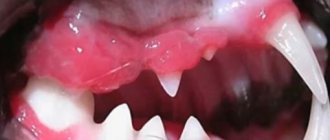
What causes cats to bleed from the mouth? — about this in the article Bleeding from the cat’s mouth.
Clinical analysis
This is the most informative type of research. It is often called a “complete blood count”. The composition of the blood and its physiological characteristics are determined. By deviation in one direction or another, one judges the presence of inflammatory processes, allergic reactions, blood supply disorders, and coagulation pathologies in the body. Blood norms in cats are as follows.
- A decrease in hemoglobin and hematocrit indicates anemia, infections, and intoxications.
- An increase in ESR occurs during oncology, heart attack, kidney disease, after surgery and during pregnancy.
- Leukocytes increase in inflammatory diseases, leukemia, and bacterial infections.
Leukocyte formula
In some cases, an extended clinical blood test in a cat is supplemented with a leukocyte formula.
blood parameters in cats supplemented with leukocyte formula
An increase in the blood of immature forms of leukocytes (band cells) is called a shift of the formula to the left and indicates acute inflammatory processes. An increase in the number of eosinophils may indicate an allergic reaction, and the appearance of basophils may indicate oncology, allergies or chronic inflammation in the intestinal tract.
Any laboratory analysis is not the ultimate truth. Other examination data are of great importance - clinical examination, data on the course of the disease, instrumental studies, response to prescribed treatment.
Source: https://tvoikot.ru/analiz-krovi-u-kota-rasshifrovka-analiza-krovi/
General clinical blood test (CBC)
Test material: venous blood.
Blood is taken into a clean disposable tube with an anticoagulant (EDTA) (tube with a green or lilac cap). Blood is stored for no more than 6-8 hours at room temperature or 24 hours in the refrigerator.
Factors influencing results:
- a decrease in hemoglobin and red blood cells can occur due to the action of drugs that can cause the development of aplastic anemia (antitumor, anticonvulsants, heavy metals, antibiotics, analgesics);
- Biseptol. vitamin A, corticotropin, cortisol - increase ESR.
Hematocrit
Hematocrit (Ht, HCT) is the ratio of the volumes of erythrocytes and plasma (volume fraction of erythrocytes in the blood).
Increased:
- Primary and secondary erythrocytosis (increased number of red blood cells):
- Dehydration (gastrointestinal diseases accompanied by profuse diarrhea, vomiting: diabetes);
- Decrease in circulating plasma volume (peritonitis, burn disease).
Downgraded:
- Anemia:
- Increased volume of circulating plasma (heart and kidney failure, hyperproteinemia);
- Chronic inflammatory process, trauma, fasting, chronic hyperazotemia, cancer;
- Hemodilution (intravenous administration of fluids, especially with reduced renal function).
Hemoglobin
Hemoglobin (Hb, HGB) is a protein contained in red blood cells, the main function of which is the transport of oxygen.
Increased:
Downgraded:
- Anemia:
- Acute blood loss:
- Endogenous intoxication (malignant tumors and their metastases);
- Damage to the bone marrow, kidneys and some other organs:
- Hemodilution (intravenous fluids, false anemia).
Red blood cells (RBC)
Red blood cells are blood elements containing hemoglobin.
Norm: cats - 5.3-10.0 *1012/l.
Increased:
- Erythremia - absolute primary erythrocytosis (increased production of red blood cells>;
- ventilation failure in bronchopulmonary pathology, heart defects:
- hydronephrosis and polycystic kidney disease, kidney and liver neoplasms;
- dehydration.
Downgraded:
- Anemia:
- Acute blood loss:
- Late pregnancy;
- Chronic inflammatory process;
- Overhydration.
Color index
The color indicator characterizes the average hemoglobin content in one red blood cell
Average erythrocyte volume
Mean erythrocyte volume (MCV) is a measure used to characterize the type of anemia. Normal: cats - 43 - 53 µm3.
Increased:
Norm:
- Normocytic anemia (aplastic, hemolytic, blood loss, hemoglobinopathies);
- Anemia that may be accompanied by normocytosis (regenerative phase of iron deficiency anemia, myelodysplastic syndromes
Downgraded:
- Microcytic anemia (iron deficiency, sideroblastic, thalassemia);
- Anemia that may be accompanied by microcytosis (hemolytic, hemoglobinopathies).
Average hemoglobin concentration in erythrocyte
Increased:
Downgraded:
Erythrocyte sedimentation rate (ESR)
Increased:
- Inflammatory processes and infections:
- Diseases accompanied by tissue decay (necrosis) (heart attacks, malignant neoplasms, etc.);
- Intoxication, poisoning;
- Metabolic diseases (diabetes mellitus, etc.);
- Kidney diseases accompanied by nephrotic syndrome (hyperalbuminemia):
- Diseases of the liver parenchyma leading to severe dysproteinemia;
- Pregnancy;
- Shock, trauma, surgery.
- The most significant increases in ESR (more than 50 - 80 mm/h) are observed in: multiple myeloma and malignant neoplasms
White blood cells (WBC)
Norm: cats - 5.5 - 18.5 *109/l.
Increased:
- Bacterial infections:
- Inflammation and tissue necrosis:
- Intoxication:
- Malignant neoplasms:
- Leukemia;
- Allergies;
- The result of the action of corticosteroids, adrenaline, histamine, acetylcholine, insect poisons, endotoxins, digitalis preparations.
A relatively long-term increase in the number of leukocytes is observed in pregnant women and with a long course of corticosteroids.
The most pronounced leukocytosis is observed with:
- chronic, acute leukemia;
- purulent diseases of internal organs (pyometra, abscesses, etc.).
Downgraded:
- Viral and some bacterial infections;
- Aplasia and hypoplasia of the bone marrow, metastases of neoplasms in the bone marrow:
- Ionizing radiation;
- Hypersplenism (splenomegaly):
- Anaphylactic shock:
- Use of sulfonamides.
- Aleukemic forms of leukemia, analgesics, anticonvulsants, antithyroid and other drugs.
The most pronounced (so-called organic) leukopenia is observed with: aplastic anemia: agranulocytosis: feline viral panleukopenia.
Neutrophils
Norm for a cat: band - 0 - 3% of WBC: segmented - 35 - 75% of WBC.
Increased (neutrophilia):
- Bacterial infections (sepsis, pyometra, peritonitis, abscesses, pneumonia, etc.):
- Inflammation or necrosis of tissues (rheumatoid attack, heart attacks, gangrene, burns):
- Progressive tumor with decay;
- Acute and chronic leukemia;
- Intoxication (uremia, ketoacidosis, eclampsia, etc.);
- The result of the action of corticosteroids, adrenaline, histamine, acetylcholine, insect poisons, endotoxins, digitalis preparations.
- Increased carbon dioxide concentration.
Decreased (neutropenia):
- Viral (canine distemper, feline panleukopenia, parvovirus gastroenteritis, etc.)
- Some bacterial infections (salmonellosis, brucellosis, tuberculosis, bacterial endocarditis, other chronic infections);
- Infections caused by protozoa, fungi, rickettsia;
- Aplasia and hypoplasia of the bone marrow, metastases of neoplasms in the bone marrow:
- Ionizing radiation;
- Hypersplenism (splenomegaly):
- Aleukemic forms of leukemia;
- Anaphylactic shock:
- Collagenoses:
- The use of sulfonamides, analgesics, anticonvulsants, antithyroid and other drugs.
Neutropenia, accompanied by a neutrophilic shift to the left against the background of purulent-inflammatory processes, indicates a significant decrease in the body's resistance and an unfavorable prognosis of the disease.
Eosinophils
Norm: cats - 0 - 4% of WBC.
Increased:
Basophils
Increased:
- Allergic reactions to the introduction of a foreign protein, including allergies to food;
- Chronic inflammatory processes in the gastrointestinal tract;
- Blood diseases (acute leukemia, lymphogranulomatosis);
- Myxedema (hypothyroidism):
- The result of the action of estrogens and antithyroid drugs.
Monocytes
Norm: cats - 1 - 4% of WBC.
Increased:
- Infections (viral, fungal, rickettsial, protozoal);
- Blood parasitic diseases (pyroplasmoidosis, including canine babesiosis):
- Tissue inflammatory processes:
- Granulomatosis (tuberculosis, brucellosis, ulcerative colitis, enteritis):
- Surgical interventions.
Downgraded:
Lymphocytes
Lymphocytes ensure an adequate immune response of the body
source
Treimut Love * Treimut.ru
Table of contents
Clinical (general) blood test norms for cats
WBC - white blood cells - white blood cells - Leukocytes.
Norm (x 109/l) - 5.5-18.5
Neutrophils, Eosinophils, Basophils, monocytes, lymphocytes
Absolute indicators reflect how many population cells are contained in a unit volume of blood plasma
- Lumph# lymphocytes
- Mon# monocytes
- Gran# Granulocytes (mainly neutrophils, eosinophils, basophils in a small number. Usually all the properties of Gran are determined by neutrophils
Indicators as a percentage of the total number of leukocytes
- Lumph% lymphocytes 22-55%
- Mon% monocytes 1-4%
- Gran% Granulocytes 35-75%
There should be no basophils and myelocytes
Absolute indicators
Lymphocytes (LYM ) are cells of the immune system, a type of leukocytes of the agranulocyte fraction. There are T- and B-lymphocytes. T lymphocytes are responsible for cellular immunity (contact interaction with victim cells), B lymphocytes provide humoral immunity (antibody production), and lymphocytes regulate the activity of other types of cells. Increased - viral infections, hyperthyroidism, oncological diseases of the blood and bone marrow. Decreased - bacterial infection, sepsis, treatment with corticosteroids, immunosuppressive therapy, some types of lymphomas, radiation therapy. Normal value, ×109 cells/l: cat: 1.0–7.0.
Monocytes (MONO) are granulocytic leukocytes with the ability to phagocytose foreign agents. Increase (monocytosis ) - infections of various etiologies, as well as the period after acute infections, blood diseases, phosphorus poisoning. Decreased - damage to the bone marrow with a decrease in its function (aplastic anemia, B12-deficiency anemia), radiation sickness. Normal value, ×109 cells/l: cat: 0.2–1.0 cells/l.
Band neutrophils (NEUT) are a type of neutrophil that have an S-shaped nucleus. They are young forms of neutrophils; over time, band neutrophils mature and transform into segmented neutrophils. Increased - infections, postoperative period, ischemic tissue necrosis, mercury or lead intoxication, cancer, some inflammatory processes, reaction to certain drugs. Normal value, %: cat: 0–3. Segmented neutrophils (NEUT) - perform a protective function against various bacterial and fungal infections, and also maintain a normal immune system. Increased - pneumonia, purulent inflammation, acute ischemia or tissue necrosis, extensive burn, diseases of the circulatory system, acute blood loss. Decreased - viral infections, autoimmune diseases, chemotherapy or radiation therapy, aplastic anemia, agranulocytosis. Normal value,%: cat: 35–75 . Eosinophils (EOS) are granulocytic leukocytes capable of absorbing various inflammatory mediators, thereby participating in allergic reactions. Increased (eosinophilia) - parasitic diseases, allergic reactions, lung diseases (eosinophilic pneumonia, asthma, allergic aspergillosis, pulmonary infiltrate), blood diseases, autoimmune diseases, diseases of the stomach and intestines, rheumatic diseases, taking certain medications. Decreased - B12 deficiency anemia, trauma. Often has no clinical significance. Normal value, % : cat: 2–12. Basophils (BAS) are a fraction of leukocytes responsible for delayed and immediate allergic reactions (anaphylactic shock reactions). Increased - blood diseases, chronic inflammatory diseases of the gastrointestinal tract, allergic reactions (food or iatrogenic nature), hypofunction of the thyroid gland, treatment with estrogens. Normal value, %: cat: 0–1.
Leukocyte shifts
Shift of the leukocyte formula to the left - acute infectious diseases, physical overexertion, acidosis and coma. Shift of the leukocyte formula to the right - megaloblastic anemia, kidney and liver diseases, conditions after blood transfusion.
RBC - (red blood cells - red blood cells) - Red blood cells
Norm 5-10 mm/μl
Increased incidence of blood diseases (primary erythrocytosis, polycythemia), hypoxia in lung diseases and congenital heart defects, dehydration (vomiting, diarrhea), insufficiency of adrenal cortex function.
Reduced value (anemia) - blood loss, hemolysis, deficiency of iron, vitamin B12, folic acid.
HGB - (Hb, hemoglobin) - Hemoglobin
Norm 80-150 g/l
Increased significance - congenital heart defects, pulmonary fibrosis, intestinal obstruction, cancer. It is also typical for “working” dog breeds: with constant intense physical activity, the need for oxygen increases and, accordingly, the level of hemoglobin increases.
Reduced value - blood loss, infectious and autoimmune diseases, helminthic infestations, pregnancy and lactation, impaired absorption of iron and vitamin B12, malignant blood diseases, chemotherapy.
HCT Hematocrit —
norm 27-49% (l/l)
Elevated HCT Hematocrit in a cat
- increase in the number of red blood cells;
- diarrhea;
- diabetes;
- plasma volume is reduced;
Reduced HCT Hematocrit in a cat
- anemia;
- plasma volume increased;
- the presence of an inflammatory process;
- prolonged fasting;
- presence of cancer;
- after a drip;
MCV Mean red blood cell volume
Norm
39-50
Characterizes the type of anemia
Macrocytic and megaloblastic anemia (B12-folate deficiency); Anemia accompanied by macrocytosis (hemolytic); Normocytic anemia (aplastic, hemolytic, blood loss, hemoglobinopathies); Anemia accompanied by normocytosis (regenerative phase of iron deficiency anemia, myelodysplastic syndromes; Microcytic anemia (iron deficiency, sideroblastic, thalassemia); Anemia accompanied by microcytosis (hemolytic, hemoglobinopathies).
MCH Average level of HGB (hemoglobin) in a red blood cell
Norm 14-19 pg
one of the indicators for determining the type of anemia
Hyperchromic anemia (megaloblastic, liver cirrhosis); Decreased: Hypochromic anemia (iron deficiency); Anemia in malignant tumors.
MCHC Mean red blood cell hemoglobin concentration (%)
An indicator that determines the saturation of red blood cells with hemoglobin.
Norm 300-380
Hyperchromic anemia (spherocytosis, ovalocytosis); Hypochromic anemia (iron deficiency, spheroblastic, thalassemia).
ESR (ESR) is the erythrocyte sedimentation rate
Normal 0-13 mm/hour
Increased value
- Inflammatory processes and infections
- Diseases in which not only an inflammatory process is observed, but also tissue decomposition (necrosis), blood cells, purulent and septic diseases; neoplasms; infarctions of parenchymal organs, pulmonary tuberculosis, etc.
- Connective tissue diseases and systemic vasculitis: rheumatism, arthritis, dermatomyositis, scleroderma.
- Metabolic diseases.
- Diseases of the hematopoietic and lymphatic tissues of the body
- Anemia, hemolysis, blood loss, etc.
- Hypoalbuminemia due to kidney disease, cachexia, blood loss.
- Liver diseases.
- Pregnancy, postpartum period, during the sexual cycle in females.
Reduced value
- Erythremia and erythrocytosis.
- Diseases accompanied by changes in the shape of red blood cells: sickle cell anemia, hemoglobinopathies, spherocytosis, anisocytosis.
- Hyperalbuminemia, hypofibrinogenemia and hypoglobulinemia.
- Diseases associated with an increase in bile pigments and bile acids in the blood: hepatitis of various natures, cholestasis.
- Severe circulatory failure.
- Epilepsy and neuroses.
- The use of certain medications: salicylates, calcium chloride, mercury preparations.
RDW Distribution of red blood cells by volume
Norm 14-31
Increased value
Macrocytic anemia; Myelodysplastic syndromes; Metastases of neoplasms to the bone marrow; Iron deficiency anemia.
PLT platelets
Norm 300-630 million\l
Increased significance - exacerbation of chronic diseases, viral or bacterial infections, blood or hematopoietic diseases, conditions after surgical procedures, malignant tumors, consequences of the use of certain groups of drugs.
Reduced value - idiopathic hypoplasia of hematopoiesis, tumor lesions (acute blood leukemia, cancer metastases, sarcoma, osteomyelosclerosis, myelofibrosis), intoxication, viral infections (hepatitis, adenoviruses), autoimmune diseases.
MPV Mean platelet volume
Norm
5-11
Shift to the left - old platelets predominate. Shift to the right - young.
PDW Platelet volume distribution width
10-18 Norm
PCT - or thrombocrit, is the proportion of platelets in the total volume of whole blood
Norm
0,1-0,5
Biochemical blood test norms for cats
Biochemical analysis depends on the laboratory in which the research is carried out and usually the standards are indicated on the form. I will give the standards of the laboratory in which we carry out the analysis, in general these are average values:
Total protein, G/l 54-77
Promoted
- with dehydration of the body,
- due to severe injuries, extensive burns,
- for acute infections (due to acute phase proteins),
- for chronic infections (due to immunoglobulins).
Demoted
- fasting (complete or protein fasting - strict vegetarianism, anorexia nervosa)
- intestinal diseases (malabsorption)
- nephrotic syndrome (kidney failure)
- increased consumption (blood loss, burns, tumors, ascites, chronic and acute inflammation)
- chronic liver failure (hepatitis, cirrhosis)
Total bilirubin, µmol/l. 3-12
It is a coloring substance (pigment), so when it increases in the blood, the color of the skin changes - jaundice.
Promoted
- damage to liver cells (hepatitis, hepatosis - parenchymal jaundice)
- obstruction of the bile ducts (obstructive jaundice)
- destruction of red blood cells
Demoted —
AST, IU\l 9-29
Promoted
- damage to liver cells (hepatitis, hepatosis, toxic damage from drugs, liver metastases)
- heavy physical activity
- heart failure
- burns, heatstroke
Demoted —
ALT, IU\l 19-79
Promoted
- destruction of liver cells (necrosis, cirrhosis, jaundice, tumors)
- destruction of muscle tissue (trauma, myositis, muscular dystrophy)
- burns
- toxic effect on the liver of drugs (antibiotics, etc.)
Demoted —
Alkaline phosphatase, IU\l 39-55
Promoted
- pregnancy
- increased turnover in bone tissue (rapid growth, healing of fractures, rickets, hyperparathyroidism)
- bone diseases (osteogenic sarcoma, cancer metastases to bones)
- liver diseases
Demoted
- hypothyroidism (underfunction of the thyroid gland)
- anemia (anemia)
- lack of vitamin C, B12, zinc, magnesium
Amylase, IU\l 580-1720
Promoted
- pancreatitis (inflammation of the pancreas)
- mumps (inflammation of the parotid gland)
- diabetes
- volvulus of the stomach and intestines
- peritonitis
Demoted
- pancreatic insufficiency
- thyrotoxicosis
Urea, mmol\l. 5,4-12,1
Promoted
- kidney disease
- nephrocalcinosis
- neoplasia
- urinary tract obstruction
- increased protein content in food
- increased protein destruction (burns, acute myocardial infarction)
Demoted
- protein fasting
- excess protein intake (pregnancy, acromegaly)
- malabsorption
- after administration of glucose,
- with increased diuresis,
- with liver failure.
Creatinine mmol\l. -70-165
Promoted
- impaired renal function (renal failure)
- hyperthyroidism - muscular dystrophy
Demoted
- pregnancy
- age-related decreases in muscle mass
- threat of cancer or cirrhosis
Cholesterol mmol\l. 1,6-3,7
Promoted
- liver diseases
- hypothyroidism (underfunction of the thyroid gland)
- coronary heart disease (atherosclerosis)
- hyperadrenocorticism
Demoted
- enteropathy accompanied by protein loss
- hepatopathy (portocaval anastomosis, cirrhosis)
- malignant neoplasms
- poor nutrition
Albuimn, g\l — 25-37
Promoted
- dehydration
Demoted
- hunger;
- dysfunction of absorption in the intestinal gastrointestinal tract;
- burns;
- bleeding;
- renal disorders.
GGT (gamma-glutamyltransferase) units/l 1-10
Promoted
- liver diseases (hepatitis, cirrhosis, cancer)
- pancreatic diseases (pancreatitis, diabetes mellitus)
- hyperthyroidism (hyperfunction of the thyroid gland)
Demoted —
Glucose mmol\l. — 3,3 — 6,3
Promoted
- diabetes mellitus (insulin deficiency)
- physical or emotional stress (adrenaline release)
- thyrotoxicosis (increased thyroid function)
- Cushing's syndrome (increased levels of the adrenal hormone cortisol)
- diseases of the pancreas (pancreatitis, tumor, cystic fibrosis)
- chronic liver and kidney diseases
Demoted
- starvation
- insulin overdose
- pancreatic disease (tumor of cells that synthesize insulin)
- tumors (excessive consumption of glucose as an energy material by tumor cells)
- insufficiency of the function of the endocrine glands (adrenal glands, thyroid, pituitary gland (growth hormone))
- severe poisoning with liver damage (alcohol, arsenic, chlorine and phosphorus compounds, salicylates, antihistamines)
Electrolytes (potassium, total calcium, phosphorus, sodium, magnesium, chlorine) mmol/l.
- potassium 3.8-5.4
Threatening values: concentrations less than 2.5 mmol/l (muscle weakness) or more than 7.5 mmol/l (cardiac conduction disorders).
Prolonged anorexia, vomiting, diarrhea, muscle weakness, bradycardia, supraventricular arrhythmias, oliguria, anuria and polyuria, hypoadrenocorticosis, diabetic ketoacidosis, urethral obstruction, when using diuretics.
Promoted
Fasting, Convulsive syndrome, Hemolysis of red blood cells, Dehydration, Acidification of the internal environment of the body (changes in acid-base balance), Adrenal dysfunction, Excess potassium in feed.
Reduced Increased physical activity, Stressful situations, Dyspepsia, Hypoglycemia, Hyperhidrosis.
- calcium 2-2.7
The main component of bone tissue. Participates in water-salt metabolism, blood clotting, muscle contraction, and the activity of the endocrine glands.
Hypo- and hyperparathyroidism, hypo- and hypervitaminosis D, bone tumors, lymphoma, leukemia, sarcoidosis, Itsenko-Cushing syndrome, heart failure, chronic renal failure, pancreatitis, osteomalacia, use of anticonvulsants, diuretics, phenobarbitals.
Hypercalcemia
Oncological bone destruction, Thyrotoxicosis, Kidney pathology, Gout, Hyperinsulinemia, Excessive intake of vitamin D.
Hypocalcium and
Impaired function of bone tissue formation in young animals, Inflammatory and degenerative processes in the pancreas, Magnesium deficiency, Impaired biliary excretion, Liver and kidney dysfunction.
- phosphorus - 1.1 - 2.3
It is necessary for the functioning of the central nervous system. Participates in many metabolic processes, its content correlates with the level of calcium (inverse relationship). The main amount of phosphorus (80 - 85%) is part of the skeleton, the rest is distributed between the tissues and fluids of the body.
Hyperphosphatemia (increased)
Treatment with diuretics and antibacterial drugs, Hyperlipidemia, decay of neoplasms and metastasis in bone tissue, kidney dysfunction, hypoparathyroidism, diabetic ketoacidosis, decreased bone mineral density.
Hypophosphatemia (decreased)
Impaired fat metabolism, steatorrhea, Inflammation of the glomerular apparatus of the kidneys, Vitamin D deficiency, Hypokalemia, Unbalanced feeding, Deposition of urate in the joints.
- magnesium 0.72-1.2
Magnesium is a vital electrolyte that works alone or together with other cations: potassium and calcium. It normalizes myocardial contraction and improves the functioning of the central nervous system. Magnesium prevents the development of calculous cholecystitis and urolithiasis. It is taken to prevent stress and cardiac dysfunction.
Increased Insufficient amount of thyroid hormones in the blood, Pathology of the kidneys and adrenal glands, Dehydration.
Reduced Fasting, Colitis, Worms, Pancreatitis, Thyrotoxicosis, Osteomalacia, Hypercalcemia.
- sodium 142-165
Systemic diseases characterized by vomiting, diarrhea, polydipsia and polyuria, muscle weakness, abnormal behavior, seizures, edema, pleural or peritoneal effusion, dehydration. Kidney diseases, heart.
Increased Excess salt in feed, Long-term hormone therapy, Pituitary hyperplasia, Adrenal tumors, Endocrinopathies.
Reduced Dehydration, Hyperthermia, Impact doses of diuretics, Hyperglycemia, Hyperhidrosis, Hypothyroidism, Nephrotic syndrome, Heart and kidney diseases, Polyuria, Liver damage.
- chlorine 107-123
Found in the body mainly in the form of sodium, calcium, and magnesium salts. Diseases characterized by vomiting, diarrhea, polydipsia and polyuria.
Increased (hyperchloremia) Dehydration, Alkalosis, Kidney pathology, Excessive functioning of adrenal glandular cells.
Decreased Vomiting, Hyperhidrosis, Treatment with large doses of diuretics, trauma. Animals with hypochloremia (reduced) may experience hair loss and loose teeth.
- iron 20-30
Iron is an electrolyte that ensures the transfer and delivery of oxygen to cellular elements and tissues.
Increased
Hemochromatosis, Hypo- and aplastic anemia, Lack of B vitamins, Impaired hemoglobin synthesis, Inflammation of the glomeruli of the kidneys, Hematological pathologies, Lead intoxication.
Downgraded
Iron deficiency anemia, Vitamin deficiency, Infections, Oncopathologies, Massive blood loss, Gastrointestinal dysfunction, Long-term use of glucocorticosteroids, Prolonged or frequent stress.
This is interesting:
- Standards for general and biochemical analysis for dogs Clinical (general) blood test norms for dogs WBC - white blood cells - white blood cells...
- Ceftriaxone for cats and dogs, dosage, how to dilute Ceftriaxone is a third-generation drug from the “Cephalosporins” group of antibiotics. Ceftriaxone is inexpensive, effective and quite…
- Calcivirus in cats, treatment, symptoms, prognosis Feline calcivirus is a dangerous virus that affects all cats, regardless of breed and…
General blood analysis
The diagnostic value of tests in veterinary medicine is very great. The animal cannot tell what exactly is hurting it, so tests are important for the doctor to form a complete picture of the disease.
First of all, you need to pass general clinical and biochemical blood tests . These studies will show the general condition of the animal’s body and organs.
The body is always exposed to various environmental factors and gives a specific response to stimuli. Each blood cell performs its functions to protect the body. When the number of certain cells increases or decreases, we can talk about a possible cause of the disease.
A general clinical blood test can show the degree of development of the inflammatory process, whether there is anemia, dehydration, and whether there are neoplasms in the blood system or not. Also, one should not forget about hidden (chronic) infectious, invasive or any other non-infectious processes in the body, which can also be detected by blood testing as one of the diagnostic methods.
A general blood test does not require any special preparation, however, in rare cases, the doctor may ask you to take the test on an empty stomach. The sample itself is taken from peripheral veins.
In our veterinary clinic, a general blood test is performed using an Exigo EOS (VET) automatic analyzer.
It only takes 10 minutes from blood sampling to the results being ready!
Interpretation of a general blood test
Based on the general analysis, the main blood parameters are determined, which are deciphered by the doctor. In your personal account (on our website), the result of the analysis will be published in the form of a schematic table containing the values of blood parameters and the reference range.
Let's look at these indicators and their normal values. It should be borne in mind that deviations from the norm do not necessarily indicate pathology - many of them can be explained by various factors.
Red blood cells (RBCs) are “red blood cells” that contain hemoglobin. The main function is to deliver oxygen from the lungs to the tissues of the body and carbon dioxide from the tissues to the respiratory organs.
Increased (erythrocytosis) - blood diseases (primary erythrocytosis, polycythemia), hypoxia in lung diseases and congenital heart defects, dehydration (vomiting, diarrhea), insufficiency of adrenal cortex function.
Decreased (anemia) - blood loss, hemolysis, deficiency of iron, vitamin B12, folic acid.
Normal value, ×10 12 cells/l:
Hemoglobin (HGB) is a red iron-containing blood pigment that performs the function of transporting oxygen and carbon dioxide and regulating the acid-base state.
Increased : congenital heart defects, pulmonary fibrosis, intestinal obstruction, cancer. It is also typical for “working” dog breeds: with constant intense physical activity, the need for oxygen increases and, accordingly, the level of hemoglobin increases.
Decreased - blood loss, infectious and autoimmune diseases, helminthic infestations, pregnancy and lactation, impaired absorption of iron and vitamin B12, malignant blood diseases, chemotherapy.
Hematocrit (HCT) is the volume fraction of red blood cells in whole blood (the ratio of the volumes of red blood cells and plasma).
Increased - hypoxia, kidney neoplasms with increased erythropotin, polycystic and hydronephrosis of the kidneys, decrease in circulating blood volume (burn disease, peritonitis, etc.), leukemia.
Decreased - anemia, pregnancy, overhydration.
Erythrocyte indices:
Mean erythrocyte volume (MCV) is an indicator characterizing the type of anemia.
Mean erythrocyte hemoglobin concentration (MCHC) is an indicator that determines the saturation of erythrocytes with hemoglobin.
The mean erythrocyte hemoglobin content (MCH) is one of the indicators for determining the type of anemia.
Red blood cell distribution width (RDW) is a measure of how much red blood cells vary in size.
Platelets (PLT) are the formed elements of blood involved in the process of blood clotting.
Increased - exacerbation of chronic diseases, viral or bacterial infections, blood or hematopoietic diseases, conditions after surgical procedures, malignant tumors, consequences of the use of certain groups of drugs.
Decreased - idiopathic hypoplasia of hematopoiesis, tumor lesions (acute blood leukemia, cancer metastases, sarcoma, osteomyelosclerosis, myelofibrosis), intoxication, viral infections (hepatitis, adenoviruses), autoimmune diseases.
Normal value, ×10 9 cells/l:
The analyzer can also calculate mean platelet volume ( MPV ).
Leukocytes (WBC) are “white blood cells” that have a nucleus. The main function is to protect the body from various pathological agents, as well as from internal typical pathological processes accompanied by severe inflammation.
They are divided into two fractions: granulocytes, or cells with granularity in the nucleus (neutrophils, basophils, eosinophils), and agranulocytes with a monochrome, non-granular nucleus (lymphocytes and monocytes).
Normal value, ×10 9 cells/l:
Leukocyte formula (leukoformula) is the percentage of different types of leukocytes, determined by counting them in a stained blood smear under a microscope.
Granulocytes (GRAN) is the total number of indicators such as band and segmented neutrophils, eosinophils and basophils.
Band neutrophils (NEUT) are a type of neutrophil that have an S-shaped nucleus. They are young forms of neutrophils; over time, band neutrophils mature and transform into segmented neutrophils.
Increased - infections, postoperative period, ischemic tissue necrosis, mercury or lead intoxication, cancer, some inflammatory processes, reaction to certain drugs.
Segmented neutrophils (NEUT) - perform a protective function against various bacterial and fungal infections, and also maintain a normal immune system.
Increased - pneumonia, purulent inflammation, acute ischemia or tissue necrosis, extensive burn, diseases of the circulatory system, acute blood loss.
Decreased - viral infections, autoimmune diseases, chemotherapy or radiation therapy, aplastic anemia, agranulocytosis.
Eosinophils (EOS) are granulocytic leukocytes capable of absorbing various inflammatory mediators, thereby participating in allergic reactions.
Increased (eosinophilia) - parasitic diseases, allergic reactions, lung diseases (eosinophilic pneumonia, asthma, allergic aspergillosis, pulmonary infiltrate), blood diseases, autoimmune diseases, diseases of the stomach and intestines, rheumatic diseases, taking certain medications.
Decreased - B12 deficiency anemia, trauma. Often has no clinical significance.
Basophils (BAS) are a fraction of leukocytes responsible for delayed and immediate allergic reactions (anaphylactic shock reactions).
Increased - blood diseases, chronic inflammatory diseases of the gastrointestinal tract, allergic reactions (food or iatrogenic nature), hypofunction of the thyroid gland, treatment with estrogens.
Lymphocytes (LYM) are cells of the immune system, a type of leukocytes of the agranulocyte fraction. There are T- and B-lymphocytes. T lymphocytes are responsible for cellular immunity (contact interaction with victim cells), B lymphocytes provide humoral immunity (antibody production), and lymphocytes regulate the activity of other types of cells.
Increased - viral infections, hyperthyroidism, oncological diseases of the blood and bone marrow.
Decreased - bacterial infection, sepsis, treatment with corticosteroids, immunosuppressive therapy, some types of lymphomas, radiation therapy.
Normal value, ×10 9 cells/l:
Monocytes (MONO) are agranulocytic leukocytes with the ability to phagocytose foreign agents.
Increased (monocytosis) - infections of various etiologies, as well as the period after acute infections, blood diseases, phosphorus poisoning.
Decreased - damage to the bone marrow with a decrease in its function (aplastic anemia, B12-deficiency anemia), radiation sickness.
Normal value, ×10 9 cells/l:
Leukocyte shifts
Shift of the leukocyte formula to the left - acute infectious diseases, physical overexertion, acidosis and coma.
Shift of the leukocyte formula to the right - megaloblastic anemia, kidney and liver diseases, conditions after blood transfusion.
Do not forget: only a veterinarian can take into account all the nuances of clinical blood test data. The values of indicators that are considered to be the “norm” are averaged. Depending on many characteristics of the animal: age, gender, size and even its diet, medications taken and past diseases - normal values can differ significantly.
If you have any additional questions, the veterinarians at the Averia Clinic are happy to help you 24 hours a day!
source
What are neutrophils and their functions in the body
Neutrophils are a type of white blood cell, i.e. blood cells that perform a protective function. Neutrophils are modest in size - no more than 9–12 microns. Their growth and maturation occurs in the red bone marrow, where most of the neutrophils reside until markers of inflammation appear in the bloodstream. Only after this do they enter the general bloodstream, heading towards the developing focus of inflammation.
The function of neutrophils is to protect the body from pathogenic particles that have entered it. But that's in general. The main target of this type of leukocytes are pathogenic and conditionally pathogenic microbes and fungi. Neutrophils practically do not react to viral invasions (in case of viral pathologies, their number practically does not increase); they do not participate in protecting the body from helminths at all. Both of these facts determine the value of a general blood test: if there are a lot of neutrophils, then we are almost certainly talking about a bacterial (most likely) or fungal disease.
The protective mechanism in their case is based on the ability of neutrophils to phagocytose (i.e., absorb and “digest” the pathogen). Since these cells are small, they cannot capture large particles. At the same time, the ability for phagocytosis in neutrophils is not so perfect that they can intercept and destroy viruses.
It may seem that the role of neutrophils in the body of cats (and other mammals, including humans) is not so great. But this opinion is fundamentally wrong. They have several very important features:
- They are massive “shock units” of the immune system. Usually their concentration in the blood is low, but in the presence of inflammation, hundreds of thousands of new leukocytes are removed from the bone marrow in the shortest possible time.
- Neutrophils are able to very quickly make their way directly to the site of inflammation, regardless of the location of the latter.
- They can penetrate almost any organs and tissues, due to their modest size, passing through intercellular spaces.
- The most important mechanisms of the immune response are “tied” to them.
All these nuances only emphasize how important neutrophils really are.