Color: normal from rich yellow to orange or even light brown
Shades of yellow and orange are given to urine by the end products of the biotransformation of hemoglobin and other blood pigments: urobilin, urochrome A and B, urorosein, etc. Urine can become brownish as a result of the oxidation of these urinary pigments during long-term storage. The color of urine largely depends on its density. Urine can become colorless (for cats this is almost always a pathology) due to diabetes mellitus and diabetes insipidus, heavy drinking, impaired renal concentration function (including CKD) and the use of loop and osmotic diuretics. Urine and urinary foam acquire a saffron-yellow and greenish-brown color with an increased concentration of conjugated bilirubin in it. Red color of urine is observed with gross hematuria, the cause of which may be acute and subacute (malignant or rapidly progressive) glomerulonephritis, babesiosis, venereal sarcoma, autoimmune hemolytic anemia, urolithiasis, neoplasms of the bladder and urethra. Urine acquires the color of meat slop with a suspension of dirty brown flakes in acute pyelonephritis, hemorrhagic and acute urocystitis. With urinary tract infections caused by Pseudomonas aeruginosa, the urine usually turns a dirty green color. Rifampicin (contained in the animal drug Dorin) turns urine brick-colored, and the endogenous interferon inducer Cycloferon, widely used in domestic veterinary medicine, and its chemical formula Anandin, which is similar to it, give urine an opalescent bluish-violet or amethyst hue.
Transparency: normal clear to cloudy
In healthy animals, urine should be clear immediately after collection. Its slight clouding when viewed in transmitted light is possible due to the presence of a small amount of epithelial cells, hyaline casts or mucus. At the same time, the presence of a large amount of salts in the urine of cats normally causes it to become cloudy during storage and transportation, especially at low temperatures. If cloudiness of urine is associated with aggregation of urates and uric acid, then heating it to 60–70 ° C, as a rule, leads to clearing. If the turbidity is associated with the presence of a large number of phosphates, then the same effect can be achieved by adding 10% acetic acid to the urine, and if oxalates, then hydrochloric acid. If the urine remains cloudy after all of the above measures, this may indicate bacteriuria. Pathological turbidity of urine usually occurs with leukocyturia and/or bacteriuria (urocystitis, pyelonephritis, pyonephrosis), hematuria (urinary and nephrolithiasis, acute pyelonephritis and glomerulonephritis, malignant neoplasms of the kidneys, bladder and urethra, prostatitis, babesiosis, hemolytic anemia), severe proteinuria (chronic pyelonephritis and glomerulonephritis, amyloidosis, diabetic nephropathy) and a large amount of epithelium in the urine (pyelonephritis, urocystitis).
Physical properties
These include color, quantity, clarity and specific gravity.
The amount of urine excreted per day in cats is determined in a hospital setting. The owner of the animal can only subjectively judge this indicator, unless the pet goes to the tray without filler, when it is possible to measure the volume by pouring the contents into a measuring cup. A healthy animal “drains” an amount of liquid that is approximately equal to the volume of water drunk per day.
- Increased urination is observed in diabetes, inflammatory processes, and chronic renal failure.
- Reduced diuresis is characteristic of shock and acute renal failure.
Color ranges from light yellow to yellow. The color largely depends on the type of food and the amount of water you drink during the day. With increased diuresis, the fluid is very light; with low diuresis, it is dark.
The coloring is affected by taking medications, as well as pathological conditions:
- with hematuria, the urine will be reddish due to the admixture of blood;
- with increased release of bilirubin - very dark, reminiscent of beer;
- with hemoglobinuria, a black color is observed;
- the presence of leukocytes will give a milky color.
An experienced doctor may suspect the presence of congestion, burns, vomiting or diarrhea by the dark color of urine. Too pale urine indicates diabetes.
Transparency . Normally, regular medium-sized typographic font can be easily read through liquid.
Turbidity appears when excreted in urine:
- microbes - indicates the presence of inflammation in the kidneys;
- salts during the development of urolithiasis;
- leukocytes - this indicator indicates some kind of inflammation in the kidneys themselves, the bladder or the ureters.
The urine reaction in cats, like carnivores, should be slightly acidic (less than 7, but not more than 6). The type of nutrition plays a significant role in the pH value:
- with pure meat it will be sour;
- for vegetarian (if this is even possible) – alkaline.
A prolonged shift in one direction or another leads to urolithiasis due to the formation of uric acid or phosphate stones.
Important! It is better to check the reaction before sending it to the laboratory using a litmus strip (can be purchased at a pharmacy). The fact is that with prolonged standing, urine becomes alkalized and the indicator may be assessed incorrectly.
- Acidic urine is characteristic of diabetic coma, kidney failure, nephritis, and kidney congestion.
- An alkaline reaction occurs with increased excretion of protein, leukocytes, and bacteria in the urine, when urea decomposes to ammonia.
Density or specific gravity must be determined to evaluate the ability of the kidneys to concentrate urine. The norm for cats is 1.020-1.035.
- A low rate occurs with diabetes insipidus.
- High density occurs with dehydration and acute glomerulonephritis.
- Sharp fluctuations from side to side indicate renal failure.
Specific gravity: norm 1030–1035 (1085) (depending on the volume of liquid consumed)
An indicator assessing the ability of the kidneys to concentrate urine. It depends on the amount of substances dissolved in it: urea, uric acid, creatinine and salts. Glucosuria, leukocyturia, proteinuria and bacteriuria, as well as the administration of large doses of drugs that have preferential elimination through the kidneys, can increase urine density, and the use of loop and to some extent osmotic diuretics significantly reduces this indicator (other diuretics have little effect on urine density, since they affect areas of the tubules and/or collecting ducts, the reabsorption capabilities of which are insignificant). However, it must be taken into account that with many glomerulonephritis, for example, proteinuria, on the one hand, increases the density of urine, and the pronounced tubular component, which is an integral part of it, reduces it. Hyposthenuria (decreased urine density) is also a consequence of diabetes insipidus (deficiency of antidiuretic hormone), chronic kidney disease (due to dystrophy and atrophy of tubular epithelial cells and a decrease in the amount of aquaporin protein in the ascending segment of the loop of Henle), heavy drinking or infusion therapy. In dogs, hyposthenuria or hypersthenuria can be established only if 2–4 portions of urine have been examined over several days. In cats, 1–2 samples are usually sufficient for this.
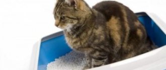
How to collect urine
There is no specific time, day or year required to take a sample. But the analysis should be submitted within the first 2 hours after collection. The frequency of delivery depends on the veterinarian's instructions. As a rule, urea is not collected for preventive purposes. Only on the recommendations of a specialist if you suspect a particular disease.
The minimum amount of urea suitable for analysis is 20 ml. At home, you can get by with a regular tray. First, we remove the litter and wash the cat litter without household chemicals (it will significantly distort the results). To disinfect, rinse the tray with boiled water.
Experienced breeders recommend using filler, but placing a plastic bag or cling film on top of it. Indentations are first made so that urine accumulates in the improvised holes.
The method is effective, but, as practice shows, in most cases the cat ruins plans. The bag will be dented or torn, and the urine will be absorbed into the filler. It’s better to do without it and do it another way.
We carry out the standard disinfection of the tray described above. We invite the pet to the toilet. Place a low object on one side of the tray. For example, an old saucer, a piece of linoleum, etc., so that the pet does not wet its paws and contaminate the sample. When the cat has done its business, we simply pour the urine into a plastic glass. It is better to buy it in the store and not use baby puree bottles or yogurt jars.
What should you do if your cat won't walk without litter? Wait until your pet settles into the tray. Approach it, lift the back and place the jar for analysis. It is better to do the fence together with a partner. One way or another, the best option would be a professional collection at a veterinary clinic.
Urine microalbumin, mg/l: normal 0–30 Protein, mg/dl: normal - negative.
Urine containing a large amount of protein foams heavily, and the foam persists for a long time. Normally, during high physical activity, hypothermia, allergic reactions and some other conditions, a larger amount of albumin may be present in the urine (functional hyperalbuminuria). In addition, proteins produced by tubular epithelial cells can also be detected in the urine. The cause of persistent hyperalbuminuria requires clarification (especially with concomitant persistent hyposthenuria), since this condition usually serves as one of the first markers of the development of severe nephropathies (diabetic nephropathy, primary chronic glomerulo- and tubulointerstitial pathies). Globulinuria (proteinuria) is always a pathology and can be associated with both infectious and non-infectious diseases of the kidneys and urinary tract.
Bilirubin, mg/dl: normal - negative.
In cats, bilirubin is not conjugated in the renal tubules. Therefore, the detection of bilirubinuria in cats (even in a mild and/or transient form) requires clarification of the causes of its occurrence. Bilirubinuria occurs in many liver pathologies. With mechanical (prehepatic) jaundice, bile with the bilirubin it contains enters the intestinal lumen in small quantities. For this reason, the bulk of bilirubin remains in the blood and is excreted from the body in urine, which gives it shades of color ranging from saffron-greenish to greenish-brown. At the same time, the content of urobilinogen and urobilin in the urine is significantly reduced, or these substances are not detected in it at all. In hemolytic jaundice caused by intensive breakdown of red blood cells (including babesiosis), urine, on the contrary, contains large amounts of urobilinogen and stercobilinogen, and bilirubin is usually absent in it.
Normal blood glucose levels in cats and deviations
Glucose is a substance that is a source of energy for the body. The result of a complex biochemical process - glycolysis - is the breakdown of glucose into simple molecules. This produces the energy necessary for metabolic processes in the body. This process is controlled by the brain, which ensures energy production when needed.
The pancreas is responsible for distributing glucose throughout the body, producing and releasing the hormone insulin into the blood. It ensures the delivery and absorption of glucose into cells: sugar is processed in cells, its amount in the blood decreases, and as a result, energy is produced. A decrease or increase in blood sugar levels indicates malfunctions in the functioning of internal organs and body systems.
Important!
A blood sugar test is performed on an empty stomach; the norm is considered to be a glucose level ranging from 3.4 to 6.1 mmol/l.
One-time changes in indicators should not cause alarm, and in case of prolonged hyperglycemia (increased glucose levels), additional examinations of the pet should be carried out.
Causes of hyperglycemia
The level of glucose in a cat's blood can increase under the influence of various factors:
- stress;
- use of hormonal medications;
- dysfunction of the endocrine system.
The following diseases of the endocrine system affect blood glucose levels:
- acute and chronic diseases of the pancreas;
- liver inflammation;
- disturbances in the functioning of the central nervous system;
- malignant tumors of the thyroid gland and other pathologies.
Exposure to negative factors leading to elevated blood glucose levels over a long period of time leads to diabetes mellitus. Characteristic symptoms of the disease:
- weight gain;
- polydipsia (increased uncontrollable thirst);
- polyuria (frequent urination);
- bad breath;
- changes in behavior - apathy, depression;
- changes in the appearance of the coat - it looks unkempt, matted, dull;
- impaired coordination of movements.
Decreased glucose levels
Low blood sugar levels (hypoglycemia) also negatively affect the functioning of the vital systems of the pet’s body. A decrease in glucose levels is provoked by the following factors:
- disruption of the endocrine system (pancreas, pituitary gland, thyroid gland);
- feeding errors (deficiency of carbohydrates entering the body with food);
- pathologies of the small intestine;
- kidney diseases;
- adrenal dysfunction;
- severe helminthic infestations;
- excess insulin.
Symptoms of the pathology are:
- constant feeling of hunger;
- nausea;
- vomit;
- decreased activity;
- lethargy;
- decrease in body temperature.
Acute hypoglycemia is a life-threatening condition. Lack of glucose in the body causes weakness, constant drowsiness, fatigue, and loss of appetite. A sharp drop in blood sugar (to 1.3-1.5 mmol/l) causes convulsions, impaired coordination of movements, muscle tremors, and can lead to hypoglycemic coma. Low glucose levels are much more dangerous than high glucose levels.
If your pet shows the first signs of illness, you should immediately contact a veterinary clinic. Without timely assistance, death is possible.
Urobilinogen, mg/dl: normal - Traces
A colorless substance, a product of bilirubin reduction, formed in the intestines, including. and under the influence of bacteria. Urobilinogen is formed from direct bilirubin that enters the intestine as part of bile and is partially absorbed back into the blood. Oxidation of urobilinogen in urine leads to the formation of urobilin. This reaction causes urine to darken during storage. With hemolytic jaundice, toxic and inflammatory liver lesions, enteritis and constipation, the level of urobilinogen increases significantly. When the bile duct is blocked (obstructive jaundice), urobilinogen is usually absent in the urine.
Urine chemistry
Protein . Normally, it should not be present, although a value of up to 0.3 g per liter is allowed. The appearance of protein in the urine indicates the presence of a pathogenic process, but which one is determined by additional research. Thus, protein can appear in biological fluid:
- with infection;
- anemia;
- pyelonephritis;
- urolithiasis;
- cystitis, urethritis;
- pyometra.
Glucose is another indicator that is not detected in the urine of healthy animals. Most often, the appearance of this carbohydrate indicates diabetes. But it can also be released during stress or acute renal failure.
Physiological glucosuria occurs when there is an excessive intake of carbohydrates into the body, due to the administration of drugs (steroids, cardiac glycosides, adrenaline).
Ketone (acetone) bodies . Their detection in urine indicates ketonuria or acetonuria. Normally, this phenomenon should not be observed. The presence of ketone bodies indicates:
- about diabetes mellitus with simultaneous detection of glucose;
- if there is no glucose, then most likely the cause of the excretion of acetone in the urine was fasting, prolonged eating of fatty foods, diarrhea or vomiting, or poisoning;
- about fever.
Bilirubin is a bile pigment. Detection of it in urine says:
- about liver problems;
- disruption of the outflow of bile due to blockage of the bile ducts;
- development of hemolytic jaundice.
Nitrite testing is not mandatory. This indicator is usually assessed when a bacterial infection is suspected. The fact is that microbes are able to convert nitrates, which are always present in urine, to nitrites.
Blood and hemoglobin in the urine are an alarm bell of a serious pathology. Blood in its pure form is found:
- in case of injury to the ureters or bladder during the passage of kidney stones;
- jade;
- tumors in the organs of the urinary system.
Coffee staining indicates an admixture of hemoglobin, which occurs in cases of poisoning, burns, and some infections.
Glucose, mg/dl: normal - negative.
Any level of glucosuria requires clarification and further examination of the patient, since it can be one of the symptoms not only of diabetes mellitus or Cushing's syndrome, but also of TIN and CGN with a tubular component. Normally, the tubular epithelium completely reabsorbs glucose from primary urine, and glucosuria not associated with diabetes mellitus indicates its severe damage. Glucosuria can be transient in nature during pregnancy and excessive feeding of various sweets to dogs, and especially cats, whose insulin reserves are small.
Red blood cells (reaction to hemoglobin), units/µl: normal - negative.
Microhemoglobinuria is characterized by the content of red blood cells in the urine up to 100 cells per field of view and is not reflected in its color. The cause of urine staining in a red-brown color of varying intensity - macrohematuria - may be hemoglobinuria (if the pathological process that led to it is sufficiently severe, the blood plasma also turns red, clinical symptoms of oxygen deficiency increase, but red blood cells are not detected in the urine sediment), erythrocyturia (the blood plasma remains straw-yellow, and fresh or leached (dysmorphic) red blood cells are found in the urine sediment), as well as myoglobinuria, the cause of which is most often rhabdomyolysis. During the day, 2–3% of red blood cells are destroyed in the body. Even if for some reason parts of the hemoglobin protein and its compounds enter the primary urine, they are completely metabolized and reabsorbed. This oxygen-consuming and highly energy-intensive function is one of the priorities for tubular epithelial cells and, when its physiological capabilities for its implementation are exceeded, it begins to be carried out even to the detriment of its own interests of metabolism and energy. Therefore, any conditions characterized by massive, and especially prolonged, breakdown of erythrocytes (blood parasitic infections, transfusion of incompatible blood, extensive hematomas), as well as muscle fibers during rhabdomyolysis, for example, can result in degeneration and atrophy of tubular epithelial cells, which, in turn, turn, can lead to TIN. In this regard, it is nephropathy, and not liver damage, as is usually believed (hepatocytes, unlike tubular epithelial cells, are capable of intensive regeneration after the disappearance of the damaging factor), are a very significant complication, for example, of babesiosis, and conditions characterized hemoglobinuria or hematuria, necessarily require clarification of its occurrence and, if necessary, subsequent nephroprotective treatment. In addition, hematuria is characterized by many kidney lesions of an autoimmune (most GN) and toxic nature, as well as infectious and oncological diseases of the urinary system and urolithiasis. The presence of acanthocytes (red blood cells with uneven contours, resembling a maple leaf) in urine sediment is a highly specific sign in determining glomerular hematuria. And the presence in the urine, in addition to acanthocytes, also agranulocytes, as well as leukocyte and granular casts, gives good reason to give the patient a clinical diagnosis - chronic GN. Hypocoagulable hematuria is also identified, which is not associated with diseases of the MVS organs.
Cat urine tests
Urine tests are an indispensable research method in diagnosing most pathologies of the urinary system and can help clarify the diagnosis for many other diseases.
For a long time, most kidney diseases in animals occur without any clinical manifestations or changes in blood tests. Changes for a long time can be present only in urine tests. Therefore, urine examination is the only method for early diagnosis of kidney disease in dogs and cats.
INTRODUCTION
Urine is a biological fluid that is continuously formed in the kidneys as a result of complex interconnected processes of filtration of the liquid part of the blood in the glomeruli and reabsorption of substances necessary for the body in the tubules. During the formation of urine, about a quarter of the blood from the minute volume of cardiac output passes through the kidneys, which means that this paired organ is even better supplied with blood than the heart muscle.
Urine formation is the process by which many end products of metabolism and breakdown of substances, excess fluid and salt, as well as hormones, enzymes, vitamins and other biologically active substances are removed from the body of animals. In addition, most drugs used to treat animals are either biotransformed in the kidneys or excreted from the body in urine.
By analyzing the composition and properties of urine over time, it is possible to obtain information about the state of the patient’s various organs and systems, assess the severity of the disease, and also draw a conclusion about the effectiveness of the treatment.
PICKUP, STORAGE, TRANSPORTATION
The information content and clinical objectivity of urine tests largely depend on how correctly its collection, storage and transportation were carried out.
The container for collecting urine for analysis should be well washed (it is better to use special containers or 5-20 cc syringes) to avoid the appearance of otherworldly impurities in it.
If not all of the urine obtained is delivered for examination, then before draining a small part (especially after storage), it is necessary to shake it well so that the sediment containing the important constituent elements of urine is not lost.
Before collecting urine, the genitals and perineal area must be cleaned of dirt and washed with an antibacterial detergent. However, complete sterility is not required for a general urine test.
In cats, urine can be collected for analysis at any time of the day (in this type of animal, natural daily fluctuations in urine composition due to physiological characteristics are minimal). To do this, you need a clean, dry and, if possible, a new tray (since the used one, even after rinsing, can retain residues of substances that contribute to the decomposition of fresh urine).
If the animal refuses to urinate without litter, a special urine collection kit for cats may be indispensable, including a package of non-absorbent neutral polymer granules, a pipette for collecting samples and a disposable plastic container for storing them.
It is necessary to store and transport urine, especially in cats, only at positive temperatures. Otherwise, due to active adhesion and precipitation of salts, it may become cloudy, which will make it difficult or even impossible to carry out some laboratory tests. Since a high content of salts in the urine is the norm for cats, their detection in urine tests cannot be the only basis for making a diagnosis of “urolithiasis”. To accurately determine this pathology in a patient, it is necessary to conduct an ultrasound of the urinary system.
It is advisable to deliver urine for testing no later than 2-4 hours after receiving it. This is due to the fact that this biological fluid in animals, as a rule, is significantly contaminated with foreign bacterial flora, the active reproduction of which is associated with a significant change in its composition.
Microorganisms also consume glucose intensively, so if diabetes is suspected, underestimated or even false-negative results can be obtained. It should also be taken into account that bile pigments that appear in the urine during liver diseases are destroyed in sunlight.
In addition to incorrect collection, storage and transportation of urine for analysis, the following factors can affect its composition:
- estrus and disease of the female genital organs (endometritis, vaginitis);
- inflammation of the preputial sac and glans penis and prostatitis in male and female cats;
- catheterization of the bladder, carried out for various indications;
- giving certain medications (diuretics, multivitamins, ascorbic acid, etc.).
GENERAL (CLINICAL) URINE ANALYSIS
General urine analysis is a laboratory method that includes the study of the physical, chemical, and some biochemical properties of urine, as well as the study of its sediment. It can be indispensable not only when a patient is suspected of having pathologies (primarily due to a shortage of highly qualified laboratory technicians), but also in the primary diagnosis of diseases of other systems and organs. A general clinical urine test allows the doctor to identify a number of hidden diseases, adjust subsequent diagnostic measures, and also evaluate the effectiveness of the treatment.
A general urine test includes a study of the color, odor, transparency, specific gravity and acidity of this biological fluid. In addition, the presence in the urine of protein, fats (in cats, the presence of fatty droplets in the urine is normal), glucose, hemoglobin, salts and some other indicators, as well as such formed elements as red blood cells, leukocytes, various renal cylinders, etc. . (See the article What does a urine test tell you)
The qualitative and quantitative composition of urine (volume, acidity level, density, content of salts and pigments) in dogs may change during the day. In domestic cats, the ability of the kidneys to concentrate urine is much higher, and its daily volume per kilogram of body weight is less than in dogs. Therefore, fluctuations in the composition of urine in cats are not as pronounced. However, a single urine test is usually not sufficient to detect organ disease or lack thereof in both dogs and cats. Sharp deviations from the norm in urine tests may be the result of violations during its collection, storage and transportation and, as a rule, need to be checked.
In cats suffering from diabetes, the most significant complication and the most common cause of death is the so-called. diabetic nephropathy. The first sign of this disease is the appearance of an increased amount of protein in the urine.
In cats, certain kidney diseases, called glomerulonephritis, occur many times more often than in dogs, since they are a common complication of both acute and chronic, and slow (i.e., characterized by viral carriage without any clinical manifestations) viral infections in this type of animal.
Special risk groups include:
- show and breeding cats, as well as animals kept in large groups (due to the constant exchange of viral agents, which are the main trigger (namely the trigger, not the direct cause) leading to the development of the disease);
- cats who, for one reason or another, were prescribed certain types of antibiotics (gentamicin, kanamycin, neomycin, monomycin, tobramycin and other aminoglioside antibiotics) or painkillers from the group of non-steroidal anti-inflammatory drugs (ibuprofen, diclofenac, ketoprofen, etc.).
For this reason, a general urinalysis is recommended for all cats, regardless of age or health status.
The need for regular urine testing in dogs and cats is also due to the fact that for a long time, most kidney diseases occur without any clinical manifestations or changes in blood tests.
Pathological abnormalities may be present only in urine tests for a long time. This is due to the colossal compensatory capabilities of the kidneys, in order to preserve their functions, they begin to work extremely hard, to the detriment of their own well-being.
Therefore, both the diagnosis and treatment of many kidney diseases must begin long before the appearance of clinical signs of trouble, since the appearance of the latter, among other things, significantly reduces the possibility of drug intervention, or even completely eliminates it.
Roman Leonard, President of the Russian Scientific and Practical Association of Veterinary Nephrologists and Urologists, Head of the Ural Center for Veterinary Nephrology and Urology, Chelyabinsk.
pH: normal - acidic
In cats, as obligate carnivores, urine is normally acidic. The predominance of plant components in the diet of dogs can change the pH of urine in the alkaline direction. The pH of urine is also affected by the level of acid-base balance in the blood, since the kidneys actively participate in its maintenance and remove excess hydrogen ions through the filtration process. Pathological causes of urine acidification (pH<5): respiratory or metabolic acidosis, hypokalemia, profuse diarrhea, anorexia, diabetes mellitus, prolonged hyperthermia. Long-term use of aspirin and methionine can also lead to urine acidification. Pathological causes of alkalinization of urine (pH>7): severe chronic renal failure, when the kidneys are no longer able to remove excess hydrogen ions from the blood (tubular acidosis), hyperkalemia, compensated or uncompensated hyperphosphatemia, hyperplasia of the parathyroid gland (hyperparathyroidism), prolonged vomiting, cancer kidneys or bladder.
Nitrites: normal - negative.
The appearance of nitrites in urine is possible under the influence of enzymes of certain bacteria that enter it both from the kidneys and urinary tract, and when it is contaminated from the outside. Therefore, nitrituria in MJ is only indirect evidence of infectious diseases of MW. On the other hand, not all uropathogens are capable of fermenting nitrites from nitrates. Therefore, if nitrites are not detected even in long-term stored urine samples, this does not necessarily exclude a urinary tract infection.
Leukocytes, units/μl: normal - negative.
Leukocyturia, especially weakly or moderately expressed, does not always (primarily in cats) indicate an infectious disease of the urinary system, although it is the most common reason for the unjustified prescription of antibiotic therapy. Most chronic GN and CKD are characterized by intense focal and/or diffuse infiltration of the renal parenchyma by agranulocytes, which are involved either in autoimmune inflammation or in the processes of destruction of sclerotic areas of the parenchyma. On the other hand, infectious (septic) leukocyturia is a characteristic sign of acute pyelonephritis. However, during remission or during a latent course of chronic pyelonephritis, it may be absent. Therefore, in most cases, an infectious disease of the urinary system is recognized only if a urine sample correctly obtained by transperitoneal urocystocyntesis during bacteriological examination reveals a microflora that is pathogenic and not saprophytic for the skin or gastrointestinal tract. However, when assessing negative results of bacterial culture (urine is sterile), one should take into account the intermittent (variable) nature of bacteriuria in chronic pyelonephritis (this is not observed in pyonephrosis), as well as difficulties in identifying certain forms of pathogenic bacteria.
Cat urine analysis: how to store and transport urine for research
The best option is to conduct urine tests on cats no later than half an hour after receiving a portion of urine.
But, more often than not, this is impossible and a large amount of time must pass before the examination.
Therefore, the study is carried out later, and the collected urine should be stored in a container in a cool place
.
This can be explained simply. 2 hours after collection in urine
the growth of pathogenic flora, including staphylococcal flora, begins.
In addition, after such a period of time, the acidity level in the urine changes, the cellular inclusions of the sediment are destroyed, and other biochemical and chemical changes occur that distort the results and the interpretation will be incorrect. If the urine is too cooled before the test, this will lead to the phenomenon of crystallization in vitro , that is, it will significantly enhance the process of crystallization of the cat’s urine.
To preserve urine for a long time before analysis, you need to add a special preservative to it.
It can be taken from a laboratory for the study of biomaterials and has a very low cost, accessible to almost everyone.
It is necessary to tell the owners of clawed pets that it is recommended to take tests regularly
at least once every six months. This is due to the fact that cats suffer from certain diseases with subtle symptoms. For example, a neutered cat has an increased risk of developing urolithiasis. And this disease must be prevented by changing and adjusting the diet. And other diseases, more often of a metabolic nature, occur more often in castrates. This fact is a reason to think carefully before deciding to castrate your pet.
In kidney diseases, the presence of staphylococcus is most often noted, and urine analysis shows
how severe the disease is. There are a huge number of diseases identified by urine. And only one analysis can give a clear picture of the state of the body of your beloved cat.
At the Emergency Veterinary Care Center for animals, urine will be quickly and painlessly taken for general and/or detailed analysis, and urinary parameters will be examined and studied. The tests are deciphered in our Center within a few hours.
If you find an error, please select a piece of text and press Ctrl+Enter.