Fungal diseases are a real scourge in both medical and veterinary practice. They are often difficult to identify, and many problems arise with treatment. One of the classic pathologies (even if rare) is Malassezia in cats. In this article, we will tell you in detail about this disease, its manifestations and treatment methods.
Malassezia, or malassezia, is a skin disease of cats caused by the yeast-like fungus Malassezia pachydermatis.
What to do in such a situation? To get started, we recommend reading this article. This article describes in detail methods of controlling parasites. We also recommend that you consult a specialist. Read the article >>>
Prevention
To prevent the development of Malassezia in cats, the following rules should be followed:
- carry out deworming and treatment of the animal against ectoparasites in a timely manner;
- exclude from the diet foods that cause allergy symptoms;
- monitor the condition of the coat and skin;
- vaccinate your cat in a timely manner;
- Be examined by a veterinarian 2 times a year.
Proper care of your cat will protect it from many diseases.
These recommendations will help you avoid many troubles with your pet’s health, and will also prevent the appearance of Malassezia and other serious pathological processes in the body.
The Malassezia fungus lives on the skin of cats all the time, and if circumstances favor it, it can lead to inflammatory processes on the skin. As a rule, they begin in the area of the external auditory canal, around the anus, in the lips and interdigital space.
In addition to these places, although somewhat less frequently, it can also affect other surfaces of the skin. Malassezia is a concomitant agent that causes dermatitis in cats in the presence of primary skin damage by some other factor. Malassezia pachidermatis is often isolated from the affected part of the skin of the auricle and causes external otitis and generalized skin lesions.
Studies of the degree of colonization of this pathogen on the skin have found a significant increase in their number (sometimes several hundred times) on damaged skin.
In addition, it was found that the number of malacesia on the skin of those breeds of cats that are more prone to dermatitis is noticeably higher than on the skin of cats of other breeds.
This fact clearly indicates that some cat breeds are more susceptible to this disease than other breeds. This may indicate that some cat breeds have disorders in terms of resistance in general and the skin in particular. Some cats with atopy generally have abnormally high populations of M. pachidermatis in both affected and unaffected areas of the skin.
In addition, in cats with fungal dermatitis caused by Malacesia, concomitant increases in skin bacterial microflora (usually Staphylococcus intermedius) are often found, but since this pathogen is also present in the norm, we can talk about the fact that these individuals have a general tendency to reduction of the protective functions of the skin.
There is still no clear understanding of the reasons that determine the transition from carriage of commensals to opportunistic infection. Sometimes other skin diseases are discovered. For example, there may be keratinization defects or hypersensitivity, but the immunological or chemical abnormalities that allow the yeast to grow have not yet been elucidated.
How contagious is the fungus in Malassezia?
When doctors use the term “malassezia,” they mean a fungal organism of the yeast species. It is slightly convex in shape and has a cylindrical structure, smooth, soft, loose, and small (if translated into microns, it is two and a half microns). Looking at it, you may think that you are seeing an ordinary nut. Malassezia crops are characterized by an unusual smell with a “fruity” tint.
When you see someone with Malassezia dermatitis, you don’t have to run away in fear or hide your pet away. The idea that Malassezia fungus is contagious is completely false. Since it is one of the components of the microflora, which tends to be present on the epithelial tissue of the animal throughout its life.
In everyday life, the fungus causes absolutely no harm. Excessively fast and large reproduction begins after any incidents that create an excellent environment for this. Most often this is provoked by various changes in the microclimate of the skin (humidity, warmth, advanced inflammation).
Prevention of ear disease
Prevention of ear problems includes cleaning with substances prescribed by your veterinarian. This type of cleaning must be carried out continuously. Do not forget that the process of cleaning the ears should not cause pain to the animal. The entire procedure is done carefully so as not to injure the cat’s hearing organs. To avoid any unpleasant moments, you need to know what the prevention of ear disease in cats is. To clean your ears you need to:
- prepare tools, substances;
- will make sure that the cat is calm and not alarmed;
- wrap the animal in a towel to protect yourself from scratching with claws;
- It is necessary to drop a substance or lotion into the ear;
- after instillation, you should not pinch your ear too much and make massaging movements;
- After this manipulation, you can take a cotton swab and remove excess liquid.
When the treatment and cleaning of the ear is done correctly, the cat owner does not need to worry that the pet may develop complications. Teach your animal from childhood to constant ear examinations. Do this delicately, combining it with gentle stroking.
It is necessary to periodically prevent ear diseases
Ear washing lotions - “Otifri” and “Epi-otik”, as well as “Bars” lotion, in addition to 2 products from the German company Beaphar - can be obtained at any veterinary store. An interesting solution is the “Miss Kiss” sticks - these are ordinary sticks soaked in a medicinal agent.
Reasons for the development of the disease
The disease is caused by the yeast fungus Malassezia pachydermatis, which is often present on the skin of animals, in the ears, and also on the surface of mucous membranes. It interacts with the no less common staphylococcus, and in the process of their joint life activity a lesion occurs called malassezia dermatitis. Yeast and bacteria, complementing each other, promote mutual growth and active development.
The transition of this fungus to pathogenic status is due to many different factors . In the vast majority of cases, this disease is secondary, provoked by hypersensitivity, endocrine diseases, and keratinization disorders. It can easily develop against the background of almost any dermatological problem, which results in damage to the upper layers of the skin.
Ear hematoma in cats
With a hematoma, blood flows from the blood vessels of the auricle into the tissue under significant pressure, pushing these tissues apart and forming a cavity. The size of the hematoma depends on the strength of blood pressure in the damaged vessel, as well as on the degree of compliance of the tissues located near it.
A hematoma occurs quickly and its volume increases until the pressure from the stretched tissue becomes equal to the pressure in the damaged blood vessel. After this, the spilled blood clots, and a blood clot forms in the blood vessel.
Most often, hematomas in cats occur on the inner surface of the ear and much less often on the outside. The damaged ear increases in size, hangs down, and the swelling is painful and hot to the touch. If the hematoma is not treated, the pain only increases, and the hematoma itself can become infected with secondary microflora, which can ultimately lead to necrosis of the ear cartilage.
During a clinical examination of such a cat, a veterinarian notes the following symptoms:
- We observe anxiety and nervousness in the cat.
- The cat almost constantly shakes its head from side to side.
- He constantly scratches his damaged ear with his paws.
- When trying to stroke the cat's head, it becomes aggressive.
Treatment. Treatment of auricular hematoma is not very difficult. If no more than 48 hours have passed since the formation of the ear hematoma, then the cat owner fixes the ears with a bandage on the back of the head and applies cold. In the future, in order to resolve the hematoma, it is necessary to use heat and apply locally irritating ointments.
If the hematoma cannot be cured at home, the owner needs to contact the nearest veterinary clinic. In a veterinary clinic, the veterinarian will open the resulting hematoma, remove blood clots from it, wash the resulting cavity with a solution of novocaine with antibiotics and give recommendations to the owner so that the hematoma resolves safely.
Characteristic symptoms
Malassezia dermatitis affects dogs regardless of breed. However, at the genetic level, basset hounds, dachshunds, shih tzus, American cocker spaniels and English setters are most susceptible to this disease. Typically, Malassezia skin lesions occur in humid and warm weather, in spring and early autumn. During these periods, animals also often suffer from allergic reactions to pollen and molds, which greatly increases the risk of developing dermatitis as a secondary reaction of a weakened body.
In 70-80% of dogs suffering from Malassezia dermatitis, concomitant dermatoses, pyoderma, and other reactions caused by the body’s special sensitivity to external irritants were also identified. The main symptom is severe itching, which haunts the animal and occurs in almost every case of lesions.
There are certain signs that can help recognize that your pet has Malassezia dermatitis:
- In many cases, dogs experience an increase in oily skin and coat.
- Symptoms often include the development of dry and wet seborrhea and the formation of crusts.
- Quite often the course of the disease is accompanied by an unpleasant odor reminiscent of rancid fat.
- If dermatitis is chronic, it may well be accompanied by manifestations such as lichenification and hyperpigmentation of skin areas affected by the Malassezia fungus.
- Classically, local lesions are located in the ears, skin folds, armpits, groin area and on the dog’s face.
- Paronychia, which is characterized by deformation of the color of the fur and claws - they become a rich reddish-brown color.
- Often, in parallel with Malassezia disease, the animal develops external otitis and interdigital furunculosis.
In a considerable number of cases, up to 40%, this problem is accompanied by the development of a disease such as pyoderma; dermatoses of other types can also occur.
The lesions can spread over the surface of the animal's skin and merge with each other, affecting a considerable area of the body.
This type of dermatitis is quite rare in cats. The symptoms and main lesions are not different from Malassezia disease in dogs. This disease is not contagious, and appears solely due to individual disturbances in the healthy balance of the fungus on the skin of the sick animal.
Malassezia pathogen
The yeast-like fungus Malassezia pachydermatis is an opportunistic microorganism that is constantly present on the skin of healthy cats, but does not cause disease. Increased proliferation of fungi and the appearance of the clinical picture of malassezia occurs with a sharp decrease in the body's resistance, provoked by:
- poor diet;
- exhaustion;
- injuries or large blood losses;
- infectious and invasive pathologies;
- stress;
- allergic reactions;
- poisoning;
- scratches, burns, cuts and other skin damage.
The localization of lesions is due to the fact that lipids are required for the life of these microorganisms. Therefore, they are actively developing in the areas of:
- external auditory canal;
- interdigital and perianal folds;
- neck;
- belly.
The clinical picture of the disease varies somewhat depending on the location of the pathogen.
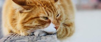
The first stage of the disease is characterized by the appearance of erythema, which later becomes covered with scales. This phenomenon is accompanied by severe itching, causing anxiety in the animal and leading to scratching.
Next, exudative diathesis develops, which is accompanied by:
- thickening of the epidermis;
- the appearance of acne;
- unpleasant odor;
- alopecia.
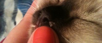
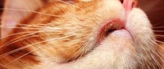
On the chin, Malassezia causes folliculitis, accompanied by copious outflow of serous exudate, maceration and exfoliation of areas of the epidermis. With the development of auricular Malassezia, the external auditory canal becomes covered with a black coating that has a specific sour odor. Intense formation of serous exudate leads to its leakage from the ears. The cat often shakes its head, bends it low, trying to get rid of the itching, and hits itself with its paws.
In veterinary practice, isolated cases of the development of the Malassezia fungus on the mucous membranes of the oral and nasal cavities, eyes and genitals have been recorded.
Treatment
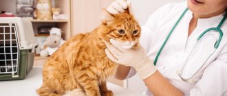
To successfully eliminate malassezia, a comprehensive treatment is required that eliminates both the symptoms and the root cause of the disease. Therefore, contacting a veterinarian in this case is mandatory.
The skin should be treated daily with a 2% chlorhexidine solution, applying antifungal ointments (Lamisil, Exoderil, Clotrimazole and others) on top. In case of a generalized course of the disease, it is more advisable to use a specialized shampoo (Mycozoral). Among antimycotic oral agents, many experts give preference to Ketoconazole and Fluconazole.
Ear fungus is treated according to the following scheme:
- 2 times a day, removing plaque with an ear stick soaked in 0.05% Chlorhexidine;
- instillation of Otibiovin or Otibiovet ear drops according to the instructions;
- use of oral Ketoconazole in the dosage prescribed by your doctor.
Clinical picture
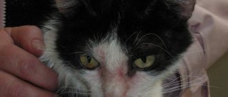
What are the symptoms? Some animals do not show any symptoms throughout the course of the disease, with the exception of constant “over-grooming” - they constantly lick themselves, causing their fur to literally stick together and become shiny with saliva. But this still rarely happens: more often the infection makes itself felt through patches of inflamed, reddened and flaky skin that appears in different parts of the body of a sick animal. Generalized forms of fungal dermatitis are relatively rare, and this can only be seen in the case of weak, emaciated and seriously ill animals, whose immune system cannot resist the action of pathogenic fungi.
Typically, classic Malassezia dermatitis develops in animals between one and four years of age. Let us remind you once again about the long-suffering sphinxes - in their case, it is very easy to see the onset of the disease. A thin brownish sebaceous film appears on the animal's skin. There is especially a lot of it in the interdigital spaces. And from this, cats, by the way, begin to constantly chew and lick their paws. In addition, the accumulation of sebaceous exudate occurs especially actively in the groin area. The consequences are the same - a sick animal constantly licks and chews these places, trying to get rid of the burning and itching.
It is important not to miss this moment. If at this time you start bathing your pet regularly, once every two to three days, using a 0.2% fluconazole solution, the disease can be defeated in just a couple of weeks. Unfortunately, in representatives of long-haired breeds, the external manifestations of the disease are not so obvious, and therefore the disease gradually progresses.
Malassezia dermatitis
Malassezia dermatitis - What is it?
Malassezia pachydermatis
is a common yeast fungus that is normally found on the skin and ear canal of healthy dogs and cats in small quantities. Under certain conditions, it begins to multiply and can lead to illness. The fungus is not contagious to people or other animals. Since it is part of the normal flora of the skin, it begins to multiply intensively only when the conditions necessary for this arise. Increased reproduction of this fungus is usually secondary to some other disease that has changed the microclimate of the skin. It generally likes moisture, warmth and inflamed skin. Dogs of “folded” breeds are more predisposed to this disease. Primary ailments that predispose to yeast infection are allergies, hormonal disorders, and decreased immunity.
Malassezia dermatitis can be localized or generalized, and is usually accompanied by itching. The degree of itching can vary from mild to very severe. The affected skin usually turns red and often thickens and darkens. The most common areas for this disease are the ears, groin, interdigital space, neck, chin, axillary areas, skin folds (muzzle, tail). An unpleasant odor may be noticeable from the affected skin.
Malassezia dermatitis is diagnosed using cytology, a test of stained samples from the skin or ears, which reveals an increased number of these fungi on the skin. A few individual fungi in a sample are normal, so culture has no diagnostic value. More than 1-2 fungi in the field of view of the microscope indicate an increased number. Sometimes you can find dozens in one field of view.
Treatment of Malassezia dermatitis or otitis is carried out using local and systemic antifungal agents. Some cases are difficult to treat
It is important to continue treatment until the cytology shows no fungi. Usually the course of treatment lasts 30-45 days, but in some cases healing may take longer
We must try to find out the main cause of the disease. In cases where this cannot be done, or the underlying disease does not respond to treatment, a relapse of Malassezia dermatitis may occur. In such cases, long-term maintenance therapy may be required.
The main provoking factors of Malassezia
In order to protect your dog from the disease and, if necessary, take immediate action, you need to know what factors may favor the active growth of fungus on the skin and diagnose possible concomitant diseases.
It is reliably known that the disease is not contagious. It is not transmitted from dog to dog. The disease has internal causes. Malassezia canine fungus is also not contagious to humans and other animals.
Many years of experience in breeding dogs allowed experts to draw a conclusion about the main reasons for the active proliferation (spread) of fungus on the skin of animals, including:
- weakened immune system;
- infectious diseases;
- chronic diseases;
- allergic reactions of various etiologies;
- any skin diseases that violate the integrity of the skin;
- taking hormonal drugs and antibiotic therapy;
- too frequent use of shampoos and other detergents;
- poor care for thick, long hair.
Weakened immunity
The main cause of the disease is weakened immunity. Low resistance to skin infections may be hereditary or may be a chronic disease. A hereditary disease is diagnosed very early, when skin diseases begin to attack the puppy at a very tender age.
Chronic diseases
Chronic diseases are no less dangerous, as they cause a persistent decrease in immunity. Constantly progressing, they create favorable conditions for the growth of colonies of the fungus - the invader. The dog's skin becomes a pathogenic environment for them, which they try with all their might to suppress.
Infectious diseases
Skin infectious diseases have the same result - immunodeficiency. In addition, many of them are characterized by severe, unrelenting itching. Skin scratched until it bleeds is a gateway for pathogens. And pathological organisms for the fungus are a sign of rapid reproduction in order to fight “for territory.”
Allergic reactions of various etiologies
An allergic reaction on the skin creates the same conditions (scratching, violation of the integrity of the skin, decreased immunity) that encourage microorganisms to actively multiply.
Treatment with hormonal drugs and antibiotics
Hormone replacement therapy in most cases has side effects. In particular, this has an adverse effect on the organs of the endocrine system. Their functions may be impaired, causing the production of natural hormones to suffer. All this does not have the best effect on the condition of the skin. And this is a direct path to activating fungal growth.
Taking antibacterial agents also has its downsides. By actively suppressing the growth of pathogenic bacteria, they at the same time deal a significant blow to the body’s protective functions. Therefore, each intake of antibiotics must be accompanied by a parallel intake of antifungal and immunostimulating agents.
Frequent use of detergents
One of the main factors of protection against fungal infection is maintaining the integrity of the skin. Frequently used shampoos and detergents can artificially disrupt this integrity. They wash away the protective layer of the epidermis, and the skin becomes dry and susceptible to various infections.
But at the same time, careful ear care is necessary. There is a favorable environment for the development of Malassezia - a warm and humid environment. The ear canals should be promptly cleaned of wax secretions, wiping them dry each time.
Poor grooming
Long, poorly combed hair creates an obstacle to good air exchange and ventilation of the skin. Humidity and high temperature of the skin make it possible for fungal infections to quickly multiply on it.
Diagnostics
One of the main diagnostic methods is cytological examination of the affected skin area. This may be a surface scraping, a biopsy, or an imprint on adhesive tape.
If the mucous membranes are damaged, scrapings or washings are taken from them.
Diagnosis of mold fungi, unfortunately, is only possible through a postmortem examination. In the cutaneous form of this mycosis, a biopsy of nodules with histological and microbiological examination is used.
Differential diagnosis from lichen is carried out using a Wood's lamp examination.
If there is a suspicion of lung damage, then a chest x-ray is performed. However, this pneumonia is no different from bacterial pneumonia, which makes diagnosis and subsequent treatment difficult.
A general urine test for genital candidiasis shows an increased content of bacteria.
It is also recommended to take a general blood test and biochemistry to determine the general condition of the cat.
Therapeutic measures
Let us note that long-term treatment of fungal diseases never leaves without consequences for the animal’s body: with inappropriate dosage and uncontrolled use of all antifungal drugs, you can easily poison your own pet. For non-capricious cats, treatment is the simplest: they are bathed twice a week using Malaseb medicated shampoo or something similar. In about four to six weeks the animal recovers completely.
If it constantly itches and licks itself, there are signs of a secondary bacterial infection, and you will have to resort to more drastic methods. In such cases, in addition to purely antimicrobial drugs, it is recommended to use the antibiotic Cefovicin. Under no circumstances should you prescribe anti-inflammatory corticosteroids - yes, they relieve inflammation, but they reduce immunity, and in case of fungal infections this is deadly.
Oral and parenteral administration of antifungal drugs is resorted to when regular washing does not help. have proven to be especially effective in treating cats . Alas, feline species of the pathogen are very resistant to these drugs (unlike dogs), and therefore the duration of the therapeutic course can exceed a month.
Source of the article: https://vashipitomcy.ru/publ/zdorove/bolezni/malassezioznyj_dermatit_u_koshek_osobennosti_zabolevanija_i_neobkhodimaja_terapija/15-1-0-1879
Diagnosis and treatment
One of the most accurate methods of “identifying” the pathogen in this case is microscopy. But growing yeast on a special nutrient medium is much more accurate. Since Malassezia can be present in healthy animals, an accurate diagnosis depends on whether the animal responds to treatment. By the way, how is Malassezia treated in dogs?
The main task is to create an environment that is hostile to the pathogen. To do this, they use drugs based on chlorhexidine, sulfur and others. Interestingly, some shampoos for people (Head & Shoulders) can be unexpectedly effective: such treatment with “folk remedies” significantly alleviates symptoms. Veterinarians also prescribe special ointments, which should be used at least twice a day, the total duration of treatment is at least several weeks.
Ketoconazole, fluconazole and similar drugs are prescribed orally. What to do if the infection is in the ears? In this case, you will have to clean them, and this must be done at least two to three times a day. Ordinary boric alcohol works well. After the ear is dry, nystatin, thiabendazole or clomitrazole are used. Here's how to treat Malassezia.
It is important to remember that treatment of this disease (like all fungal pathologies) takes more than a couple of days! You better get ready for several weeks of intensive therapy right away. Moreover, the dog must always be under the supervision of an experienced veterinarian, since the antifungal drugs used for this disease are far from harmless
If you see your dog vomiting or exhibiting inappropriate behavior after using them, call your veterinarian immediately!
How is Malassezia pachydermatis treated?
The dosage of antifungal agents is different for animals and humans. The treatment regimen is determined by the veterinarian; self-medication can aggravate the condition. Treatment for Malassezia in cats includes antifungal medications such as:
- "Ketoconazole";
- "Itraconazole";
- "Chlorhexidine";
- solution of sulfur and salicylic acid;
- piroctone olamine;
- selenium sulfide;
- "Enilconazole".
Before starting antifungal therapy, it is necessary to cure the concomitant ailments that caused Malassezia.
You can increase your cat’s immunity with vitamin complexes:
- "Canvit Multi";
- "Kanvit Nutrimin";
- "Natur Protection Daily Vitamin Formula";
- "Beafar."
Balancing your diet will help speed up the healing process. A balanced diet with medicinal food is also recommended to restore the structure of the coat and skin. These include:
- Royal Canin Intense Beauty;
- "About Plan Derma Plus";
- "Brit Car Kat Sunny Beautyful Hair";
- "Trainer Staff Adult Beauty."
Classification of fungal diseases
Diseases caused by pathogenic fungi are divided into 2 groups depending on the location of the lesion:
- superficial mycoses, the causative agents of which parasitize the skin and mucous membranes of animals;
- systemic mycoses that cause pathological changes in internal organs.
Diagnosis of the second group of diseases is difficult due to the lack of clinical symptoms characteristic of common fungal infections. Therefore, even signs of a cold or intestinal disorder should be a reason to contact a veterinary clinic, where an experienced doctor will be able to recognize the exact cause of the disease.
The most frequently reported fungal diseases include:
- ringworm caused by parasites of the genus Trichophyton and Microsporon;
- Malassezia, caused by the ear fungus Malassezia;
- aspergillosis, caused by saprophytes of the genus Aspergillus;
- candidiasis provoked by yeast fungi of the genus Candida;
- cryptococcosis caused by the saprophytic fungi Cryptococcus.
Once pathogenic microbes have reached the skin surface, they immediately begin to harm the health of the cat (by releasing toxic toxins and enzymes).
Over time, this can lead to the death of the epidermis from the upper skin and the development of inflammatory processes.
After all, without proper care, the fungus will penetrate deeper into the pet’s body, damaging the fur structure and hair follicles.
What signs can be used to determine the presence of harmful parasites? First of all this:
- Loss of damaged pieces of fur;
- The presence of empty bald spots on the cat with clear boundaries in the head, ear area, and also on the paws;
- Restless behavior (the animal runs from corner to corner, itches and often rubs against objects);
- Scabies;
- Bad smell;
- The presence of a strange, pinpoint plaque in the ears;
- Formation of tubercles on the skin surface;
- Frequent sneezing;
- Bloody runny nose.
If the situation is neglected, the initial symptoms can develop into a severe form of the disease, which will lead to anemia, pneumonia, and complications in the gastrointestinal tract.
If the cat is still young or weakened (for example, due to lack of food), this can be fatal.
Ringworm vaccines for cats
Today, Russian veterinary medicine has four vaccines in the form of injections, which are prescribed for lichen in cats, dogs and fur-bearing animals.
Vaccerm Vaccine for cats
The biological product is a dry or liquid mass with inactivated spores of dermatophytes (fungi of the genus Microsporum canis, Microsporum gypseum and Trichophyton mentagrophytes).
The drug Vakderm was created for the immunization of healthy cats, dogs, rabbits and fur-bearing animals, as well as for identifying infected animals. If the cat has previously been in contact with a shingles animal, but clinical signs have not yet appeared, then after vaccination the process will accelerate. But the vaccination will not harm a completely healthy cat and no signs of disease will appear.
According to the manufacturer of the Vakderm vaccine:
- the vaccination has a therapeutic effect, if it is given three times (1 time to detect the disease, 2 and 3 times to speed up healing when lichen has been detected), then after 15-25 days the animal will recover;
- After immunization, animals become immune to lichen for 1 year.
Recommended scheme for administering injections against lichen for cats:
- The first vaccination of kittens is permissible from 30 days of age, adult cats - at any age;
- repeat injection - after 14 days and wait 25 days until stable immunity is developed.
Injections are given intramuscularly at any time of the year.
You cannot vaccinate cats with Vakderm in the second half of pregnancy.
Possible reactions after vaccination: drowsiness of the cat for 2-3 days and painful swelling at the injection site (goes away after a few days).
Important! If a dry vaccine is purchased, the drug should be used within half an hour after dilution.
Vakderm F
This vaccine is exclusively for cats because it consists of inactivated feline ringworm spores. The drug is completely similar to the Vakderm vaccine in terms of the principle of action, regimens of use, and contraindications. It differs only in that it has a mono composition, while Vakderm consists of a mixture of lichen spores from different species of animals.
Vaccine Polivak-TM
This is a shingles vaccine for cats that includes 8 varieties of mushrooms. Specially adapted for domestic cats. The vaccine is produced by (Narvak).
Manufacturers recommend starting vaccination of healthy kittens at the age of 1-3 months, adult cats - at any age, twice, with an interval of 14 days. After 30 days from the last injection, cats develop immunity to microsporia and other forms of lichen, which lasts for 12 months.
If the animal lives in an unfavorable environment, then a year later a revaccination is carried out, which must be repeated annually.
When treating lichen, sick cats are vaccinated three times with an interval of 2 weeks. After 1 month the animal recovers.
According to the stated principle of action, Polivac is no different from Vakderm, only the manufacturers are different.
Microderm
The vaccine is produced by Microderm. The drug is more expensive in cost than previous drugs.
Microderm is intended for the treatment and prevention of dermatophytoses (from microsporia and trichophytosis) in cats, dogs, rabbits and nutria.
The method of administration of this vaccine is similar to analogues: a healthy kitten is vaccinated twice and repeated annually. Sick cats are injected with Microderm once into the muscle. After 2 weeks, a pronounced therapeutic effect is observed, which is manifested by the rejection of epithelial scales and the growth of new hairs.
Prophylactic use of the vaccine does not cause the development of the disease in healthy animals. Immunity develops approximately 3-4 weeks after the last vaccination.
Allergic skin diseases
Many breeders associate the word “allergy” with a runny nose, watery eyes and other minor troubles. This is partly true, but allergic reactions in practice are much more dangerous. In particular, they are considered the main cause of incurable and deadly diseases, but even in “moderate” cases of pathology, these pathologies can cause a lot of trouble.
Allergic dermatitis
Dermatitis is a pathology accompanied by inflammation of the skin. Accordingly, in this case, the inflammation is caused by a serious allergic reaction.
Firstly, the latter leads to a catastrophic weakening of the skin’s protective mechanisms, which is why it can no longer resist the action of pathogenic and conditionally pathogenic microflora.
Secondly, a much worse option is possible: due to increased vascular permeability, lymphocytes begin to migrate en masse into the thickness of the epithelium. This effect is called lymphocytic or neutrophil infiltration and, by the way, it often leads to the development of autoimmune inflammation. Stress factors accelerate and intensify the pathological process.
Symptoms of allergic dermatitis in cats
Unlike “simple” inflammations, the symptoms of which develop over a long period of time, allergies are lightning fast. Just yesterday, your cat could just be itching, and by morning, large areas of her skin would become inflamed. It becomes swollen, turns red, and acquires a slightly “watery” consistency of dense jelly.
Another sign of the allergic nature of the disease is always itching. It is so strong and exhausting that the cat can tear off shreds of skin by scratching it furiously. All this leads not only to the formation of many scratches and even wounds on the skin, but also to its purulent inflammation (due to the ubiquitous pathogenic microflora).
Treatment
First, it is necessary to stop the allergic reaction, which is quite enough to treat mild and unadvanced cases.
The animal is prescribed loading doses of antihistamines (which include the widely used Diphenhydramine).
To prevent the development of secondary bacterial infections, broad-spectrum antibiotics are prescribed.
To eliminate diffuse areas of inflammation, anti-inflammatory corticosteroids are needed. Their advantage is that they significantly reduce the risk of developing autoimmune pathologies. The most common and effective medication is prednisolone. Its average dose is 0.1 mg per kilogram of live weight (once a day).
Often these drugs have to be used for a long time, several weeks. To shorten the treatment period, it is much more practical to simultaneously administer dexamethasone. This remedy allows you to reduce the dose of corticosteroids and reduce the period of their use.
Milliary dermatitis
One of the varieties of the previous pathology. However, this disease has specific features that make it possible to distinguish it into a separate subsection.
Symptoms
The disease also begins with severe itching and swelling, but edematous phenomena are rare, and instead of them numerous nodular rashes appear on the skin. Hair falls out en masse on the affected areas of the body, and the pet’s resting places are simply littered with it.
Treatment is similar to that for ordinary allergic dermatitis.
Allergic eczema
In many ways, this disease is similar to the previous one, but still it differs in a number of specific aspects, which is why these pathologies should not be considered comprehensively.
Symptoms
- It all starts with an itch.
- Soon, slight redness appears on the skin of the sick animal.
- Red spots turn into ulcers and erosions. They are instantly contaminated with secondary bacterial microflora, resulting in purulent inflammation with itching.
- The inflamed areas are very painful, the sick cat’s condition quickly deteriorates, and he refuses to eat.
- The fur on the affected areas partially falls out and partially sticks together due to pus.
- The skin along the boundaries of healthy and inflamed areas macerates and swells.
Treatment
Similar to the treatment of allergic dermatitis. It differs only in large doses of corticosteroids, the mandatory prescription of large quantities of antihistamines (to quickly relieve itching) and dexamethasone.
Additionally, broad-spectrum antibiotics, without which it will not be possible to cope with bacterial inflammation.
Alternatives to steroids in the treatment of atopy
Most owners are referred to a veterinary dermatologist when steroids are no longer effective or an underlying medical condition prevents their use (eg, diabetes, heart disease). Since house dust mite allergy appears to be a common allergen that exists year-round, steroids are not an option.
In seasonally affected animals, occasional steroid injections or oral prednisone may be helpful. Long-term use should be avoided as there are other options.
Although not intended for use in cats, cyclosporine has become a good alternative to steroids in the treatment of atopic cats. The drug has been used since the 1970s for kidney transplants, even at higher doses than in dogs. (7.5-10 mg per kg per day).
For cats with atopic dermatitis, I recommend using 5 mg/kg or even lower to start. I start with a low dose to avoid the gastrointestinal side effects of vomiting or diarrhea. Either cyclosporine capsules or cyclosporine modified oral liquid 100 mg/ml are used. And we start with 0.1 ml.
The disadvantage of oral liquid is that the 50 ml bottle must be used within 60 days. The advantage, in my opinion, is less gastrointestinal upset. Be sure to check for toxoplasmosis status before use. Use of cyclosporine may cause relapse of the disease.
It may be wise to avoid cyclosporine in outdoor cats given the possibility that they are eating raw meat. Some have been observed to develop mild neutropenia. Therefore, it is important to check a complete blood count and serum profile before and regularly during use.
After success in taking cyclosporine daily, switch to taking it every other day or even less. It has the advantage that it can be administered orally and does not require several months to be effective, unlike immunotherapy.
Atopic dermatitis in cats is a reality and difficult to treat, let alone diagnose.
Luckily, there are more options than just steroid injections, which were previously considered safe for felines.
We now know that diabetes, demodicosis and heart disease are sometimes associated with hormone use. Since atopy is lifelong, it is preferable to use the alternatives available to them, which are much safer.
To receive detailed advice or qualified assistance, contact me for help in any way presented on the site. More information on the page: treatment of skin diseases in cats
Causes of ear fungus
Once again, the fungus will not “break through” the immunity of a healthy cat. In cases of fungal pathologies, you always need to think about what exactly was the root cause:
- Allergic reactions.
- Parasitic infestations.
- Diseases of bacterial etiology.
- Foreign bodies entering the ear canal.
- Injuries.
- Anatomical factors (for example, congenital anomalies of the ear canals).
- Hereditary pathologies that reduce immunity.
- Diseases of the endocrine glands.
Finally, we should not forget about long-term use of antibiotics or corticosteroids, but we have already written about them above.
Characteristics of predisposing factors
Allergic reactions. Allergies very often contribute to the appearance of fungal pathologies. Moreover, sometimes “fungal” inflammation in the ears appears even before the first signs of allergy appear. There are two problems here: firstly, allergic reactions themselves greatly weaken the immune response (since the body is busy destroying itself). Secondly, with these pathologies, the environment inside the ears changes significantly, which also has a beneficial effect on the growth and development of fungi (and yeast in particular).
Parasites. Ear mites, Otodectes cynotis, are the scourge of many cats. Mites cause chronic inflammation, and their waste products not only seriously change the pH inside the ear canals, but also act as real “compost” for pathogenic fungi and “conditionally symbiotic” yeast. In addition, due to constant itching, animals regularly injure their ears, which also does not end well.
Bacterial infections. Streptococci, staphylococci and other pathogenic bacteria not only cause severe inflammation, but also greatly impair the immune system, which is why fungal microflora also comes into play. The ears of a healthy animal are protected from this scourge, but any pathologies significantly weaken the barriers.
Foreign bodies and injuries. The stubble of old (last year's) grass is especially dangerous. Firstly, it perfectly injures the ears on its own. Secondly, old grass particles contain a huge number of fungal spores. The combination of trauma and a foreign body = almost unambiguous development of a bacterial or fungal (or fungal immediately after a bacterial) infection. If your cat regularly walks outside, check the condition of his ears just as regularly.
Hormonal pathologies. Deficiencies or excesses of various hormones are a very dangerous cause of fungal pathologies. Thyroid hormones, glucocorticoids synthesized by the adrenal glands and sex hormones - all of them not only directly affect fungal ear infections (more precisely, they increase the likelihood of their occurrence), but also on the health of the entire skin.
What it is?
Malassezia pachydermatis is a type of yeast.
In the vast majority of cases, they are part of the normal microflora of the skin and can be found in any animal. But sometimes something happens, after which harmless yeast suddenly becomes active and causes a lot of problems. Typically, these fungi are found in the external auditory canal; they are found in the anal sinuses, vagina and rectum. Malassezia can affect animals of all breeds and ages; gender differences also do not play any role. Why can this microflora become active? In general, the reasons in this case are similar for all fungal infections. Any chronic and, in particular, hereditary autoimmune diseases that lead to a prolonged decrease in immune status are classic predisposing factors. Cats suffering from any bacterial infection, allergy or seborrhea almost always have inflamed and irritated skin. Perhaps a better “springboard” for mushrooms cannot be found.
It should be noted, however, that this kind of infection in cats is very rare, but the most common of them is Malassezia in the ears of a cat. This is due to the fact that the parasite feels most comfortable inside the ear canal.
General information
This is the name of an inflammatory skin disease caused by yeast fungi of the genus Malassezia. The uniqueness of these microorganisms lies in the fact that they can be found literally everywhere. They are found on soil, grass, and are easily found on the skin of all animals and humans. That is, initially these fungi are not pathogenic. Even they are not always opportunistic - not all weakened animals develop Malassezia dermatitis. So the study of the pathogen continues to this day.
Particularly high levels of Malassezia yeast can be found on the skin of Sphynx and Devon Rex dogs. No correlation between infection and food, living conditions and genetics of individual breed lines is yet known. Of course, a pet that is poorly fed and kept in a damp and dark room has a much higher chance of getting sick, but these are extreme cases.
Yeasts of Malasezzia species, in addition to dermatitis, can cause feline paronychia (inflammation of the base of the nail), seborrheic dermatitis in Sphynxes, Devon Rexes, and external dermatitis in all cat breeds. This yeast is also suspected to be associated with idiopathic facial dermatitis in Persian and Himalayan cats. In cats, the disease is most often caused by the following types of pathogenic fungi:
• Malassezia pachydermatis. • Malassezia obtuse. • Malassezia nana. • Malassezia globose. • Malassezia slooffiae. • Malassezia sympodialis. • Malassezia furfur. • Malassezia restricta.
Therapy
Treatment of Malassezia in cats can be carried out in several ways. But let us warn you once again that before starting therapy it is very important not only to identify the pathogen, but also to make sure that it is the one that caused the disease. If treatment over a long period of time does not produce a positive effect at all, a full examination of the animal should be carried out in order to identify the true root cause of the pathology.
To create an unsuitable environment for the fungus to live, it is necessary to remove excess fat from the cat's skin. For this, both specially developed shampoos and more conventional products can be used. A 1% solution of chlorhexidine has proven itself well. In severe cases, the concentration may be increased. Products containing benzoyl peroxide and sulfur are also used.
Elimination of parasites and diseases
Before proceeding with any diagnosis for atopy, it is important to rule out ectoparasites. Flea allergic dermatitis and Heyletella mites cause similar clinical signs to atopic dermatitis. If you do not have information about fleas or Cheyletiellosis, it is still a good idea to empirically treat ectoparasites and evaluate them, followed by treatment for other pets.
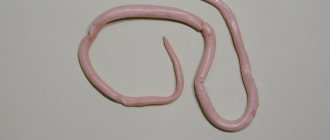
There are asymptomatic carriers of Cheyletiella mites. These parasites are detected by scratching, livestock testing, or observation of larvae (eggs) in fecal samples that resemble hookworm eggs, except for the large ones.
It is best to use selamectin every 15 days in three doses for all dogs and cats in the family, as well as for environmental disinfection. At the same time, especially for pets who do not suffer from seasonal illness, it is a good idea to start a hypoallergenic diet and continue it for up to eight weeks.
I prefer to use canned hypoallergenic diets without grains, grains or cheese. This eliminates allergies to mites when storing food. Most atopic cats are allergic to house dust mites and are not seasonally affected. Therefore, food allergies should be ruled out.
Remember that the only valid method for diagnosing food allergies is to eat a hypoallergenic diet for up to 8 weeks. Hypoallergenic means a diet without any ingredients that the cat has previously eaten (such as corn, wheat, eggs, beef, chicken, soy, dairy, fish or lamb).
The biggest problem with special diets for cats is getting them to eat new foods. It's best to get several different cans (or bags) of dry food in different flavors to see what your pet prefers. Unfortunately, home-cooked food is dangerous due to taurine deficiency.
Demodicosis in cats appears to be caused by two types of Demodex mites: Demodex cati and Demodex gatoi. The latter is contagious to others. The first tends to point to underlying internal medical problems: immunodeficiency virus, feline leukemia virus, etc.
Some acquire Demodex gatoi due to excessive use of systemic steroids, likely prescribed due to an underlying allergy. Both types are treated locally with lime sulfur rinses every 5-7 days.
Ivermectin for demodicosis is dangerous due to potential neurological side effects, so I prefer to avoid it. If the cured animal still has itching, you should try to determine the cause. Sometimes Demodex cati appears only with otitis media. I recommend a few drops of lime sulfur 2-3 times a week in the ears, and ask owners to report to me any possible increase in inflammation.
Other less common differentiations of feline atopic dermatitis include pemphigus foliaceus. It usually includes the nose, ears, nail beds, and nipples. Blisters (pemphigus is Latin for "blister") usually appear as a crust, often associated with fever. Diagnosis is made using a skin biopsy. Primary Malassezia dermatitis is rare in cats. But it is often observed in some cats and dogs, accompanying atopy. Diagnosis is made using skin cytology , and treatment includes antifungal drugs: ketoconazole, itraconazole.
Methods of combating the disease
In case of such skin disorders, the animal can and even should be bathed in a timely manner in order to reduce the concentration of the pathogenic fungus on the surface of the skin. The only condition is that this must be done with the use of specialized products that contain antifungal components. These include Nizoral, Sebazol, Mycozoral and other similar shampoos.
It is also recommended to use a special lotion that contains 2% enilconazole. Treatment of Malassezia dermatitis is also recommended using local remedies, which include vinegar in a concentration of 2%. It helps create an acidic environment, which is unfavorable for the fungus that causes dermatitis. It is enough to use such compositions no more than three times a week to achieve positive results.
Shampoos containing chlorhexidine provide double benefits, as they have not only antifungal, but also antibacterial effects. In most cases, this lesion is associated with the development of bacterial dermatitis, so it is important to fight both diseases at the same time.
Establishing diagnosis
Malacia dermatitis can be suspected if the skin disease is accompanied by itching, redness, inflammation and an unpleasant odor and localization. Clinical signs can complicate and vary keratinization defects and allergic skin diseases.
When making a diagnosis for dermatitis caused by Malacesia, special attention is paid to persistent clinical manifestations and, first of all, to an increase in the population of M. pachidermatis on the damaged skin and a positive response to adequate antifungal therapy.
Typically, fungi are identified by cytological methods, although biopsy and culture may be more effective in providing information.
Yeast is isolated from the skin, as a rule, by taking smears, although the quantitative method is more informative. The contact method is most suitable for clinical practice. To do this, small agar plates should be pressed onto the affected skin for about ten seconds and then incubated for 3.7 days at 32 to 37 degrees Celsius to count the colonies.
Malacia dermatitis is diagnosed by contact method
In most cases, if the cat is healthy, the amount of yeast in the groin and armpits is usually less than one colony-forming unit per 1 square cm. however, in the interdigital spaces and lip folds their number may be higher.
In addition, “breed” differences in population size can normally be observed. The population density in the armpits of some healthy representatives of some breeds exceeds the above-mentioned norm by ten times! This emphasizes how important response to treatment is as a diagnostic criterion, which is the most evidence-based.
To successfully treat feline Malassezia, it is necessary to make a correct diagnosis. This can only be done in a clinical setting, taking into account the history, clinical examination and laboratory results.
A scraping is taken from the animal at the border of the affected and healthy skin. The discovery of a large number of Malassezia yeast spores in it suggests that this particular pathogen is the cause of the development of the disease. To accurately determine the species of the pathogen, cultures are done on nutrient media.
Laboratory tests are required to make a diagnosis
When the diagnosis of Malassezia in a cat is confirmed, the veterinarian determines the root cause that led to a decrease in immunity and develops a comprehensive treatment.
What can lead to the formation of fungus
If we consider the picture as a whole, we can say that the appearance of nail fungus in cats (and not only it) occurs when the pet has physical contact with other representatives of its species.
This is especially true in rainy weather or in a house where there is a constant humid environment. In addition, you should be wary of minor injuries, diaper rash, etc.
The following factors can lead to the appearance of fungus:
- A young and fragile body;
- The period of lambing in a cat, when the body’s defense systems weaken;
- Decreased immunity due to the activity of a dangerous virus;
- Poor feeding;
- Carrying out immunosuppressive therapy;
- The influence of other serious diseases such as diabetes.
In some cases, diseases of this type may be asymptomatic, so they can only be detected after the hosts themselves have become infected. Very often children become the first carriers, as they most often play with pets.
Help the site, share with friends