As every pet lover probably knows, for our smaller brothers, the range of all kinds of “sores” sometimes causes real shock. Parasitic diseases, fungi, injuries... It’s impossible to keep track of everything! Fortunately, not all things on this short list pose a truly serious danger to animals. A sad exception is the same necrosis: in cats, this pathology often causes very serious consequences, sometimes leaving the cat disabled.
What it is?
Necrosis in medical and veterinary practice is a pathological process in which necrosis of a certain part of cells or entire tissues is observed. A special feature is the complete or partial preservation of the viability of the entire organism. It should be noted here that tissue necrosis in a cat, which develops against the background of deep degenerative processes (diabetes mellitus), is usually called necrobiosis. This pathology is much more severe and often ends in death.
Tissue necrosis
A type of necrosis in which dead tissue changes under the influence of environmental factors (air, microorganisms, moisture, etc.) (drying out or putrefactive decay) is called gangrene.
In practice, necrosis of bones, tendons, ligaments, and fascia is usually called necrosis, and necrosis of skin, muscles, auricle, tail, finger and udder is gangrene.
Frequent causes of necrosis are mechanical damage (crushing, squeezing, pinching, ruptures, bruises, wounds), thermal and chemical effects (burns, frostbite, the action of alkalis, acids), infectious diseases ( necrobacteriosis , gas gangrene, canine distemper, erysipelas and leptospirosis of pigs and other diseases), intoxication ( poisoning ), circulatory disorders with tissue nutritional disorders (embolism, thrombosis), neurotrophic disorders due to damage to the nervous system.
Tissue necrosis is observed in many pathological processes: inflammation, wounds , ulcers, fistulas, tumors .
The development of necrosis is facilitated by exhaustion and cooling of animals, blood loss, weakening of the cardiovascular system, penetration of pathogenic microbes into tissues, and other possible influencing factors.
dry gangrene on the ridge due to fungal poisoning (horns)
The pathogenesis of tissue necrosis is based on the processes that cause the death of cells and tissues:
1. direct effect of injury on cells, tissues and organs; 2. disruption of tissue nutrition, especially oxygen, as a result of blood and lymph circulation disorders; 3. accumulation of toxic substances (poisons, toxins) in the body disrupts the regulatory influence of the central nervous system on tissues and organs.
Tissue necrosis
Traumatic necrosis occurs as a result of tissue destruction under the influence of mechanical force (crushing, ruptures, etc.) or as a consequence of significant disturbances or complete cessation of blood circulation. Exposure of tissue to temperatures above 60C or below -15C leads to their rapid death. Strong acids, under the influence of which cell proteins fold, cause dry necrosis. Strong alkalis, dissolving proteins and breaking down fats, cause wet necrosis. Necrosis, in infectious diseases, is caused by the action of toxins on tissues, the accumulation of exudate or gases in tissues (during an anaerobic infection), which compress the paths of blood inflow and outflow.
Tissue necrosis due to horn (mushroom) poisoning occurs as a result of prolonged vasospasm and the action of toxic substances on cells and tissues. Thrombosis and embolism of blood vessels leads to disruption of tissue nutrition and subsequent necrosis.
There are dry and wet gangrene.
Dry gangrene develops slowly. In this case, the affected tissue first turns pale, its sensitivity and temperature decreases. Subsequently, as necrosis progresses, the affected tissues dry out, become hard, insensitive and cold. After some time, a demarcation line appears in the form of a pink border at the border between dead and healthy tissue. Then the dead tissue gradually falls off, and the resulting defect heals with scarring.
Dry gangrene is usually not accompanied by symptoms of intoxication, since microorganisms in dry tissues develop poorly, there is practically no decay of dead tissue, which means that toxic substances are not absorbed into the body. Therefore, the general condition of sick animals with dry gangrene changes little.
Dry gangrene occurs in animals in the area of the auricle, tail, in places of bone protrusions, on the crest and lateral surfaces in case of poisoning with horns. Wet gangrene is characterized by softening and putrefactive decay of dead tissues; it may be a consequence of containing a large amount of fluids (blood, lymph) and the entry of putrefactive bacteria into the tissues.
Locally, wet gangrene is accompanied by rapidly increasing swelling and coldness of the affected tissues and organs, and their complete loss of sensitivity. Then the affected tissues soften and disintegrate, and putrefactive exudate begins to be released. A demarcation line with wet gangrene usually does not form, as a result of which the process of necrosis progresses.
The general condition of a sick animal with wet gangrene is, as a rule, severe; the disease is accompanied by depression, loss of appetite, and a sharp increase in general body temperature.
Wet gangrene of the skin and tissues lies deeper, in the area of the lower extremities, and is observed in animals with necrobacteriosis. In general, this type of necrosis is typical for internal organs (lungs, intestines), which contain a lot of fluid.
What causes it?
There are many reasons. Thus, intestinal necrosis may well develop against the background of chronic enteritis or colitis, if the animal has not been treated at all. Very often, a strong helminthic infestation can lead to a similar outcome: the worms “do not stand on ceremony,” often feeding on intestinal tissues, so that severe inflammation and death develop extremely quickly. We should not forget about negative environmental factors. For example, necrosis of the paw pad is practically guaranteed for a domestic cat, which, due to an oversight of the owners, ended up outside on a cold winter day. However, the same result can be observed if an animal steps on something, the wound becomes infected, and the tissue begins to die en masse.
Treatment
Gangrenous stomatitis requires long-term treatment. In severe cases, it is necessary to resort to surgical intervention: damaged areas on the teeth are cleaned, completely damaged teeth are removed, and necrotic soft tissues are excised. In mild forms of the disease, it may be sufficient to take measures to eliminate inflammation and thoroughly disinfect the oral cavity.
To suppress the pathogenic microflora that causes inflammation of the mucous membrane, the cat is prescribed a course of therapy with bactericidal antibiotics. It can be:
- penicillins;
- macrolides;
- chloramphenicol;
- tetracyclines;
- cephalosporins.
Such drugs are available in the form of tablets, injection solutions, as well as aerosols or gels for external use.
It is mandatory to treat the animal's oral cavity with antibacterial solutions. It should be carried out after each feeding of the pet. First, you should carefully clean the cat's teeth and gums from food debris, then irrigate the oral mucosa with a warm solution of medicine or a decoction of herbs. This is not an easy procedure because it is painful and unpleasant for the cat, and she will most likely resist. Therefore, it is better to carry out such sanitary treatment together.
Medicines for antibacterial treatment can be used:
- 3% hydrogen peroxide solution;
- 1% sodium bicarbonate solution (baking soda);
- furatsilin solution (1 tablet per 100 ml of water);
- potassium permanganate solution (1-2 crystals of potassium permanganate per 50 ml of warm water).
To prepare herbal decoctions that can be used to irrigate the cat’s oral mucosa, plants that have anti-inflammatory, astringent and antibacterial properties are suitable: calendula, chamomile, oak bark, sage, string.
Important! Under no circumstances should a cat be treated for stomatitis with medications intended for humans. “Human” drugs have a different composition from veterinary drugs, and can cause serious complications in the animal or act as a poison.
A cat suffering from gangrenous stomatitis should be fed warm semi-liquid or pureed food until all the wounds in the mouth have healed. The water in the drinking bowl must be clean and changed every day.
Classification
In recent years, a clearly defined classification has been formed, which includes almost all types of necrotic processes encountered in veterinary practice:
- Dry variety (coagulation type). As the name suggests, the cells dry out in this pathology, and the body “fences itself off” from them with a clearly defined demarcation line of the inflammatory process. It is this type of necrosis of the occipital-spinous ligament that develops, leading to the appearance of a well-defined, extremely unpleasant fistula.
- Wet type. In this case, the disease is much more severe than in the previous case, since the cells and tissues turn into a kind of swollen, sticky mass of a very unpleasant appearance and with a disgusting smell of rot. This type of skin necrosis develops in cats when the cause of the lesion is the action of some pathogenic fungus or pyogenic microflora. It should be noted that the formation of a demarcation line in this case is very slow, and in some cases there is none at all, since the tissues are affected faster than the body can respond to it.
- Gangrene. Perhaps the most terrible type of this pathology, the photos of which alone can cause horror. With a 99% probability it leads to amputation of the entire limb or part of it, and is also guaranteed to lead to the development of sepsis and severe intoxication, which often cause the death of a cat. Its treatment should begin at the slightest sign of necrosis, since otherwise it will be very difficult to save the animal!
Necrosis
— necrosis of a part of the body (cells, tissues) while preserving the life of the entire organism.
The process of slow death of tissues and organs against the background of ischemia and deep degenerative changes is called necrobiosis. The main etiological factors, the action of which leads to the occurrence of necrosis, are: bruises, crushing, compression, high and low temperatures, electric current, radiant energy, strong acids and alkalis, their salts, toxic substances; pathogens of anaerobic and specific surgical infections; disorders of the circulatory system, endocrine and autonomic nervous systems, leading to impaired trophism. Necrosis often occurs under the influence of several factors.
Tissue necrosis occurs mainly due to cessation of nutrition or direct damage. Depending on the underlying cause, tissue necrosis can develop quickly (burns) or slowly, for example due to compression. Dead tissue areas become foreign to the body, from which it is freed through demarcation inflammation.
With aseptic necrosis and its deep location, small areas of dead tissue undergo lysis, phagocytosis, followed by resorption and replacement of the defect with scar tissue, or the necrotic tissue is encapsulated. When an area of necrosis becomes infected, purulent inflammation develops in the surrounding living tissues with the formation of a granulation barrier around the necrosis area, followed by the formation of an abscess. After opening it, a fistula is formed. In cases where dead skin and underlying tissue are sloughed off, an ulcer forms. Often, with necrosis due to the absorption of decay products and the vital activity of microbes, sepsis occurs.
The following main types of necrosis are distinguished:
Coagulative, or dry, necrosis. It is characterized by coagulation and compaction of the protoplasm of cells and interstitial matter, followed by drying. This is due to the cessation of blood flow, bleeding and release of moisture into the environment surrounding dead tissue, or exposure of tissue to high temperatures, strong acids, formaldehyde, heavy metal salts, toxins and other factors.
With dry necrosis, demarcation inflammation develops relatively quickly, since dead tissues, deprived of some moisture, have a less toxic effect on the living tissues surrounding them. Externally, the area of necrosis is characterized by a sharp outline, compaction, protrusion above the surface of the organ, it is colored gray-yellow or clay-yellow; the cut surface is dry, the tissue pattern is unclear or the tissue is a curdled mass. Later, along the periphery of the necrotic tissue, signs of demarcation inflammation and rejection of the dead area are clearly expressed. With necrosis of bones, cartilage of the auricle, and in horses the soft cartilage, a purulent fistula is formed.
Liquation, or wet, necrosis. With it, the affected tissues swell, soften, and undergo decay, forming a shapeless mushy mass. It occurs when dead tissues have excess moisture, and its release into the environment is delayed, or the tissue itself, due to the hydrophilicity of its colloids, absorbs moisture. With wet necrosis, the formation of a demarcation shaft is slow due to the toxic effect of decay products on the surrounding living tissue.
Prevention measures and therapy
It should be remembered that any disease can be relatively easily prevented. To prevent the development of the necrotic process, in many cases, basic care measures will be sufficient: examine your cat from time to time, check for severe bruises, wounds, and bites. Treat your cat promptly for helminths, fleas and ticks, and feed him good food. If there are sources of potential danger in the house, such as a hot stove, provide protective screens. This will be all the more useful since all these things are also dangerous for small children.
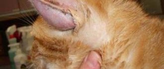
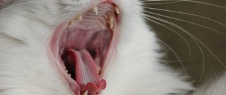
Symptoms of gangrenous stomatitis
Although cats do not express that they are in pain through sounds, it is not difficult to distinguish an animal suffering from stomatitis. A cat suffering from gangrenous stomatitis suddenly loses activity and stops eating. At first, a hungry animal tries to take food from the feeder, but immediately throws it away, as if the food was hot.
Trying to eliminate pain in the mouth, the cat rubs its muzzle on the floor or furniture. He stops taking his toys into his mouth and does not lick the fur, because such movements cause severe pain. Another typical sign of inflammation of the oral mucosa is excessive salivation and fever. In most cases, there is an unpleasant, putrid odor from the mouth.
If you try to open the cat's mouth, she will resist in every possible way, even to the point of displaying aggression, but an examination needs to be done. With gangrenous stomatitis, the oral cavity (palate, tongue, inner surface of the cheeks, and especially the gums) becomes swollen and red. Necrotic areas similar to bleeding wounds are visible on the mucous membranes. In severe cases, the submandibular lymph nodes may be enlarged.