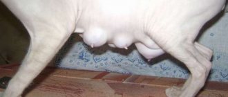
The appearance of a lump: what does it mean?
Note that the appearance of a slight swelling after surgery in itself is quite common and should not be immediately considered a pathology. But this only applies to cases when a couple of days have passed since sterilization. At this time, post-traumatic edema is “relevant”, due to which the swelling is formed.
But in cases where several days have passed since the operation, and the lump still does not seem to resolve, a veterinary examination is necessary.
The statement is also quite true for situations where the tumor somehow bothers the animal, or its size (for example, a tumor the size of an orange) does not allow the swelling to be considered a consequence of post-traumatic edema.
What does it mean? In such cases, we can confidently speak about either a hernia or a serious abscess. Both of these pathologies do not lead to anything good, and therefore they should be eliminated as quickly as possible. However, other reasons are also possible.
Remember! That the animal should in any case be examined by an experienced veterinarian, there is no need to independently engage in diagnostics without having the appropriate knowledge and skills.
The lump is on the seam
So, a lump has formed on the seam. To understand the reasons for its occurrence, you need to evaluate the appearance and size of the tumor:
- It is necessary to palpate the lump and evaluate the texture of its surface, as well as the degree of softness/hardness, the presence or absence of pain in the animal at the time of palpation.
- The size of the tumor is very important. The rule here is simple: the larger it is, the worse the situation is, and the sooner it is necessary to show the pet to the veterinarian.
We list the most likely reasons for the appearance of a lump:
Bacterial contamination of the postoperative suture. It is indicated by increased local and general temperature, redness and soreness of the tissues around the lump. The swelling itself has a hard consistency in the first days, then softens; when palpated, you can feel transfusions and fluctuations of purulent exudate.
If quite a lot of time has passed since the operation, the tumor “ripens”, an abscess appears, and the tumor festers. The process is accompanied by copious discharge of purulent exudate.
Cones of bacterial origin are very dangerous, as sepsis can develop at any time. One of the distinctive signs of an “abscess tumor” can be considered its rapid development: often the lump appears within three or four days from the moment of the operation.
It happens that when removing stitches, the veterinarian (or the owner himself) leaves one or a couple of loops in the skin. As a rule, there is nothing wrong with this, but sometimes such forgetfulness results in the growth of a connective tissue capsule on the loops.
This nature of the pathology is indicated by the high density and even rigidity of the tumor (it is very hard to the touch, the texture resembles a piece of wood), as well as its long development. As a rule, such bumps may appear on the suture line no earlier than a couple of weeks after surgery.
Sometimes swelling on the suture line is a consequence of individual intolerance to the drugs used either during anesthesia, or during the preparation of the surgical surface immediately before surgery.
Simply put, swelling is a consequence of an allergic reaction. An allergic etiology is indicated by severe (and sometimes painful) itching, as a result of which the animal, even without really recovering from anesthesia, constantly tries to scratch its belly, or even frantically gnaws at it.
If the lump has formed under the seam
Note that in this case the pathology is much more dangerous. If the appearance of a lump is due to the same purulent inflammation, and the swelling itself is a classic abscess, then it can “explode” at any moment. The cat, accordingly, will most likely die from peritonitis.
The examination of your pet should be approached with the utmost care and utmost caution:
- It is necessary to carefully feel the seam line, avoiding strong pressure. An alarming sign is pain, as well as swelling and redness of the external tissues. If the suture directly above or near the lump is inflamed and noticeably swollen, you should inform your veterinarian as soon as possible.
- The general condition of the animal is of great importance. If the cat is lethargic and refuses food and food, then it must be taken to the clinic immediately. Such signs never mean anything good.
The reasons are almost the same as in the previous case, but there are several features:
- If there is a lump under the stitch, then it may well be a hematoma. However, distinguishing it from an abscess based on indirect signs is not an easy task. As a rule, with a hematoma, the local body temperature in this place remains normal, and pain is usually not observed.
Remember! You should feel the suture line as carefully and delicately as possible, as there is a risk that the hematoma (or abscess) will burst, which will lead to new problems.
- The lump may be an abscess. It is formed only when during the operation the basic rules of asepsis and antisepsis were very grossly violated.
- An ordinary bruise. In cats, after surgery, coordination of movements (due to general anesthesia) is often severely impaired. It should be taken into account that with strong impacts, bumps often appear under the skin, and not deep in the seam. Their appearance can be associated with both traumatic edema and hematomas.
- A hernia is the most serious cause of a lump appearing directly below the suture line. And even an owner with minimal experience can identify it. The tumor in such cases is very soft, voluminous, and in the thickness of the hernial sac (in the most severe situations) the intestinal loops can be easily felt. If these signs are detected, the animal must be immediately taken to the clinic, since hernias are easily strangulated, and they lead to intestinal necrosis, fecal peritonitis and other pathologies, often resulting in the death of cats.
What to do if a lump forms on your stomach
Help for the animal may be required in the event of a prolapsed hernia, suppuration of the suture, or infection of the wound. If the swelling does not subside for several days, increases in size, festeres, or your pet feels unwell, be sure to contact your veterinarian to avoid serious complications. The veterinarian, having established the cause, will prescribe symptomatic treatment aimed at normalizing the general condition of the operated animal.
When a postoperative hernia forms, if there is no pinching, the problem is solved by repeated sutures or reduction of the hernial protrusion. If the process is started, surgery may be required.
If the tumor is malignant or benign, the veterinarian, after tests, decides on the choice of treatment. The animal is prescribed maintenance therapy or surgery.
In any case, the choice of treatment methods for the appearance of a lump on the abdomen, above or below the suture, depends on the root cause, the general condition of the cat, and diagnostic results.
Do not self-medicate so as not to harm your mustachioed pet. All manipulations and therapy should be prescribed by a veterinarian.
What to do: first aid and elimination of pathology
In all of the above cases, we would advise you to first contact a veterinarian. Even when the lump is on the surface of the seam, it can be dangerous to the health and even the life of the animal.
Lump on the seam
Everything is quite simple here:
- If the lump festers, the local and general temperature is elevated within three days after the operation, and the site of the swelling hurts more and more (which is easy to determine by palpation), then there is only one way out - an immediate visit to the veterinarian.
- The owner will not be able to cope with purulent infections after surgery on his own.
- If you suspect the formation of a connective tissue sheath around sutures forgotten during removal, the approach is the same. Visit to the veterinarian. The pathology is not particularly dangerous, but can only be eliminated surgically.
- Finally, drug intolerance and allergic reactions. At home, you can give your cat about half or ¼ of a diphenhydramine tablet, but in cases accompanied by severe itching, we would also advise calling a veterinarian immediately.
Lump under the seam
Often the reasons for the formation of a tumor in these cases are more serious, and therefore in any case you will have to contact a veterinarian. There are, of course, some nuances:
- If a hematoma is suspected, cold can be applied to the abdomen for 10-15 minutes. Low temperature helps to compress blood vessels, which reduces the intensity of pain. The same can be done for purulent inflammations, but in any case, the cat must be shown to a veterinarian. If the animal has a very high local or general body temperature, you should not delay a visit to a specialist.
- Cold also helps with bruises, and heat (warm lotions) on the second day. The problem is that when falling, the cat could receive not only bruises, but also more severe injuries, the consequences of which often manifest themselves later.
Formation of a hernial sac = immediate visit to the veterinarian. The hernia cannot be eliminated at home; delay is fraught with compression and necrosis of the intestinal loops.
Treatment
Sometimes a small bump under or above the stitch indicates tissue growth due to healing. An experienced veterinarian will always recognize this phenomenon and say that there is no need to worry. This swelling does not require treatment and goes away on its own after 3-4 weeks.
Infection and abscesses are treated with antibiotics. In severe cases, it may be necessary to open the abscess.
For bruises and hematomas, the doctor may prescribe the application of cold and treatment of the affected area with special gels and ointments.
For inflammation of the lymph nodes, the veterinarian prescribes antiviral and anti-inflammatory drugs. If the animal's body temperature is not elevated, dry heat is recommended.
Prevention
In many cases, the bump is the “handiwork” of the owner himself. More precisely, the result of his failure to comply with the basic recommendations of the veterinarian.
Accordingly, prevention is quite simple:
- For 12 hours before surgery and the same amount after it, you cannot feed the animal. Neglecting this rule can lead to the formation of a hernia.
- After anesthesia, the cat's coordination of movements is significantly impaired. There is no need to place it on a bed or ottoman. The ideal option is a separate basket in an isolated room.
- If the animal is overly active and constantly tries to scratch or lick the seam, it is necessary to put a surgical collar on it.
Failure to follow these rules is the main reason why a cat develops a lump after sterilization.
Sterilization of cats is a long-known and widely used surgical operation in veterinary medicine. Its popularity is facilitated by the fact that sterility, even surgical, rarely results in the development of complications in the operated pets. But this doesn't always happen. Unfortunately, a hernia in a cat after sterilization is a rare phenomenon, but still not unique.
Preventing the formation of lumps under the skin
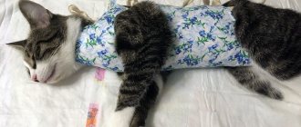
Preventing postoperative complications consists of following the veterinarian's instructions. The first days after the operation, the pet is given rest, does not play, and is not allowed to go for walks. The surface of the seam is treated with an antiseptic wound-healing spray in accordance with the instructions of the veterinarian. After the operation, a blanket is put on the animal so that the healing suture, which itches, does not tear. If the cat manages to remove the blanket, a cervical collar is put on it.
The protective dressing is removed after the sutures are removed. However, it is advisable to do without active games for a month.
In the first days after surgery, the cat is given adequate food with a wet consistency. During this period, avoid foods that cause flatulence or constipation. Adherents of natural nutrition will have to come to terms with the need to transfer their pets to ready-made comas for sterilized cats.
Hernia in a cat after sterilization: what is it?
Without going into details, a hernia is a pathological phenomenon, the meaning of which is that part of the internal organs gets under the skin. Those. a kind of “bag” forms on the cat’s stomach, inside of which (in most cases) there are intestinal loops, sometimes there is also a piece of the liver or other internal organs. They get under the skin through the hernial ring.
Roughly speaking, this is a hole in the muscle membrane. A hernia, even a small one, should never be taken too lightly:
- Such neoplasms always show a pronounced tendency to increase in size. In advanced cases, a hernia may grow on the cat’s stomach, the size of which can compete with a large orange.
- The insides located in the dangling hernial sac are in great danger. A cat can hit itself at any time, and the consequence of the blow is often a rupture of the intestinal loops located in the hernia itself.
- The hernial ring often has an unpleasant habit of shrinking. The result is compression of the internal organs passing through it. This leads to necrosis, deterioration of local blood circulation and other unpleasant consequences.
It happens, however, that there is nothing inside the hernia except a small amount of subcutaneous fatty tissue. Such neoplasms are considered relatively safe, and there seems to be no need to urgently eliminate them.
In any case, the animal must be examined by a veterinarian. Only he can determine the real degree of danger of a hernia.
Treatment of umbilical hernia in a cat
After diagnosing and making an accurate diagnosis, it is necessary to begin treating a hernia in the cat’s stomach.
Important! It is forbidden to try to treat an animal yourself, without consulting a doctor. You cannot carry out thermal procedures or try to straighten protruding organs with your own hands.
Conservative therapy involves the use of ointment, the use of a special therapeutic massage of the affected area and the application of a plaster to fix the area after reduction.
In a kitten, with good treatment, the hernia can close by 6 months. In other cases, for older individuals, conservative treatment is not enough.
Folk remedies
The most effective folk method is to straighten the affected area and fix it with a plaster. Additionally, use a flat, dense object, such as a coin or button, to make the fixation more reliable. In a young pet, with a small hernia, it heals.
The preferred treatment for hernias is surgery. It is quite simple, removal is possible already on the eighth day. The rehabilitation period in most cases passes without complications, the animal quickly returns to its usual way of life.
It is advisable to put a blanket on the cat so that he does not have the opportunity to lick the seams. To prevent infection, the operated area should be promptly treated with a disinfectant solution.
Small, ungrounded and non-hazardous formations, as a rule, are not operated on. These are mainly inguinal hernias. Alternative treatment is prescribed. The veterinarian repairs the hernia and then applies a fixing blanket, which the cat will have to wear for several months (from 1 to 3, based on the individual characteristics of the cat’s health).
If the reduction procedure is successfully performed, the hernia heals. However, it should be noted that this method is not entirely convenient. The owner will have to constantly ensure that the cat does not tear off the blanket and, if necessary, adjust the fixing device.
Diaphragmatic hernia requires a different approach to treatment. Compared to other types of neoplasms, the operation is more complex, involving opening the chest and returning all incorrectly located organs to their place.
We invite you to familiarize yourself with Ringworm in Sphynx Cats. Is it Transmitted to Humans?
The veterinarian sutures the diaphragm, then performs its plastic surgery, applying a special mesh or his own tissue. If organs are injured, they are resected. At the initial stage of an intervertebral hernia, drug therapy with anti-inflammatory and painkillers cannot be ruled out.
The animal's physical activity should be limited. If surgery cannot be avoided, the veterinarian removes part of the vertebra along with the prolapsed disc and in the area of the tumor. After surgery, the cat will need complete rest. For large-scale spinal cord injuries, euthanasia is indicated.
The prognosis is favorable in most cases. The main thing is to start the treatment process in a timely manner.
In addition to the external manifestation of hernial protrusion, the pathology may be accompanied by symptoms characteristic of a violation of certain functions of the internal organs that are involved in this process. That is, if the intestines are strangulated, the clinical picture will be supplemented by indigestion, abdominal pain, vomiting, and diarrhea. If the heart or lungs are damaged, then these are signs of cardiac and pulmonary failure, etc.
Inguinal
Causes
Here are the main reasons for the development of postoperative hernias in sterilized cats:
- “Amateur performance” of an animal that managed to remove the stitches on its own. A very common and very dangerous reason. Moreover, a hernia is far from the worst consequence. If nothing is done, the pet can open the seam even further, literally “gutting” itself.
- Poor quality of operation. If the incision is longer than necessary, the weakened muscle tissue may not be able to withstand it, causing the internal organs to go under the skin on the abdomen.
- This happens when operating on too old pets. Their muscle tissue is already weakened due to purely age-related reasons; surgery can only accelerate the appearance of a hernia. Classical cavity sterilization is especially dangerous. If possible, it is much better to operate elderly pets using laparoscopic sterilization.
- Indirect consequences of anesthesia. Because of this, the animal can fall even in completely ordinary, everyday situations, which in other cases do not pose any danger. The most unpleasant thing is not even the bruises: as a result of an unsuccessful fall, a cat, which at other times has incredible plasticity, can easily break its paw (or even more than one). In addition, her stitches may come apart, and this is the direct cause of the hernial sac.
Treatment of postoperative hernias: general information
Breeders should remember that the treatment of such pathologies is exclusively surgical (as a rule), in a well-equipped veterinary clinic. If the pathology is not advanced, therapy is quite simple:
- First (using the same palpation and ultrasound), the veterinarian accurately determines the boundaries of the hernial ring and the diameter of the canal. This is necessary to determine the surgical technique, suture material and other important factors.
- After this, the specialist decides on the anesthesia technique. In some cases, when a hernia has just appeared, only local anesthesia is sufficient, but in situations where the neoplasm has already reached a large size and upon palpation the intestinal loops and other internal organs are clearly felt in it, general anesthesia is necessary.
- After administration of anesthesia and general preparation for the operation (including fixation of the animal, shaving of the surgical area and its antiseptic treatment), an incision is made.
- Through the surgical incision, the veterinarian will set the internal organs back into the abdominal cavity, after which the hernial sac will be sutured.
- If the skin at the site of the former sac is greatly stretched (which often happens), it is trimmed, the cavity is lightly powdered with veterinary powder based on streptocide, after which everything is sutured.
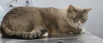
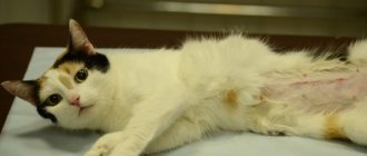
The cat has a lump under the seam
As a rule, a lump under the seam does not pose a threat and goes away on its own after some time.
A characteristic sign of the body's own defense is hyperemia and swelling of the sectional area.
It often happens that tissue swelling reaches a certain size and visually looks like a so-called lump . Such seals are not dangerous for the pet and disappear on their own after the hyperemia and swelling subsides and healing begins. For complete confidence and distinction from the pathological process, a consultation with a veterinarian is necessary.
Proliferation of granulation tissues
Another factor in the appearance of compactions is the proliferation of granulation tissues in the operated area.
Such growth is also not a dangerous symptom and does not require a special approach, but, as in the first case, it requires professional advice.
Suture dehiscence after surgery
A dangerous sign that requires careful inspection and elimination is the separation of internal seams, when the external seams are intact and it is difficult to visually determine the internal problem.
This complication will be indicated by the appearance of a seal that protrudes through the outer layer. Often such a hernia appears through a hole in the external suture, usually a piece of an internal organ - the omentum or intestinal loop. With gentle pressure on the hernia, the protruding part easily disappears.
What to do after surgery: rehabilitation and care
So what should the owner do? When the animal recovers from anesthesia (and this takes up to five or more hours), it should be given a little drink from a syringe (no more than 10 ml of water). The cat must be placed in a room protected from drafts, isolated from access to other pets.
Of course, the postoperative wound (like the animal itself) needs care. The owner of the animal does not have to do anything particularly difficult:
- You definitely need to get hold of a surgical blanket! This simple device prevents licking and scratching of the surgical suture (which inevitably leads to the development of complications, including purulent inflammation). The blanket needs to be changed as it gets dirty, but at least once a day.
- During the first week, you need to treat the seam once a day. For this purpose, miramistin, levomekol, chlorhexidine are used.
- It is necessary to ensure that the operated pet does not pretend to be a squirrel, constantly playing and jumping. It will take at least a week for her body to endure physical activity without consequences. Otherwise, the suture can easily come apart (which can lead to a new hernia or something worse).
- For the first four days, the cat should be fed with rich chicken broth, from time to time flavoring it with meat and vegetable puree (from baby food). This is necessary so as not to overload the digestive system of the pet, weakened after two operations.
A hernia in a cat is a fairly common surgical pathology. It is a kind of protrusion on the body, consisting of a hernial sac and its contents.
The hernial sac is usually a part of the peritoneum or its fascia, and the contents are various internal organs (intestines, bladder, part of the stomach) or the omentum. The protrusion is usually clearly visible on the body under the skin, so it cannot go unnoticed.
The situation is separate with intervertebral and diaphragmatic hernias. In the first case, there is no hernial sac as such with prolapsed internal organs; there is extrusion of the intervertebral disc, and the more it protrudes beyond the intervertebral space, the worse it is for the animal.
With diaphragmatic pathology, the organs of the thoracic cavity fall into the abdominal cavity into the formed hole in the diaphragm or, vice versa, depending on in which part of the body the pressure increases. There is usually no formed hernial sac either.
If the contents of the hernia can be reduced well, then it is called reducible. If it is not possible to set it, it is irreducible. If there are signs of an inflammatory process in the area that fell out, it is pinched.
It is important to understand that in the absence of timely assistance in case of infringement, the compressed organ area dies due to impaired blood circulation in this area and its trophism (nutrition). There is a significant risk that the animal will die. Finding yourself on the operating table in this state, not a single surgeon will give any predictions until the operation is over, and the veterinarian sees with his own eyes what is inside.
Important: any strangulated hernia must be operated on immediately!
Hernia symptoms
Diagnosis is carried out using an external examination. The hernia is always visible; it looks like a new growth in the navel area. It is also necessary to determine the type of disease. A reducible hernia is movable when pressed and may disappear when the pet changes body position. The cat feels good due to the absence of pain.
The irreducible one has a convex shape that increases in size over time. It is hot to the touch and causes a lot of discomfort in the animal.
Care after hernia removal
To diagnose the pathology, the veterinarian needs to conduct a visual examination of the animal, palpation for the presence or absence of pinching (in case of external protrusion of the neoplasm).
If an internal form of hernia, characteristic of diaphragmatic and intervertebral pathologies, is suspected, diagnostic studies such as ultrasound, MRI, X-ray with barium contrast, and myelography are performed. In some cases, electrocardiography is prescribed (to monitor heart function).
Depending on the location and volume of the hernia, there may be variable symptoms. If a hernia has formed anywhere in the abdominal cavity, this leads to the formation of a protrusion at the site of the hernial ring with prolapse of the omentum or abdominal organs. If organ infringement occurs, symptoms of intestinal obstruction may occur - profuse vomiting, lethargy, refusal to eat.
If a diaphragmatic hernia occurs, then external protrusions do not appear, but due to compression of the lungs by the abdominal organs, respiratory failure often occurs, accompanied by shortness of breath, and in severe cases, cyanosis of the mucous membranes. If the intestines are strangulated, it may be accompanied by symptoms of intestinal obstruction.
Intervertebral hernias cause compression (compression) of the spinal cord, which is anatomically located above the spine and serves as a conductor of nerve impulses from the brain to the organs of movement and internal organs. Therefore, if an intervertebral hernia develops, the motor function of the limbs may suffer, even to the point of complete paralysis.
When a hernia appears in a cat, you can additionally notice that the animal has become lethargic and refuses to eat. Due to pain, the pet moans, screams, and experiences constant severe anxiety. The formation changes color, becomes red or black, increases in size, and becomes denser.
A hernia in a cat is accompanied by the same symptoms as in a cat. Namely:
- when touching the formation, the animal is hurt;
- the temperature rises;
- the pet’s behavior changes, he becomes irritable;
- changes in breathing are observed, it becomes difficult, frequent;
- Appetite decreases, the animal drinks little and quickly loses weight.
Types of hernias
The following types of pathologies are distinguished in cats:
Umbilical
occurs most often and in 99% of cases is congenital and genetically determined. This means that if the parent cats had this pathology, then most likely it will manifest itself further in the kittens.
Occurs when the omentum or intestinal loops (which is less common) “fall through” into the umbilical ring. May be infringed. Most often reported in small kittens whose umbilical cord was incorrectly bitten/cut or too close to the abdomen. Umbilical hernia in cats also sometimes occurs after the cat is spayed when the internal sutures come apart.
Inguinal
such a hernia appears due to the peculiarities of the development of the inguinal ring, where the bladder, intestines, uterus or omentum prolapse (if there are signs of obesity) against the background of increased intra-abdominal pressure. It can be reduced, but there is a high risk of strangulation due to the peculiar location and increased physical activity of cats by nature.
Diaphragmatic
Mustachioed pets may already be born with this pathology, but in most cases they are acquired as a result of serious injuries, falls from a height, blows, etc. Due to the increase in intra-abdominal pressure, the intestines begin to put intense pressure on the diaphragm, breaking through it more and more and putting pressure on the organs of the chest cavity. It is very difficult to diagnose, because... Due to the peculiarities of the anatomical structure of cats, even on an ultrasound and x-ray, the hernial sac is not clearly visible, except for the darkening of the general plan. To make sure that the cat has a diaphragmatic hernia, they try to feed it with a contrast agent (porridge with barium) and take an x-ray at the right time. A dangerous type of pathology that can lead to the death of a pet.
Perineal
appears when the peritoneum stretches so much that a protrusion forms between the rectum and uterus (and in cats, between the large intestine and bladder). The main provoking factors for the occurrence of perineal hernias in cats are various types of intestinal problems (diarrhea, constipation with excessive straining, inflammatory processes).
Intervertebral
Between the vertebrae of the spinal column there is a certain fibrocartilaginous tissue that acts as a kind of padding and shock absorber between them. In case of injury, when excessive pressure occurs between the vertebrae, this tissue begins to literally be squeezed out of the intervertebral space and pinching occurs (almost always). This protrusion is called an intervertebral hernia in a cat. It can be in one place of the spinal column, or in several places (multiple). Usually registered in old cats over 15 years of age.
Scrotal (scrotal)
is a kind of inguinal. More precisely, they are practically the same thing, only the entire contents of the hernial sac will fall out not into the groin area, but directly into the scrotum. It is rare and occurs in very large cats with large testes.
Pericardial-peritoneal
in fact, this is the same diaphragmatic hernia, with the only difference that the hole is so large that the intestinal loops directly fall into the chest cavity with their entire mass, putting very strong pressure on the heart and lungs. In practice, it is extremely rare, difficult to diagnose and usually posthumously, because signs of heart and pulmonary failure develop very, very quickly.
Surgical reduction of traumatic hernias
When purchasing a puppy, all veterinarians recommend paying attention to the presence or absence of a hernia. So what are hernias, what are they, where do they come from, what to do with them?
These are the questions that potential owners and breeders ask themselves if they are faced with hernias in one way or another.
Hernia
is a temporary or persistent prolapse of internal organs through a natural or pathological opening with protrusion of the membrane lining the anatomical cavity. A hernia has its own structure: a hernial ring (opening), through which a hernial sac protrudes, consisting of the peritoneum, transverse abdominal fascia and hernial contents (intestines, omentum, stomach or other organs, tissues).
What types of hernias are there?
Based on their origin, hernias can be congenital or acquired. Congenital hernias develop due to too wide anatomical openings, or due to a defect in the abdominal wall (Fig. 1).
Figure 1 – Congenital hernia in a puppy.
Acquired hernias
are most often formed due to excessive physical tension of the muscles (Fig. 2).
Figure 2 – Acquired abdominal hernia in a horse.
A hernia in which the contents are freely reduced is called reducible.
, if reduction is impossible due to adhesions, it is called irreducible; in the presence of an acute inflammatory reaction, it is called strangulated. Sudden strangulation of a hernia can occur after sudden movements and jumps due to a sharp increase in pressure in the abdominal cavity. Some part of the intestine gets into the hernial sac, and as a result the loop becomes pinched from the outside. According to the location of the hernia, there are the following: umbilical, inguinal, perineal, traumatic, abdominal, diaphragmatic, etc. UMBILICAL HERNIA.
If an umbilical hernia is present in a kitten or puppy, a small, soft to the touch, and painless swelling in the navel area is most often detected. Upon palpation, the contents can be easily reduced into the peritoneum and the hernial ring is clearly palpated. The contents of the hernial sac in an umbilical hernia are most often the omentum, sometimes intestinal loops.
Figure 3 - Umbilical hernia in a kitten.
This type of hernia occurs quite often, due to insufficient closure of the abdominal wall near the navel. Most often, an umbilical hernia is a congenital pathology and is a defect in the intrauterine formation of the umbilical ring. Less commonly, in the presence of some anatomical predisposition of the animal, an umbilical hernia occurs due to excessive stress, tension in the abdominal wall and increased intra-abdominal pressure, for example, due to excessive flatulence, severe and constant overeating, and other factors.
Why are umbilical hernias dangerous?
As the animal grows, the hernial sac can increase in volume, while remaining narrowed in the area of the hernial orifice. In most cases, the hernial contents are the omentum, which, however, is sometimes reduced, and sometimes not reduced, due to the fact that it is attached to the hernial sac. Thus, the so-called strangulated hernia can occur, when intestinal loops filled with gas or contents fall into the hernial sac and are strangulated by the hernial ring, intestinal motility is disrupted, obstruction of the gastrointestinal tract develops, which leads to inflammation, further to poor circulation and necrosis (death). Strangulated hernias without surgical intervention are fatal.
ATTENTION!!!
Do not try to reduce a strangulated hernia yourself and under no circumstances use a heating pad! Hernias do not resolve on their own; gluing nickels and the like to the hernia site does not lead to the desired results! What kind of help do animals with an umbilical hernia need?
If an umbilical hernia is strangulated, then immediate surgery is required, but is it worth bringing your pet to this state?! If the hernia is small, no more than a few millimeters, it may well close on its own without surgical intervention. Those puppies and kittens that have a hernia at birth need to be constantly monitored, i.e.
Reduction of a non-strangulated umbilical hernia is a planned operation. It is performed under general anesthesia or local anesthesia. Don't wait until the hernia is strangulated, have surgery right now.
The operation is performed in the dorsal position; local anesthetic is injected around the base of the hernia into the abdominal wall from four points (Fig. 4). In this case, the solution is first injected into the subcutaneous tissue, then the needle is penetrated into the dorsal wall of the rectus abdominis vagina, and, piercing it, the needle is immersed in the retroperitoneal tissue, continuing to inject the solution.
Figure 4 - Infiltration of tissues surrounding the hernia.
Having completed anesthesia, the skin is cut with a longitudinal incision in the middle of the hernia. Then, using a gauze swab, a piece of peritoneum is dissected from the skin to the umbilical ring and inserted into the abdominal cavity. A loop-shaped suture made of absorbable material is placed on the hernial ring. It is recommended to scarify the edge of the hernial ring in order to cause a more dense fusion in the future.
For an irreducible hernia, the serous sac is opened. If, upon examination of the hernial contents, a commissure is found between the intestine and the peritoneum, it is necessary to excise the adherent area of the serous sac, leaving it on the intestine. When there is adhesions connecting all the loops of intestines that were in the hernia cavity into a common mass, it is necessary to expand the umbilical ring with a button scalpel, and then push the intestines into the abdominal cavity. The umbilical ring is closed, as in the treatment of a reducible hernia.
In case of a strangulated hernia, as in the previous case, the serous sac is dissected, the strangulated intestine is removed onto a sterile napkin and its condition is determined. In case of swelling and hyperemia, the intestine must be moved into the abdominal cavity. If it is impossible to straighten it with little effort, it is necessary to expand the umbilical ring under finger control with a button scalpel.
In some cases, when the hernial ring is very wide and the aponeurosis of the abdominal muscles is weak and there is a risk of a hernia forming again, plastic surgery using a polypropylene mesh is used. The polypropylene mesh grows into the tissues of the body and animal and remains in the body for life.
All of the above operations for umbilical hernias end with the removal of excess skin and closure of the skin wound with an interrupted suture.
INGUINAL HERNIA.
The hernial opening in an inguinal hernia is an unnaturally wide gap (the inguinal canal) or a rupture in the tissue of the abdominal wall. Any hernial opening in animals tends to expand over time. The most important anatomical element in an inguinal hernia in animals, which determines the formation of an inguinal hernia, is the natural connection of the cavity of the common vaginal membrane with the abdominal cavity.
It communicates through the vaginal canal, which remains after the testis descends into the scrotum. The hernial sac is formed by the parietal peritoneum with the underlying fascia. With prolonged existence, the sac grows with scar tissue, contributing to the formation of adhesions, bridges and the formation of communicating chambers.
Inguinal hernias can be congenital or acquired. Acquired hernias are formed due to mechanical damage to the abdominal wall and prolonged straining. An inguinal hernia in a kitten can form due to increased intra-abdominal pressure, when weak spots in the abdominal wall are stretched (the inguinal canal). According to the condition of the contents, there are: reducible, irreducible and strangulated inguinal hernia. Most often, an inguinal hernia is left-sided.
A hernia in which the intestinal loop or omentum (bladder) is located in the vaginal canal should be called a vaginal canal hernia. If the same organs lie in the cavity of the common tunica vaginalis, such a hernia should be called intravaginal. If a new hernial sac is formed, lying completely separately in the scrotum - between the skin, Cooper's fascia, on the one hand, and the common vaginal membrane, on the other, then the hernia should be called true scrotal.
We invite you to familiarize yourself with Fluffy cats: the most beautiful breeds with long thick hair, description and photo
In cases where a rupture of the wall of the inguinal canal occurs and the peritoneal hernial sac with its contents lies outside the vaginal canal, i.e., in the inguinal canal, a true inguinal hernia is formed (Fig. 5). This hernia can easily become a true scrotal hernia if the hernial sac descends under the influence of the contents and intra-abdominal pressure to the base of the scrotum.
Figure 5 - Inguinal hernia in a male dog.
An inguinal hernia in females may contain: intestines, bladder or uterus. The first signs of an inguinal hernia are the presence of a small swelling in the groin area on one or both sides, spherical or elongated (Fig. 6.). If the contents of the hernia are a pregnant uterus, then as it grows, the hernial sac increases (in this case, only surgical treatment is meant, with the removal of the fetus).
Figure 6 - Inguinal hernia in a bitch.
ATTENTION!!!
All types of inguinal hernias require surgical intervention. The intervention technique may vary, depending on the sex of the animal and the owner’s wishes to remove or preserve the pet’s testicle. As a rule, in male dogs, the operation is performed to remove the testis. If the testis is left, there is a risk of re-formation of a hernia or necrosis of the testis, that is, another surgical intervention.
ABDOMINAL HERNIA.
The causes of the formation of such hernias are injuries to the abdominal wall with the formation of a muscular or aponeurotic defect. The contents of the hernia, depending on its location, can be any movable organ of the abdominal cavity. Hernias are located in the area of the ilium, xiphoid cartilage, navel, hypochondrium.
Symptoms and treatment
In addition to external signs of protrusion, the condition is accompanied by general signs of malaise, depending on which organs were compressed:
- nausea, vomiting, lack of appetite;
- diarrhea, followed by constipation and complete absence of stool;
- pain in the abdomen/groin/scrotum;
- back pain accompanied by impaired motor activity up to paw failure;
- pulmonary and/or heart failure.
The general treatment regimen usually follows two directions:
- surgical intervention;
- non-invasive treatment (without scalpel intervention).
If the protrusion is not pinched and can be reduced, then the animal is put on a special surgical anti-hernia blanket with a multi-tiered lining, which is worn for several months. During this time, the internal organs from the protrusion have time to fall into place, and the hernial “window” closes on its own. This bandage is not applied to all types due to the peculiarities of the anatomical structure of the body of cats and their natural mobility and plasticity. The blanket copes well with umbilical hernias and very rarely with inguinal and perineal hernias.
- Symptoms
In the area of the same name, a certain protrusion is detected. Usually soft and painless. When pinched, pain, local redness, swelling are observed, not only the local temperature rises, but sometimes the general temperature as well. In addition to local symptoms, nausea, vomiting, and signs of indigestion (diarrhea, constipation, or no bowel movement at all) may be observed. The diagnosis must be made by a specialist, because there is a high probability of confusing a hernia with an abscess.
- Treatment
In the initial stages, wearing a blanket (up to six months) is possible without infringement. If conservative treatment does not produce results, they resort to herniotomy - removal. If the hernia is reducible, then all its contents are reduced, and the edges of the umbilical ring are sutured. If it is irreducible and there is strangulation, then the operation is performed immediately, the bulging area is usually removed (usually against the background of necrosis, the area of the strangulated intestine and/or omentum can no longer be saved), the hernial ring is sutured.
Inguinal and scrotal
- Symptoms
Pathologies of the same type, with the only difference that a soft and painless protrusion is found in the same areas - in the groin or scrotum. A scrotal hernia is usually irreducible, because this is difficult to do due to the presence of the testis in the scrotum; the inguinal can be reducible, but this is pointless, because It is almost impossible to put on a blanket; organs will continue to fall out. They are often discriminated against. Symptoms: in addition to visual protrusion, the cat may limp, the limb protrudes to the side due to the inconvenience of walking, and when the bladder is pinched, there may be involuntary urination. When pinched, body temperature may increase.
- Treatment
Special bandages for inguinal, scrotal and perineal conditions are not used in practice. It is much more effective and safer for the health of the animal to perform the operation immediately. The subtleties of surgical intervention are similar to those of the umbilical. If it can be adjusted, everything is adjusted, the inguinal opening is sutured.
If it is impossible to reduce, after the incision the feasibility of preserving the strangulated area of the prolapsed organ is determined, the hernial sac is eliminated, the remains are reduced, and the hole is sutured. Particular attention should be paid to the operation if there was prolapse of the bladder and uterus during pregnancy. In the second case, the organ may have to be removed along with the offspring, taking into account the strength and duration of the infringement. If necrosis of the bladder occurs, then the animal is offered to be euthanized, because the process of urination becomes impossible.
Removal surgery only makes sense when it reaches a decent size and begins to cause discomfort to the cat. In all other cases, a wait-and-see approach is chosen, the pet’s mobility is limited and all attention is focused on the general condition of the animal.
- Symptoms
It is impossible to notice such a pathology externally. It is also not always possible to look at X-rays and ultrasound. The main signs: shortness of breath, impaired heart function, the cat rarely lies down and more often on its side, it is clear that the animal is in pain due to the pain that the hole in the diaphragm causes when it contracts on the compressed organs.
- Treatment
Treatment is only surgical, and the earlier the diagnosis is made, the higher the likelihood of saving the pet. Prolapsed intestinal loops not only affect the heart and lungs, but they themselves are most often pinched due to the increased elasticity of the diaphragmatic openings.
The organs are set back in place, they are completely inspected, and at the same time, supportive therapy for the heart and lungs is required. The “hole” in the diaphragm is sutured. In rare cases, plastic surgery may be required to transplant fragments onto the diaphragm due to the hole being too large.
- Symptoms
A soft and painless protrusion is found near the anus (in cats) or around the internal genital organs, slightly under the vagina (in cats). It can only be on one side, in rare cases it can be bilateral. There are no infringements. If you hold the animal by the front paws, the protrusion will become larger, and if you lift it by the hind paws, it will hide. This is a kind of test to clarify the diagnosis. Usually there are no additional symptoms and no signs of discomfort. But if the size is too large, it can put pressure on the bladder and internal genital organs.
Treatment is the same as for inguinal.
- Symptoms
It manifests itself with all the signs of pulmonary and/or heart failure, which increase at such a speed that most often they do not have time to help the pet. Shortness of breath and irregular heart rhythm are accompanied by complete apathy and refusal to eat and drink. Possible development of pulmonary edema (severe wheezing, coughing, sneezing with mucus). Murka should be taken to a specialist immediately.
- Treatment
The condition is practically incurable, because... the diagnosis is usually already made at the stage when it is no longer possible to compensate for pulmonary and heart failure. Moreover, most often animals die before a diagnosis is made (i.e., pathology is determined only at autopsy). The likelihood that a pet with this diagnosis will survive is very low.
- Symptoms
Here, all symptoms are based on damage to motor activity, because The lumbar spine is usually affected. Coordination of movements is impaired, there is no stable support on the limbs (usually the hind limbs), the cat cannot jump somewhere, paralysis and muscle atrophy may develop. With deep degenerative processes, signs of damage to the central nervous system may be observed - convulsions, uncontrollable twitching of the limbs, etc. If there are signs of damage to the central nervous system, treatment becomes inappropriate.
- Treatment
No one will perform the operation right away. If motor activity is slightly impaired, then NSAIDs (non-steroidal anti-inflammatory drugs), steroids and painkillers are prescribed. If there is no result and there is at least some chance of help through surgery, an operation is performed. If motor activity is severely affected, drug therapy has not produced results, and surgery is not possible, then it is proposed to euthanize the animal, because it will no longer be able to live a full life.
General recommendations
Blanket (bandage)
After sterilization, the abdomen should be protected with a special blanket (bandage) with ties on the back. The protective bandage is worn all the time until the stitches are removed, plus another day or two, so that the cat does not lick small wounds from the threads.
The people were taken aback! Joints will recover in 3 days! Attach...
Few people know, but this is exactly what heals joints in 7 days!
A cat blanket is usually made of natural cotton fabric and does not cause physical discomfort to the animal after surgery. But due to the peculiarities of tactile sensitivity, the cat may be lazy to walk in it or have a strange gait. When the bandage is removed, everything falls into place.
You need to be careful that the cat does not cling to anything with the blanket and does not get stuck anywhere.
Eyes
If the cat is handed over to the owners after the operation in a state of anesthesia, then the process of blinking for the pet falls on them. Cats under anesthesia often have their eyes open. To prevent the cornea from drying out, you need to periodically cover your eyes (blink) or drip artificial tears or 0.9% saline solution onto their surface.
General condition of the house
Upon arrival home, the cat should be placed warmly (for example, on a warm heating pad) and on soft bedding, because After anesthesia, the process of thermoregulation is disrupted. Be sure to place it on the floor so that during the awakening process the animal does not fall from a height. It is necessary to lay down a well-absorbent diaper, because... Until recovery from anesthesia, the cat cannot control the physiological needs to empty the bladder and rectum. There may be trembling throughout the body (general tremor) or vomiting.
You need to place the cat on its right side to reduce the load on the heart.
At first, the cat should not actively play, jump, or climb on pieces of furniture. If sterilization was carried out after childbirth, and there are kittens left in the house, you need to limit their contact for the first time. 2-3 month old kittens can suckle a cat for a long time, and this is fraught with injury to the postoperative wound. Sterilization of a nursing cat is carried out for emergency reasons, because... postoperative complications in the mammary gland are possible.
- On the first day after surgery, the cat must definitely go to the toilet - it is important not to miss stagnation in urination and bowel movements after anesthesia.
- For the first two days, you may need pain relief. Pain is indicated by increased aggressiveness, apathy, meowing, refusal to eat, dilated pupils and reluctance to move.
- Antibiotic therapy is not required if the operation is planned and carried out taking into account the rules of asepsis and antisepsis.
- Vitamin preparations and restoratives are prescribed only to old and weak cats who have undergone a difficult operation.
- In case of urgent need (the suture is bleeding or internal bleeding has been detected), hemostatic therapy may be prescribed.
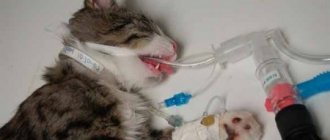
Coming out of anesthesia
Three types of anesthesia are usually used. After each type of anesthesia, the cat comes to its senses differently.
- Muscle relaxants + analgesics. The most effective mixture, used most often. The main disadvantage is the difficult recovery from such anesthesia: from 5-6 hours to a day.
- Muscle relaxant + epidural anesthesia. The mixture is slightly toxic, the cat easily tolerates it and quickly recovers from such anesthesia (up to 8 hours maximum). But there is a high probability of complications if the injection into the epidural space is carried out incorrectly - full sensitivity and motor activity in the hind limbs can return up to 2 days. When performing such anesthesia, the qualifications and experience of the surgeon are very important.
- Gas (inhalation) anesthesia. A very effective and minimally toxic method, but it is rarely used due to the lack of special equipment and mixtures of substances for anesthesia. The cat comes to its senses almost immediately as soon as the anesthesia machine is turned off.
When recovering from anesthesia, the cat will be out of control for some time, coordination will be impaired, and the cat’s behavior after sterilization may seem inadequate. There are attempts to get up, run somewhere, perhaps meowing, and an obvious lack of understanding of what is happening around. It is important not to let the animal hide in the far dark corner, so that there are no difficulties in removing it. For some time, the cat may not respond to the name, walk slowly, unsteadily and unsteadily. The main thing is that the pet is visible all the time for the first day!
Food and drink
On the day of the operation, the cat does not need to be fed, only watered as soon as it begins to rise after recovering from anesthesia. In the first hours after waking up, it is better to do this through a syringe. Be sure to monitor whether the animal makes swallowing movements so that it does not choke.
Start feeding from the second day with 1/3 of the usual portion. The cat should drink on its own. Food should be easily digestible and low-fat. On the 3rd day, the cat should begin to show independent interest in food, i.e. start asking. All food should be balanced. Portions are increased gradually, but not to the maximum - sterilized cats should be protected from obesity.
There are cases that a cat does not eat after sterilization for 2-3 days, but only drinks. If the lack of appetite is no longer associated with any additional symptoms, this phenomenon can be considered as an individual variant of the norm.
Processing and removal of seams
Throughout the entire postoperative period, the suture should be clean and dry. Any suppuration, inflammation or bloody wetting is a reason to contact a veterinarian.
Depending on the type of operation, the stitches may be on the abdomen along the linea alba, on the side, or in the form of punctures if the operation was performed using a laparoscope.
Sutures are placed on the muscle layer and on the skin (if on the stomach) or only on the skin (with an incision on the side, the muscles are not cut, but moved apart, and during laparoscopy small punctures are made). Internal sutures are applied with self-absorbing threads, external sutures are applied with surgical thread (it must be removed over time).
Until the stitches are removed, the wound is treated daily with any antiseptic solution, but without alcohol, so as not to cause skin burns. There is an opinion that seams do not need to be processed, but to do this, the possibility of contamination of the seam must be completely eliminated. For convenience, it is better to carry out the treatment together: one person holds the cat standing on a horizontal surface, carefully picking it up under the front paws, the second person rolls up the blanket and processes the seam.
There are sprays with which the seam is treated immediately after surgery, and then every few days (for example, Aluminum spray or Chemi spray), and this is an almost 100% guarantee that no infection will get into the wound.
On days 7-10, the sutures are removed (after the 12th day, the threads will begin to grow into the skin). This can be done at a veterinary clinic or on your own.
Seams are made in two ways: stapled or continuous. The staple stitch consists of several threads tied into knots at equal distances. It is enough to pull the protruding threads, separate the knot from the skin, cut the loop and pull out the thread. If the seam is continuous, then the knot is cut off on one side, the seam fastening is on the other, the thread is pulled through the middle and the whole thing is pulled out. When can you remove a cat's blanket? On the same day as the removal of the suture, it is allowed to remove the bandage, but if the cat does not lick the wound, or when the wound is already densely overgrown with hair, which prevents irritation of the wound by the rough cat's tongue.
Prevention of occurrence
To prevent the occurrence of hernias in a cat, it is enough to follow simple recommendations (unless, of course, the pathology is congenital):
- monitor the condition of the gastrointestinal tract, prevent the occurrence of chronic constipation, diarrhea and any kind of inflammation in the intestines;
- minimize the animal’s self-injury, do not allow it to jump often and far/high, windows should be secured to avoid falling from high floors;
- do not let your pet give birth more than once or twice a year (this is the ideal frequency);
- contact a veterinarian in the slightest situation when the condition of a mustachioed pet begins to confuse you;
- monitor your pet after sterilization. If there is a risk of divergence of deep postoperative sutures, it should be shown to the doctor who performed the operation to take action;
- help break the umbilical cord of old giving birth cats;
- It is forbidden to heat any neoplasms on the body until the diagnosis is made;
- if a herniotomy operation has been performed, you should listen to all the recommendations of the veterinary surgeon to avoid relapses.
Grinchuk Ekaterina Andreevna veterinarian
Clinical picture
Each of the above types of hernias has characteristics of its course, by which it will not be difficult to determine the pathology. At the initial stage, the hernia is very small, soft, invisible and practically does not manifest itself at all. The cat leads a normal lifestyle and does not experience discomfort. If the hernia can be diagnosed early in the disease, it can be repaired.
In the future, as the disease develops, the cat may experience hyperthermia, weakness, and lethargy. Experiencing discomfort, the animal constantly licks the area with the tumor. An overgrown hernia can become inflamed.
When the intestines are strangulated, constipation, weight loss, and dehydration are observed. If the bladder is affected, urination may be uncontrollable and difficult, and abdominal pain may appear.
An intermediate hernia does not cause any inconvenience to the pet, the animal’s behavior does not change. You can detect it visually: if you hold the cat by the front paws, the hernia will protrude.
Intervertebral hernia has a pronounced clinical picture. The owner notices that the cat has poor coordination of movements. The animal shows less activity, it is difficult for her to walk and jump. In severe cases, complete or partial paralysis of the limbs, damage to the spinal cord tissue and central paralysis may occur.
With a diaphragmatic hernia, which makes breathing difficult and causes oxygen starvation, the animal’s mucous membranes turn blue. In severe cases, pulmonary edema is diagnosed.
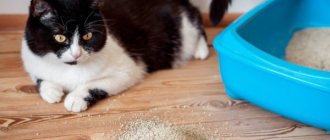
Constipation
If everywhere is clean, but the cat's tray is still empty, you should pay attention to the behavior and general condition of the pet. Constipation in cats after surgery can be very painful. The cat takes a pose characteristic of symptoms of constipation: the animal screams pitifully, the tail trembles. If you experience constipation after surgery, you should not use any laxatives without consulting a doctor. You can help with constipation by giving your animal one tablespoon of Vaseline oil.
It is perfectly absorbed by the body and does not harm the general condition of the animal at all. For constipation after surgery, this drug can be given three times a day. You need to take a syringe without a needle and inject the mixture into the cat’s cheek. If constipation does not go away after 24 hours, be sure to call the veterinarian. An experienced specialist, after an examination and x-ray, ultrasound, will be able to tell you how to cure constipation in a cat after sterilization.
Sometimes a regular enema can help a cat much more than a laxative for constipation. However, such manipulations must be done by a doctor. In most cases, constipation after sterilization can be eliminated by creating a special diet and prescribing softening medications.