What are bedsores?
Bedsores are a pathological change in the skin, subcutaneous tissue, muscles, bones, and other body tissues, developing as a neurotrophic disorder, the causes of which are disturbances in the innervation, blood and lymph circulation of a local area of the body, with prolonged contact with a hard surface.
Brief description of pathological changes on the body:
- develop on the side of the body adjacent to the hard surface;
- characterized by stages of pathogenesis, beginning with stagnation of blood circulation, in the absence of treatment, ending with neurotrophic necrosis of the wet or dry type, sepsis or gas gangrene;
- develop most quickly, within 24 hours, in exhausted patients with congestion in cardiovascular failure;
- localized on protruding areas of the body, the most typical areas of damage are:
- when the patient is positioned on his back, the area is affected (sacrum and coccyx, buttocks, spinous processes of the spine, area of the shoulder blades, heels);
- when the patient is positioned on his stomach, the area affected is (knee joints, iliac crests, protruding surface of the chest);
- when the patient is positioned on his side or half-sitting, the area (ischial tuberosities) is affected;
- rarely localized on the back of the head and folds of the mammary glands.
Specific localization of bedsores on the skin: under plaster casts, in places of tight fit of materials that do not penetrate moisture (oilcloth diapers, rubber tubes), folds of bed linen, bandages, etc.
Specific localization of bedsores on the mucous membranes: under dentures, with long-term drainage of the urethra - on the urethra, with long-term catheterization of blood vessels - on the mucous membrane of blood vessels.
Bedsores rarely develop in young, conscious people without a history of chronic diseases. Typically, in this category of patients, if bedsores occur and develop gradually, there is a high probability of missing the impending pathology.
What to do if your cat is losing hair and has sores?
Parasites on a cat's body
If the owner notices sores on the pet's skin, it should be examined. Diseases do not require immediate contact with a veterinarian, and in some cases the felinologist is able to help the pet himself. Examination of the fur reveals scurrying insects, as well as their excrement in the form of black dots and whitish eggs. The most convenient option is to apply Spot-on drops to your back and let it dry to prevent licking off. To relieve itching, use antipruritic sprays Stop-Itching or Contravance.
Diet
If there are no ectoparasites, the diet should be analyzed. Stop treats, stop begging. Change the food to a hypoallergenic one, for example, dry food based on lamb and rice. If the diagnosis is correct, improvements become noticeable after 14 days. The situation is more complicated for adherents of natural products. You will have to replace all the components of the food with those that the cat has never eaten. Choose a vitamin and mineral supplement. In such a situation, it will not be possible to do without qualified help.
Animal conditions
At the same time, they analyze the conditions of detention. Place household chemicals in places inaccessible to the animal. Stop smoking in the presence of your pet, and you may have to stop using new cosmetics. Use a humidifier. They remove ornamental plants to places inaccessible to the cat or make a choice in favor of one of the two creatures. Regularly carry out wet cleaning and vacuum thoroughly.
The first signs of bedsores
- Subjective sensations that the patient can communicate to caregivers when he is conscious and has preserved pain sensitivity in parts of the body:
- tingling on the skin in places where bedsores are likely to develop, is associated with stagnation of biological fluids (blood, lymph) feeding the nerve endings;
- loss of sensation (numbness) after about 2-3 hours in this area of the body.
- Visible signs of an incipient bedsore that caregivers must know:
- stagnation of peripheral blood and lymph, initially in the form of venous erythema of a bluish-red color, without clear boundaries, localized at the point of contact of the bone and muscle protrusions of the body with the bed, intensity of skin coloring: from barely noticeable to rich;
- desquamation of the skin epidermis with or without preliminary formation of purulent blisters.
These are signs of a developing bedsore. It is urgent to take measures to prevent further deterioration of the pathology.
What to do to eliminate the first symptoms of a bedsore?
To do this you need:
- change the patient’s position every two hours; if there are no contraindications, it is recommended to use special pillows to change the position of the limbs and body relative to the surface of the bed, forming gaps between the skin and the bed;
- monitor the level of the head of the patient’s bed; the head of the bed should be lower or level with it;
- regulate the humidity of the patient’s skin with hygiene products (washing cream, foam, solution, spray, you can warm baths (it is forbidden to use hot water), do these procedures at least twice a day, in case of uncontrolled bowel movements, remove contaminants as quickly as possible;
- remove excess moisture from the skin and skin folds (water, remains of liquid food, urine, wound exudate, sweat) using special absorbent pads, diapers, napkins, towels, films;
- regularly remake the bed or change bed linen at least once a day;
- do not perform intense massage; light stroking of skin areas with signs of stagnation is allowed; carry out this procedure carefully, without friction, especially in areas with close bones;
- use anti-bedsore mattresses of balloon or cellular type, equipped with special silent compressors to maintain and change the rigidity of its base, with adjustable and programmable inflation of different areas.
- use, for patients in wheelchairs, pillows filled with gel foam, air, monitor changes in body position in the chair at least once an hour.
Diagnosis of stroke
Symptoms can sometimes be vague, especially if there are concomitant diseases. Therefore, timely examination of the animal, which includes a set of measures, is important.
Blood test
General and detailed analyzes help determine the factors that provoked the stroke and clarify the type of pathology. First of all, pay attention to hormonal levels, the presence of cholesterol in the blood, and the rate of platelet aggregation.
Analysis of urine
The condition of the body can be judged by the percentage of leukocytes and the presence of protein.
The study will reveal the presence of impurities in urine that can lead to acute ischemia.
Ultrasound of the heart and blood vessels
It is an auxiliary diagnostic method. Ultrasound waves make it possible to assess the state of the circulatory system and the functioning of the myocardium.
Why are bedsores dangerous?
Bedsores are pathologies whose treatment is best avoided. If this could not be done, then when foci of skin maceration form, pathogenesis develops very quickly, with the formation of foci of tissue necrosis and is characterized by long-term treatment of a purulent wound. The outcomes of bedsores are dangerous. In some cases, bedsores are caused by:
- extensive excisions of soft tissues and the formation of defects with impaired innervation and blood circulation of the underlying areas of the body,
- lower limb amputations;
- necrotic damage to the periosteum and bone tissue in the form of osteomyelitis, periostitis;
- depletion of the body's defenses, complicating the treatment of the underlying disease;
With the development of bedsores of the wet necrosis type, the wound becomes infected, with the development of purulent processes (phlegmon, sepsis, gas gangrene).
With the development of bedsores of the type of dry necrosis, a protracted pathogenesis develops with long periods of healing of the defect.
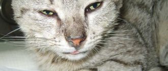
The cat has sores on the neck and itches: how to treat dry sores
If an independent fight against itchy sores and hair loss does not lead to success, seek veterinary help. The specialist conducts a clinical examination, collects anamnesis, and prescribes laboratory tests.
Tests for parasitic skin lesions and dermatophytes in cats
How else can you help an animal if nothing helps?
Depending on the final diagnosis, he develops a treatment strategy using potent medications and feed additives in addition to those discussed above:
- veterinary hypoallergenic food;
- multivitamin supplements in the case when the cat owner intends to use natural feeding;
- oral or injectable antibiotics;
- tableted antifungal agents;
- parenteral administration of an acaricidal agent;
- If ringworm is diagnosed and the listed remedies do not lead to the desired result, antifungal vaccines that have a therapeutic effect are used.
Causes of bedsores
The cause of bedsores is as follows. Our body is completely riddled with small blood vessels. Through these vessels - capillaries - blood flows to various organs of the body. If blood vessels are compressed, blood stops flowing to the tissues, as a result of which the tissues become necrosis.
If a person remains motionless for two hours, his blood vessels are compressed and blood stops flowing to certain areas of the body tissue. Therefore, bedsores form. Remember that it is very dangerous to sit or lie still for a long time.
Bedsores also form if a wet sheet is often pulled out from under a sick person. In this case, blood vessels rupture. This is completely invisible to the human eye. But after the blood vessels rupture, blood stops flowing to the tissues. Bedsores form.
Also, blood vessels can rupture if a person cannot, for example, walk and constantly slides down to take a different position.
Symptoms of diseases
ATTENTION! The same symptoms may be present in different diseases.
Therefore, it is advisable to collect a more complete picture of the animal’s condition and observe other changes in behavior:
- Increased thirst in a cat, the cause is diabetes. With such a disease, the owner cannot help but notice frequent urination, provided that the animal lives in the house and is constantly in sight of people.
- Diabetes also causes severe depletion of the animal's body.
- Thirst of an incessant nature is also inherent in a disease such as renal failure, especially at a later stage, with polydipsia. The cat will urinate frequently, but only a small amount of liquid can be seen in the litter box.
- Old cats may refuse to eat and suffer from vomiting . If the animal is also breathing heavily or experiencing difficulties with the swallowing reflex, the health condition is dangerous and qualified help is needed.
- Heavy breathing and periodic cough are symptoms of hypertrophic cardiomyopathy. The danger of the disease is that it does not appear immediately. At a late stage, in addition to shortness of breath, cyanosis of the mucous membranes and decreased activity are also noted. The animal cannot bear physical activity, sometimes even move its hind legs.
- Decreased activity, lethargy, dull coat are signs of the development of liver lipidosis . Unfortunately, these signs do not appear immediately, but only when saving an old cat is already quite difficult. The pet may even fall into a comatose state, and lethargy is possible.
- When a cat has lost a lot of weight in a very short time, and his appetite only increases, he may have hyperthyroidism. The disease develops actively, then you can observe severe thirst and frequent urination, tachycardia. The animal changes in appearance; it is not only thinner, but also loses the beauty of its coat. The cat will be restless, sometimes even aggressive.
- Cats can develop pyometry . This is indicated by spotting, high fever, and hardening of the abdomen. The cat does not eat, urinates frequently, and may suffer from indigestion.
Risk factors for developing pressure ulcers
It has been observed that bedsores develop in bedridden patients at different times. In medical institutions, to systematize the assessment of risk factors for the development of pressure ulcers, the Norton, Braden or Waterlow scales are used. At home they don't matter. Based on these criteria, risk factors associated with care errors and individual characteristics of the patient, suitable for home use, have been formulated.
Factors associated with errors in the organization of patient care:
- untidy bed, changed less than once a day;
- rare change of underwear to dry and clean ones;
- neglect of hygiene procedures (treating the body with special solutions, drying, massaging areas of the body where possible without additional trauma to the bedsore);
- hard, uneven bed surface.
Factors associated with the individual characteristics of the patient’s condition:
- elderly age;
- exhaustion or, conversely, obesity of the patient;
- diseases of the cardiovascular system;
- diseases associated with impaired innervation of the body (including strokes);
- disorders associated with changes in metabolic processes in the body (diabetes mellitus, impaired water-salt metabolism or normal drinking restrictions);
- unbalanced nutrition or lack of protein in the diet, protein dystrophies (protein metabolism disorders);
- the patient's condition (coma, dementia, etc.) in which he does not control bowel movements or urination.
In addition, factors that provoke the occurrence of bedsores include smoking, diabetes, lack of water and little nutrition, excess or, on the contrary, very low weight, urinary and fecal incontinence, dirty skin, crumbs and small objects in the bed, allergic reaction to products skin care, folds, seams, buttons on underwear, as well as injuries and diseases of the spinal cord and brain, sweating at elevated temperatures.
Why does a cat have sores on its neck and hair loss: 7 reasons
Pathological factors affect the entire body, however, skin diseases most often occur on the neck.
Hair loss in combination with sores occurs for the following reasons:
- Presence of external parasites. Blood-sucking insects irritate the skin and a rash occurs.
The affected surface itches, the cat scratches the skin until it is wounded. Hair falls out, exudate is released, it dries out, and crusts form. external parasites in cats - Allergy to food components. Most often, the skin reacts to food that is unusual for a cat - smoked, salted, fried, cheeses, sausages.
However, failure can occur when the cat is kept on a mixed diet, combining ready-made food with natural food. Allergy to dry food - Atopic dermatitis is a hypersensitive response to external irritants: the smell of tobacco smoke, perfumes, pollen, aromas of ornamental indoor plants, own hair or from other animals.
In the summer heat or winter, when the central heating is on, the skin dries out, cracks, and becomes vulnerable to microbes that constantly live on the surface of the coat. atopic dermatitis - Subcutaneous mites. Ear itches parasitize the inner surface of the shell.
Notoedrus - on the outside. With improper treatment or its absence, areas of the skin on the neck are affected, hair comes out, opportunistic microbes attach, exudate sweats out, dries out and scabs form. Subcutaneous mites in cats - Fungal infections : trichophytosis, microsporia, malassezia occur due to weakened immunity and the addition of bacteria that constantly live on the cat’s skin.
lichen on the body of cats - Stress : a cat gets nervous when a new pet appears in the apartment, a dog chases it, children torment it. Psychogenic stimuli cause itching, scratching to wounds, and hair loss.
- Side effects of medications . Your cat may have a reaction to flea drops, collars, vitamin supplements, antibiotics, or medications intended for human use.
Stages and degrees of bedsores
The pathogenesis of bedsores is characterized by stages of development. There are four stages of pathogenesis.
Stage I bedsores
Visually determined by venous erythema at the site of skin contact with a foreign surface. Venous erythema is the result of obstructed blood outflow from a local area.
How to distinguish venous erythema from arterial hyperemia and bruise?
- Difference from arterial hyperemia:
- the color of venous erythema is red-bluish, the color of arterial erythema is bright red;
- the local temperature of venous erythema corresponds to the skin temperature or slightly lower, the local temperature of arterial hyperemia is a warm area of the skin at the site where the temperature is determined.
- Difference from a bruise:
- the red-bluish color of the skin at the site of finger pressure does not change (bruise)
- the same color of the skin at the site of pressure turns pale (venous hyperemia).
Venous hyperemia (erythema) of protruding bony areas of the human body adjacent to foreign surfaces, without violating the integrity of the skin, is the most important sign of a pressure ulcer of the first stage.
Stage II bedsores
Visually determined by thinning of the epidermis - the upper layer of the skin, followed by peeling and the formation of blisters. Pathogenesis develops as follows: venous congestion provokes disruption of tissue nutrition, innervation of a body area, excess fluid in the skin, causes swelling (maceration) and rupture of epidermal cells.
Superficial damage to the skin in the form of thinning and peeling of the epidermis, loss of integrity, maceration (moistening) is the most important sign of the second stage of a bedsore.
Stage III bedsores
Visually defined as a wound with suppuration (contamination with microflora) or without suppuration.
Involvement in the pathogenesis of deep layers of skin tissue, subcutaneous tissue, muscles with a purulent type of inflammation and the beginning processes of necrosis (tissue death) is the most important sign of the third stage of a bedsore.
Stage IV bedsores
Visually it is defined as a local cavity or defect formed as a result of decay (necrosis), along the edges of the cavity there are walls of the defect on which purulent inflammation continues.
A necrotic cavity and its expansion due to inflammation of the walls is the most important sign of the fourth stage of a bedsore.
In different parts of the body, there may be different stages of bedsores.
Symptoms of bedsores in dogs
Bedsores are visually obvious. Bedsores are most common on the elbows, but they also often occur on the thighs, chest, sides of the legs, hip area, and other parts of the body. Dog owners may not notice the formation of a bedsore until the area begins to ulcerate or until the dog begins to continually lick the affected area. Dog owners may notice one or more of the following signs of bedsores in dogs:
- A bare, wrinkled, red-purple, and thickened area of skin near a pressure point (protruding bone).
- Fluid-filled area under the skin.
- Ulcer or abscess.
- Constant licking of the affected area by the dog (may be accompanied by a change in coat color).
Purulent bedsores
Suppuration of the bedsore area begins in the second stage, develops in the third and fourth, after the wound is contaminated with staphylococci, streptococci, and other pyogenic microorganisms of the area.
A common way of developing a purulent bedsore is erysipelas and phlegmon. In severe cases, a bedsore develops into sepsis or gas gangrene.
Development of purulent bedsores similar to erysipelas
Erysipelas is a local purulent inflammation in a local area of the skin. The causative agent is hemolytic staphylococcus and other pyogenic microorganisms. Erysipelas occurs in people with weakened immune systems, in the older age group, and is accompanied by toxicosis and hyperthermia.
Symptoms of erysipelas with bedsores:
- the skin at the site of the bedsore is bright red with a noticeable dense cold swelling - the main symptom;
- increase in body temperature up to 39 °C;
- headache, weakness, nausea;
- a purulent wound develops;
- exudate from the wound, without treatment, enters the bloodstream.
Development of purulent bedsores according to the type of phlegmonous inflammation
Phlegmon is a diffuse purulent inflammation without clear boundaries. The causative agent is staphylococcus, other pyogenic microorganisms, E. coli. Phlegmon can be localized under: skin, fascia, in the intermuscular space.
Symptoms of phlegmonous inflammation with bedsores:
- a glossy swelling without clear boundaries, red in color, hot to the touch - the main symptom of the onset of phlegmonous inflammation;
- body temperature up to 40 °C;
- rapid development of a fistula with purulent or putrefactive contents.
Development of bedsores according to the type of septic inflammation
Sepsis or purulent blood poisoning is a continuation of erysipelas or phlegmonous inflammation. The extreme stage of sepsis is septic shock, which often results in the death of the patient.
Symptoms of purulent septic inflammation in bedsores
Symptoms can be identified such as:
- atypicality – a variety of symptoms, the absence of the main one (pathognomonic).
- high temperature at the beginning of the process and low at the end;
- rapid development of the process.
The symptoms of sepsis begin to be combated in the early stages of a bedsore with the help of antibiotic therapy. Currently, bringing a patient to a septic state rarely happens, perhaps due to the patient’s immunodeficiency state and insensitivity to antibiotics.
Development of bedsores like gas gangrene
Gas gangrene is a severe purulent, putrefactive pathology. The causative agent is the microorganism Clostridium, most often Clostridium perfringens (soil microorganism). Infection occurs when contaminated soil comes into contact with the macerated surface of a bedsore.
Symptoms of gas gangrene in bedsores:
- sounds of crepitation (crunching) when passed over a damaged area of skin is a pathognomonic symptom.
- rapid development within six to seven hours;
- skin color gray-blue;
- the wound is dry;
- strong putrid odor.
Dangerous skin diseases of cats
Caused by internal pathologies or quickly progress to the stage of exacerbation, life-threatening.
Demodicosis
Microscopic mites are very active. Poisoning from bites spreads so quickly and abundantly that many cases of death are known.
In a mild form, white pimples with pus, partial baldness, red crusts, scratching and a small rash are noticeable on the skin. Over a long period of time, the cat easily scratches the pustules until they become ulcers, introduces infections and bacteria, becomes very weak, and loses appetite.
Demodicosis in cats
Ostiofolliculitis
Ulcers are located in hair follicles. The hair bag becomes clogged with sebum, becomes inflamed, and a staphylococcal bacillus settles inside the bag. If the disease is not treated at the beginning, it quickly progresses into acute furunculosis and peptic ulcer.
It can be detected by redness around the roots of the hairs, purulent pimples up to the size of a pea, burst blisters with crusts on top. The cat becomes restless and does not allow itself to be petted, as touching causes pain.
Ostiofolliculitis
Abscesses
Inflammation in the form of a soft cyst or lump on the skin. There is a lot of purulent exudate inside it. It starts to smell after it bursts and the liquid leaks onto the skin. The smell is especially pungent and unpleasant. Large abscesses greatly impair immunity. The cat may contract an infection during treatment. Therapy is also not fast. Drains are placed in the burst wound to remove all remaining fluid. We have to use strong antibiotics.
Abscesses on a cat's neck
Malasseziosis
Fungal infection. One of the most difficult to treat skin diseases in cats. Diagnosis is difficult and the course is protracted. Treatment requires heavy drugs that harm the rest of the body.
The characteristic smell gives off mold, spoiled cheese or milk. In the groin, near the back opening, behind the ears, near the mouth, claws and nose, you can find redness, rashes, moisture, and sores. The skin becomes painful, so the animal cannot be handled, hisses, and scratches. Relapses occur frequently and for a long time.
Malasseziasis is one of the most difficult to treat skin diseases in cats.
Ringworm
Pathology of an infectious fungal nature. Kittens and cats with oncology or weakened immunity after viruses are more often susceptible.
Fungal spores can be brought to your pet on street clothes or shoes. Spread occurs through household items and personal contact between animals.
The cat is actively losing hair on its head, limbs and tail. The skin in exposed areas peels, itches, becomes very red and pink. In these areas, pustules and crusts appear, which are easily scratched off with claws to open wounds. The disease can gain momentum for up to 2 years. With this course, the pet quickly weakens, loses weight, stops eating and may die from exhaustion. During this period, it is easiest to become infected with fatal infections, since the body does not have the strength to resist viruses. In severe cases, the ulcers spread to the paw pads and it becomes difficult to move.
Ringworm
Treatment without the participation of a doctor is impossible.
Atopic dermatitis
Chronic allergic reaction of the skin to irritants. The body reacts to dust, mold, pollen, household chemicals, and food with severe itching, rashes, hair loss, and ulcers. The cat will scratch especially a lot on the head, ears, chest, neck, lower abdomen and thighs. Dermatitis is severe and weakens the animal. Ulcers are constant and profuse. It is difficult to select living conditions and food for the safe living of a pet.
Atopic dermatitis is a chronic allergic reaction of the skin to irritants
Skin treatment for bedsores
There are three areas of skin treatment for bedsores.
- Hygienic treatment of the skin to maintain the physiological properties of the skin (humidity, skin pH, softening the integument, increasing elasticity), removing physiological secretions of the body (sweat, sebum, epidermal scales), aggressive environments of feces (urine and feces) and deodorization of the skin;
- Preventive treatment of the skin to stimulate local blood circulation, restore sensitivity, and prevent skin cracking.
- Therapeutic treatment, use of medicines in preparative forms (ointment, cream, solution, gel, spray, powder, powder, solution for external use, etc.),
Solution for treating bedsores
In modern surgery, the use of solutions in the treatment of bedsores has somewhat lost its former importance. Meanwhile, simple solutions have been successfully used for many decades in military field surgery to treat wounds and their complications.
- To prevent the development of bedsores, the following are recommended: 2% solution of camphor alcohol, 0.5% solution of ammonia, 1-2% solution of tannin in alcohol, 1% salicylic alcohol 2-3 times a day.
- For the treatment of bedsores the following are recommended:
- external solutions – 25% MgSO4 solution, 10% hypertonic NaCl solution with chymotrypsin, 05% aqueous solution of chlorhexidine bigluconate;
- parenteral solutions - intravenous, drip 0.5% metronidazole solution.
- Solutions in any concentrations are not recommended for prevention and treatment: iodine, KMnO4 - potassium permanganate, brilliant green, hydrogen peroxide.
It is important to monitor the patient's diet. Your diet should include foods that contain zinc, iron and other beneficial microelements. Bedsores are often caused by iron deficiency. Dairy products, fish, poultry, and chicken eggs are rich in it. The patient should eat more green vegetables and fresh fruits. For those patients whose digestive system does not cope well with meat, broths are suitable as an alternative.
On topic: Prevention of bedsores
Prevention
Keeping your pet's sleeping area clean is a disease prevention measure.
Veterinary communities recommend the following to prevent bedsores:
- To sleep the animal, use a special soft, springy bedding - an anti-decubitus bed.
- If the animal is paralyzed, then it needs to be turned over every 2 hours and feces and urine removed in a timely manner.
- Monitor the condition of the bedding, its cleanliness and room temperature that is comfortable for the animal. It should not contain crumbs from food, toys or folds of fabric.
- After contact with the surface of the litter, it is advisable to wipe the skin with a disinfecting solution.
- If a cat is predisposed to the formation of bedsores due to disruption of the endocrine system or diabetes, it is necessary to conduct regular body examinations and strengthen preventive measures.
Bedsores are mainly a disease of old cats or animals with paralyzed limbs and reduced mobility due to other diseases. Their treatment takes a long time and effort. Therefore, it is necessary to carry out timely prevention against this disease in order to prevent the pet from becoming severely critical.
Bedsores on the heels, buttocks and tailbone
The usual position is for the patient to lie on his back. The most vulnerable places for the development of bedsores are the buttocks, tailbone, heels, and sometimes the area of the shoulder blades. Therefore, first of all, when performing hygienic procedures on a patient, you should pay attention to these areas of the body.
Bedsores on the heels
An unusual localization at first glance, but it occurs frequently. There are several heel diseases that, although not associated with bedsores, are evidence of the vulnerability of this area of the body, namely: heel necrosis (Haglund-Schinz disease), bursitis, heel epiphysitis, and so on.
Causes of pressure sores on the heel:
- load when lying on your back;
- thick skin, it is difficult to notice the beginning of the pathology;
- cracks in the skin of the heels, increased likelihood of microbial contamination;
- congestion of the lower extremities is a common companion for older people and occurs in some diseases (diabetes mellitus, etc.).
Symptoms of bedsores on the heels
Many sources indicate the absence of warning signs of bedsores on the heels. The appearance of a bedsore may be preceded by a white spot, tingling, or loss of sensation in the heel area.
Preventive actions
Prevention of pressure sores on the heels includes:
- use heel relief devices (wedge-shaped pillows, sheepskin, special mattresses);
- stimulate blood circulation (light massage of the calves), rub in 2% camphor alcohol, change the position of the legs every two hours;
- protect the skin of your heels from microflora, for example, using a colloidal butterfly bandage (Comfil Plus), which creates the effect of a moist, sterile chamber. The materials from which the bandage is made and the impregnating composition allow the bandage to be left on the wound for two to five days.
Treatment of bedsores on the heels
The remedies and methods for treating bedsores on the heels do not differ from the treatment of bedsores on other parts of the body. The anatomical features of the location of the bedsore are taken into account.
Bedsores on the buttocks
Bedsores on the buttocks have dangerous consequences. This area is located close to important organs (rectum, hip joint, pelvic organs, nerve nodes and blood vessels innervating the lower extremities), disruption of which significantly affects the quality of life, and in some cases, the preservation of life.
The buttocks have a powerful muscular system that should resist the formation of bedsores. However, regular contamination of the skin with urine and feces, without proper hygienic treatment, stimulates the development of bedsores. When a necrotic focus forms, extensive soft tissue defects are formed that are difficult to treat. The warning signs and symptoms of bedsores on the buttocks are typical.
Preventive actions:
Prevention of pressure sores on the buttocks includes:
- carry out regular hygiene procedures for the buttocks and perineum using washing creams, liquids, sprays; to simplify the procedures, use special latex-coated gloves that isolate the hands of the person caring for the patient and do not injure damaged areas of the patient’s skin;
- to prevent diaper rash on the skin of the buttocks and perineum, use absorbent panties, sheets, diapers, diapers, and neutral absorbent powders;
- Regularly, every two hours, change the patient's position, use anti-bedsore mattresses with programmable changes in the hardness of the bed and its different areas, use pillows, bolsters and other devices to prevent bedsores on the buttocks.
Treatment of bedsores on the buttocks
In the early stages, Russian-made multi-truss wipes or their analogues are recommended. The use of multifarm wipes is indicated for purulent processes with weak to moderate exudation. The exposure time and course frequency are indicated on the packaging.
Treatment of advanced forms of bedsores on the buttocks is carried out taking into account the anatomical features of this part of the body, similar to the treatment of purulent wounds.
Bedsore on tailbone
This area of the body protrudes somewhat and when lying down, the bones of the tailbone are in close contact with the bed of the bed. Bedsores on the tailbone are dangerous due to the small muscle layer and the presence of important nerve plexuses in it. Melting of tissues during necrosis damages nerve endings and provokes disruption of the innervation of the lower part of the body.
Symptoms of a bedsore on the tailbone
Precursors and symptoms of a pressure sore on the coccyx correspond to the classical ideas of the development of its pathogenesis. Due to the anatomical proximity of the buttocks and tailbone, preventive measures and treatment of the early stages are identical to those on the buttocks.
Treating bedsores at home
What is a bedsore?
A bedsore is damage to the skin of varying degrees, from abrasion to deep lesions down to the bone. What causes a bedsore?
In bedridden patients who, due to certain circumstances, are in a state of immobility or are inactive, a bedsore forms.
The elderly age of the patient is an aggravating factor, since in such people the skin is thinner and more susceptible to injury, so bedsores are more common in older people. Certain parts of the body are exposed for a long time to such factors as: pressure, friction, shift of the inner layers of the skin in relation to the upper layer, which is the main cause of bedsores. In places exposed to these factors, blood vessels are compressed, blood does not flow to the tissues and, therefore, the supply of nutrients and oxygen stops. From such a “hunger strike”, tissue cells begin to die (necrotize) - the skin is destroyed. Where do bedsores most often occur in bedridden patients?
Most often, in bedridden patients, depending on the body position in which the patients are most of the time, bedsores form in the areas:
- Coccyx;
- Buttocks;
- Heels;
- Shoulder blades;
- Elbows;
- Back of the head
In these places, the bones are located very close to the skin, so it is here that the negative effects of pressure and tissue shear manifest themselves to a greater extent.
The formation of bedsores, in the absence of proper preventive measures, occurs quite quickly. Preventive measures that are quite simple, but require strict and regular adherence, can reduce the risk of bedsores. What needs to be done to prevent the occurrence of a bedsore or prevent its further development?
The main measures should be aimed at:
- something that will help eliminate the causes that made the bedsore possible;
- restoration of blood flow to the places where bedsores form.
Therapy for the treatment of bedsores is similar to prevention measures, only wound care (bedsore) is added.
Important
I. Daily examination of the patient’s skin must be carried out at least once a day. This is especially true for places where the risk of pressure sores is most likely (sacrum, buttocks, hips, heels, elbows, knees, ankles, back of the head). Pay attention to changes in skin color and integrity problems such as:
- abrasions;
- scratching;
- scratches.
II. To relieve pressure on the patient's body, it is possible (necessary) to use auxiliary means that will make the load inconsistent or intermittent. We are talking about anti-bedsore mattresses, pillows and pads filled with water, gel or air. Special beds that allow you to change the position of the patient’s body will also be a good help.
Great attention should be paid to the hygienic care of your loved one. Keep the skin moderately damp. Dry skin is most susceptible to tissue shifts and the destructive force of friction. Too wet skin is no less bad - diaper rash occurs and a favorable environment is created for the proliferation of harmful microorganisms. Currently, a large number of special products have been created that will help you ease the work of caring for a bedridden patient with bedsores.
Disposable foaming mittens, sponges, or gloves will help you carry out hygiene procedures. For skin care, modern standards recommend the use of professional cosmetics: creams, foams, lotions, gels to create a surface protective film on the skin, which minimizes the impact of factors such as friction. Change bed and underwear on time and correctly. If necessary, use diapers and absorbent diapers.
It is important to ensure your loved one is receiving proper nutrition. (Balanced nutrition - more protein, mandatory presence of vegetables and fruits in the diet, adherence to food intake, drinking regime). If the patient is not completely immobilized, teach self-help techniques for ambulation. It is very important to unload areas of the body that are subject to pressure; for this it is necessary to change the position of the body every 2 hours (at night, if necessary, wake the patient only to turn him over inhumanely). If possible, depending on the severity of the patient’s condition, perform breathing exercises and physical therapy.
If a bedsore occurs, we provide care (treatment) for the wound. For this purpose, various medications are used, as well as modern dressings.
. Let's talk about the possibilities of modern dressings in the prevention and treatment of bedsores at home. There are many therapeutic and therapeutic dressings for care that can help cope with the problem of bedsores. The use of modern dressings significantly reduces the treatment time for bedsores. They are easy to use, do not require special skills and do not require daily replacement. Relatives caring for a patient with bedsores can independently change the dressing. Such dressings are changed after 1–7 days, depending on the type of dressing. This allows you to disturb the wound less and create favorable conditions for healing. And for the patient, reducing the number of dressings has a positive effect. The choice of dressings depends on the condition of the bedsore (wound surface). It is important to take into account the size of the bedsore, the depth of the lesion, the presence of infection, the amount and nature of discharge from the wound (color, smell, viscosity).
Depending on the properties, wound dressings are used for:
- maintaining optimal humidity;
- removal of excess moisture and wound secretions (exudate);
- fight infection and prevent re-infection (containing an antimicrobial component);
- cleansing the wound;
- ensuring painless and atraumatic removal during dressings, etc.
Therefore, when choosing a dressing, it is important to correlate the condition of the bedsore and the properties of the dressings. A bedsore goes through 4 stages in its development and it is very important to take this into account when choosing treatment. It happens that what helps at stages 1-2 cannot or is ineffectively used at stages 3 or 4 of the development of a pressure sore. Let's try to figure out how to choose the right bandage for the treatment of bedsores of various stages.
Prevention and treatment of bedsores stage 1-2
First, let's look at what a bedsore looks like in the initial stages of development.
I. Stage
This is the initial stage of a bedsore. When examining the skin, you observe that the skin is intact, not destroyed, but there are changes in color: pallor of the skin area or persistent redness. What dressings can be used?
Aerosol spray OPSITE SPRAY
When sprayed, it forms a transparent film on the skin. We apply it to intact skin in places where the first signs of bedsores appeared or for the purpose of prevention to places at risk of bedsores - we get additional protection of the skin from friction. Provides protection for weakened skin from irritants: biological secretions, moisture; from diaper rash in the fold area. Helps prevent irritation and serious skin problems, instead of dealing with them after they appear. We apply it daily. There is no need to rinse off; the bandage peels off on its own after 72 hours.
Film dressings OPSITE FLEXIFIX, OPSITE FLEXIGRID
Transparent films with adhesive surface. Provides access of oxygen to tissues and evaporation of moisture (prevents the accumulation of fluid under the bandage), and also allows visual monitoring of the condition of the skin. They do not interfere with hygiene measures because they are waterproof. Protects the skin from bacteria, microdamages and factors that contribute to the development of bedsores (excessive humidity, incontinence, etc.) We apply a bandage to intact skin in places where the first signs of bedsores appeared or for the purpose of prevention on places at risk of bedsores. Can stay up to 7 days without a shift.
II. Stage
Redness of the skin remains, but there is a superficial violation of the integrity of the skin (abrasions, blisters, peeling, ulcers, scratching). At stages I-II, preventive measures and treatment of bedsores are aimed at eliminating tissue compression and restoring normal blood circulation in these areas. The main task is to protect the skin from friction, pressure and exposure to adverse environmental factors. For this purpose, the following are successfully used:
Hydroselective dressing CUTINOVA HYDRO
A soft, thickened film dressing actively promotes the healing process, as it retains the necessary growth factors in the wound. Self-fixing, no additional dressings (adhesive plasters, bandages) are required. The translucency of the dressing allows you to observe the condition of the wound. Can remain on the wound without changing for up to 7 days. Effectively helps prevent further development of bedsores. The bandage can be used at 1-2 stages of bedsore development.
Absorbent sponge bandage ALLEVYN
In various designs (self-adhesive, there is a special shape for the sacrum, for the heel). The bandage is intended for both the prevention and treatment of bedsores. The bandage effectively reduces the force of pressure - the effect of creating a soft pad that provides the patient with additional comfort and protection from the formation of bedsores. As a result of compression of the skin and blood vessels, stagnation and tissue death occur. When the pressure eases, the vessels dilate and blood circulation is restored. Allevyn can be on the wound without changing for up to 5-7 days.
Treatment of bedsores stages 3-4
III. Stage
Deeper damage is observed: the skin is completely damaged to its full depth and the process extends deep into the muscle layer with penetration into the muscle. As a rule, these are already quite deep bedsores; there may be copious discharge from the wound; when an infection occurs, the discharge from the wound has an odor and is of a different color (depending on the type of infection).
IV stage
These are usually deep, purulent bedsores. Death (necrosis) of all soft tissues occurs; cavities form, and there may be “pockets” along the edges. Tendon and/or bone formations may be visible at the bottom of the bedsore. At stages 3-4, an infection may occur, in which case there will be purulent discharge from the bedsore. The surrounding skin will also be affected and there may be swelling and redness. At stages III-IV, treatment of pressure ulcers is aimed at removing (rejecting) dead tissue, treating wound infection and stimulating rapid healing of the cleaned wound. At this stage, it is important to receive surgical help, since it is not always possible to cope with only medications (ointments, powders, etc.) and bandages.
Surgical treatment
— this is the surgical removal of necrosis and closure of a pressure wound, including skin grafting. The task is to clean the wound surface of the bedsore. The goal of bedsore treatment is to restore the skin in the area of the bedsore. To clean, heal and care for the wound surface of a bedsore, dressings are selected based on the condition of the wound (the presence of non-viable tissue, the presence or absence of signs of infection, the amount and nature of discharge from the wound, the viability of the wound edges). To treat stage 3-4 bedsores, bandages are used to clean the wound, stimulate the formation of new cells (granulations), and protect them from drying out and secondary infection.
It is recommended to use special dressings:
- dressings that help cleanse bedsores;
- antimicrobial dressings, which provide infection prevention and are also used to treat infected bedsores;
- absorbent dressings are capable of absorbing large amounts of wound fluid and providing moisture balance;
- dressings that provide an optimal environment for bedsore healing;
- various atraumatic (non-adhesive to the wound surface) dressings.
In order to “cleanse a bedsore” from necrotic tissues, you can use:
Modeled alginate bandage ALGISITE M
Provides effective drainage of cavitary wounds and their cleansing. Alginate fibers of the dressing, reacting with secretions from the wound, turn into a gel, which mechanically binds microorganisms on the surface of the wound. The dressing should be changed every 2-3 days.
Hydrogel with INTRASITE GEL delivery system, and modeling hydrogel bandage INTRASITE CONFORMABLE
Provides gentle, quick wound cleansing from necrotic tissue.
Stimulates autolysis (self-dissolution of cells) in the wound and promotes rapid wound healing. To enhance the effect, fix on top with OPSITE FLEXIFIX, OPSITE FLEXIGRID
or
CUTINOVA HYDRO
.
CARBONET
absorbent deodorant dressing with activated carbon . Absorbs heavy sticky exudate, absorbs unpleasant odors from the wound, absorbs bacteria and rotting decay products. In order to prevent secondary infection of bedsores, as well as on infected bedsores, antimicrobial dressings are used.
Armband with silver ACTICOAT
Thanks to the presence of silver, the dressing has an antimicrobial effect, suppresses pathogenic microorganisms, thereby not only preventing the development of wound infection, but actively fighting it. BACTIGRAS Chlorhexidine Mesh Dressing - Chlorhexidine is continuously released from the dressing, providing a long-lasting antibacterial effect (infection control). When using various ointments, for better absorption of the ointment into the affected area, absorbent film dressings OPSITE Post-Op Visible and OPSITE Post-Op are perfect as a fixing sterile dressing. The ointment must be applied to the center of the dressing (on the functional pad), and not to the bedsore itself. For bedsores with a large amount of discharge from the wound, the use of special absorbent sponge dressings (Allevin) is recommended. Effectively absorbs discharge from the wound and is able to retain it under pressure. Maintains an optimal moist environment necessary for healing. Does not require frequent changes of dressings (on the wound up to 5-7 days without changing).
When treating bedsores, modern treatment methods - negative pressure wound treatment (NPWT therapy) - can be used to remove heavy discharge (exudate). The main effects of this therapy: Stimulates blood supply in the area of the bedsore, brings the edges of the wound closer together, activates granulation (healing) processes; reduces swelling of surrounding tissues. When treating pressure ulcers at home, you can use the
PICO
Negative Pressure Wound Treatment System .
It is especially effective for the treatment of bedsores that cannot be treated with other methods. During the healing phase of a pressure ulcer, dressings can be used to provide an optimal healing environment and protect new cells. Absorbent dressings JELONET
are easily modeled on wounds of complex configuration, do not stick to the wound, and protect granulations. This is a mesh dressing impregnated with paraffin so that it does not stick, which can act as a basis for the use of medicines: ointments, powders, etc.
All of the above dressings are very effective in the prevention and treatment of bedsores, including infected bedsores. Remember!!!
When treating a bedsore at home, proper medical supervision is required. Otherwise, severe complications may occur: osteomyelitis, sepsis, toxic shock and death. A bedsore is a wound (infected bedsore-purulent wound), which can only be cured by a set of measures for the general treatment of bedsores, in accordance with the stage of the wound process. If you do not take this circumstance into account, then not even a very high-quality anti-decubitus mattress, not a single expensive bandages, plasters, ointments, etc. will be effective.
How to treat bedsores?
Treatment of pressure ulcers of the second, especially the third and fourth stages should correspond to the treatment of purulent wounds. Purulent wounds are difficult to treat, but over many years of field surgery, a standard treatment algorithm has been developed. Of course, additions and improvements to treatment are regularly made, but the goal and objectives of treatment have remained unchanged.
I. In the first phase of pathogenesis
In the first phase, when the bedsore wound is filled with pus and necrotic tissue, you should:
- ensure the drainage of pus from the wound;
- relieve swelling;
- suppress microbial contamination of the wound.
Inspection of the wound and cleaning of the edges from necrotic tissue is performed in a surgical department. To drain the pus, drainage tubes are made and they are regularly inspected.
Related: List of home remedies for bedsores
Passive drainage can be done at home:
- To do this, the wound is filled with special napkins soaked in compounds that promote the outflow of pus. The napkins are changed periodically. You can use regular bandages as napkins, the edges of which do not fall apart into threads. Solutions and ointments are used to impregnate napkins.
- Enzyme therapy is the next method of surgical treatment of purulent wounds (therapy with pus-removing enzymes).
- Antiseptic solutions for external use. furatsilin, hydrogen peroxide, boric acid (currently used to a limited extent). Modern formulations are indicated for use - 0.5% solution of iodopyrone, 1% solution of dioxidine.
- Physical methods of treatment. They use traditional methods (UHF, ultrasonic cavitation, oxygenation, vibrophonation, laser therapy and other similar methods)
Outdated medicines: hypertonic solutions of 10% sodium chloride, 3-5% boric acid solution and others. Currently, the use of such solutions is limited due to the low suction force from 4 to 8 hours.
Hydrophobic ointments (liniments, emulsions) on a petroleum jelly basis (Liniment according to Vishnevsky, synthomycin emulsion, tetracycline, neomycin and others). Their disadvantage is that they do not absorb pus; the antibiotics in their composition do not work at full strength.
Modern medicines: hydrophilic (water-soluble ointments) - Levomekol, Levosin and other water-soluble compounds. They remove pus from the wound well, within about 20-24 hours. Attention! Use hydrophilic ointments only if there is pus in the wound; in another situation (no pus), these ointments are not effective.
Proteolytic enzymes (trypsin, chymotrypsin, others). To enhance their action, use a combination of these or other enzymes with ointments, for example, a combination of enzymes and Iruksol ointment.
II. In the second phase
In the second phase, after cleaning the bedsore from pus, healthy tissue appears. A healthy scab is a thin layer of dried granulations. A purulent scab is a thick crust consisting of dried pus. Recovery under a purulent scab is impossible!
When healthy granulations appear, treatment is prescribed:
- relieving inflammation;
- protecting healthy granulations (healthy tissues) from accidental damage;
- stimulating tissue repair processes.
Most common disease
The list of diseases is long, and others may be added to it. Many can appear in small, adult young individuals, and only get worse in old age:
- Kidney failure.
- Diabetes.
- Pyometra.
- Hypertrophic cardiomyopathy.
- Hyperthyroidism.
- Oncological diseases.
- Dental diseases.
- Eye diseases.
Why do cats have the biggest health problems as they age? With each year of adult life, the body’s immunity decreases, it fights less against the onset of a particular disease. The body wears out, it is more susceptible to viruses and bacteria, and it is more difficult to endure injuries. And this once again suggests that you need to be doubly attentive to an old pet.
It is not difficult to do this; it is enough to observe the pet, analyze its behavior and note changes in it. For example, an elderly cat refuses food for a long time. This is unlikely to be due to the heat; a healthy animal cannot avoid eating for a long time . It is imperative to identify other symptoms, show the cat to the veterinarian, and tell him about your observations.