4847Administration
The bladder of all living beings is a unique organ, which is striking in its elasticity and is second only to the uterus in its ability to stretch. Every owner should know where a cat’s bladder is, since there are situations when an emergency home determination of the animal’s condition requires palpating this organ.
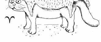
The bladder is pear-shaped, and if you understand where it should be, it can be easily felt even by a non-specialist. If the procedure is carried out carefully, it will not cause harm to the cat, but it will provide a lot of information about how the urinary system works correctly and whether the cat is sick. The filling of an organ with urine does not affect its location. Invariably, a cat's bladder is located in the lower abdomen immediately in front of the crest of the pubis in the lumen of the small pelvis.
© shutterstock
Location of the cat's bladder
The location of the bladder does not depend on its fullness - it is always in the lower part of the peritoneum, in the lumen of the small pelvis, immediately in front of the crest of the pubis.
The cat's urinary system.
At the exit, the bubble narrows and forms a neck - cervix vesicae. Easily accessible for surgical intervention and simply palpable due to its convenient location and the fact that it is not covered by the omentum. If the cavity is full, it reaches all the way to the navel, so palpation if urine stagnation is suspected should be more than careful due to the severe thinning of the walls.
Depending on the degree of fullness, it can move slightly in the pelvic cavity, but remain relatively stable in its original physiological orientation due to the fact that it is held by ligaments.
Functionality
In terms of functionality, it serves as a bag for temporarily holding urine until it is removed from the body.
The presence of urine in the body is a temporary process; it is later excreted.
Structure
- The outer side consists of fibrous connective tissue, underneath is the detrusor - a layer of muscle tissue, the smooth muscles of which move and help push out urea.
- Next is the submucosal tissue, which is a membrane of elastic fibers that performs a supporting function for the mucous membrane to keep it inside the bladder.
- The mucous membrane itself contains a transitional epithelium, consisting of special cells that subsequently form folds. These folds are formed in the absence of fluid in the cavity of the organ and allow it to subsequently expand to large volumes.
- The serous membrane covers the apex and the body of the bladder itself.
- In the lower part of the body it transforms into the umbilical fold - plica vesicoumbilicalis media and passes further towards the bone of the womb, and then to the navel itself. The difference in the genitourinary system of females and males is insignificant.
In males, the sac is located under the womb, in females - under the uterus, but in both of them it is accessible for palpation and can be easily felt.
The bladder in cats is accessible to palpation.
Determining the degree of bladder fullness
The cat's nervousness will indicate the presence of pain.
You can determine the degree of filling of the bag yourself.
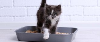
- To do this, you need to put the cat on all fours, slightly supporting it under its belly.
- The animal should be with its hind limbs closer to the owner.
- Hands should be placed on the cat's back so that both thumbs are on the animal's rump, and the rest should be palpated on the stomach closer to the urinary canal.
Nervousness and attempts by the cat to escape will indicate the presence of pain, which means immediately contacting a doctor.
Feeling the bladder
To more specifically determine the topographic location of the organ, you should place your palm closer to the base of the tail, perpendicular to the ridge. The projection of the upper border of the organ will be the side of the palm opposite to the tail. Normally, in the absence of urine, a soft, insignificant fluctuation should be felt under the fingers, evenly distributed within the boundaries of the organ, while the boundaries themselves are not felt.
Normally, in the absence of urine, the boundaries of the organ are not felt.
The presence of daily urea retention will be indicated by a compaction of soft consistency the size of a walnut.
The feeling of liquid will roll freely under your fingers. A delay of more than two days will be felt under the fingers by a compaction, the size of a tennis ball, with a consistency reminiscent of soft rubber, with a slight fluctuation.
If a larger lump with a harder consistency is felt, consult a doctor immediately. Your cat will most likely need to empty his bladder using a catheter.
If upon palpation there is a seal, the bladder should be emptied using a catheter.
Preparation
At home, the manipulation is performed in the toilet or bathroom. Before starting the procedure, the pet must be calmed in a gentle, calm voice and stroked.
Then you should carefully palpate the abdomen to determine the degree of bladder tension and allow the cat to get used to the touch.
Before the procedure begins, the male penis is examined for superficially located uroliths. The discovered stones are removed from the edge to the base of the penis, gently squeezing it with two fingers. First, fingers should be lubricated with liquid soap or petroleum jelly.
Possible causes and treatment options for bladder atony
The causes of USC are inflammatory diseases of the urinary system, mainly its lower parts - the bladder (cystitis) and urethra (urethritis), urolithiasis (urolithiasis), and much less often - tumors. The symptoms listed above are associated with inflammation of the affected organ and pain, the appearance of abnormal inclusions in the urine and often obstructions to the normal flow of urine. An animal suffering from this disease is characterized by a tense, unnatural posture, indicating impaired urination.
The urethra (urethra) is the lowest section of the urinary system of animals, through which urine, which is formed continuously in the kidneys and accumulates in the bladder, is regularly released into the external environment during urination.
Acute urinary retention occurs due to blockage of the urethra by mucus, crystals, blood clots and small stones, and occurs almost exclusively in cats, and is extremely rare in cats.
This disease can be caused by the following reasons:
- Urolithiasis disease.
- Tumors.
- Inflammation of the mucous membrane of the bladder.
- Damage to the central nervous system.
- surgical intervention;
- dysfunction of the spinal roots as a result of certain diseases, for example, advanced syphilis;
- medicinal effects on the nervous system (anesthesia, psychotropic substances);
- general intoxication of the body, poisoning or activity of pathogenic microbes;
- long-term infectious disease of the bladder (cystitis);
- colds;
- atony of skeletal muscles and muscles of internal organs as a systemic disease (weak muscles cannot support the spine and spinal cord, as a result the innervation of the body is disrupted); This point includes infancy, when the muscle frame has not yet strengthened;
- diseases of the nervous system, exposure to stress factors;
- various mechanical injuries of the spine, especially in the sacral area, spinal cord injuries, perineal injuries;
- kidney stones or sand;
- diseases of the endocrine system, metabolic disorders, excess weight;
- untimely emptying of the bladder, frequent overflow, leading to stretching of the muscle walls;
- age factor;
- a combination of factors (nervous, hormonal) in women during menopause.
Bladder atony often develops in women after childbirth (especially in the first year). This is a normal phenomenon, caused by a temporary weakening of the muscles of the bladder after an impressive load during pregnancy and childbirth. Treatment is usually not required as the disease goes away on its own. This process can be accelerated by special gymnastics that uses the pelvic floor muscles (Kegel exercises).
These symptoms allow us to confidently diagnose “bladder atony” and prescribe treatment.
We recommend!
Disturbed innervation of the bladder leads to the fact that the body is unable to control the process of accumulation and separation of urine. The muscular walls of the organ, which are normally capable of greatly stretching and holding large amounts of urine, lose their tone. The brain does not receive nerve impulses sent by wall receptors, signaling their stretching as a result of the bladder filling with urine. The so-called urinary incontinence develops, its uncontrolled release drop by drop.
These factors - individually or in combination - can cause acquired bladder atony. In addition, the disease can be congenital, caused by genetic or embryonic developmental pathologies. Treatment in this case can be difficult and limited only to neutralizing the effects of atony through the use of modern hygiene products.
The disease most often occurs in young children and the elderly, as well as in women who have recently given birth. These groups are united by one parameter: muscle weakness (all in general and specifically the muscles of the pelvic organs).
Urinary incontinence after childbirth
Examination by a veterinarian
The doctor needs to tell all the symptoms that the owner noticed. During the examination, the veterinarian will perform palpation, which can be used to identify large tumors and organ development abnormalities.
In case of incontinence and other urological disorders, a urine test will be prescribed, most often microscopy of the sediment will be required. Previously, we have already given a detailed explanation of a urine test for a cat. If necessary, ultrasound and x-rays are also performed.
Additional studies may be needed, such as a blood test, microflora culture, and analysis of renal epithelium in the cat’s urine.
Important: obvious neurogenic disorders such as injuries, tumors, spinal hernias, and urinary disorders are treated by eliminating these factors.
In cases of functional disorders, a complete and comprehensive examination cannot be avoided. Only a complete collection of all indicators will make it possible to correctly diagnose and correctly prescribe treatment.
During the examination, the veterinarian will recommend using special diapers for animals. There should always be a clean, absorbent diaper on the sleeping area. These care items are sold at any pet store.
Important: do not scold your pet for its inability to tolerate, do not punish it under any circumstances. A stressful situation will only harm the animal, but affection and care, on the contrary, will help cope with the disease.
Diseases with similar symptoms
In addition to bladder spasm, urinary retention can be caused by the following pathologies:
- Obstruction or blockage of the urethra, especially often encountered by cats who have injured the external genitalia or have had an infectious disease.
- Kidney failure, when organs completely or partially lose the ability to synthesize urine.
- Tumors or damage to the urinary tract.
- Inflammatory process or poisoning by toxins.
- Spinal cord infections, spinal column injuries.
- Uric acid crystals in the urethra (in older castrated cats), stones in the kidneys, urethra, bladder.
Most often, cats suffer from problems with urination; in cats, the channel for excreting urine is much wider. At risk are neutered pets whose owners do not pay enough attention to their diet.
Cats who have undergone surgery must eat special food that includes the necessary vitamins, minerals and supplements. The situation is aggravated by obesity, which impedes the functioning of all systems, including the excretory and endocrine ones.
At-risk groups
global $ads_google;
//data-ad-slot=”2475549904″ $ads_google = empty($ads_google) ? false : true; ?> if ($ads_google == false) {?> Urolithiasis occurs in 15% of cats. This high figure is due to the fact that there are many potential factors for the development of the disease. According to statistics, symptoms of urolithiasis are five times more likely to occur in males. This is explained by the anatomical structure of the urethra. Unlike the urinary canal of females, in cats it has an S-shaped bend and a thinner lumen. The disease is most often recorded in animals aged 1 to 6 years. Considering that urolithiasis is often fatal, owners of furry pets need to know all the risk groups. These include cats:
- With a hereditary predisposition. Experts identify a number of breeds that have a genetic tendency to urolithiasis. First of all, these are Persian, Burmese, Siamese, and Maine Coon cats.
- Obese. Excess weight is directly related to overeating. Against the background of impaired metabolism, excess minerals entering the body are deposited in the form of uroliths.
- Castrated. The operation indirectly affects the development of urolithiasis. Neutered cats become less mobile. And this in turn implies the accumulation of excess weight.
- Suffering from chronic or acute infectious diseases. Pathogenic microbes and their metabolic products are the nuclei around which salt crystallization begins.
Symptoms of atony of the bladder walls
The animal becomes lethargic, stops playing, and groans. There is a noticeable urge to urinate. Characterized by the adoption of tense, unnatural poses. The bladder is enlarged. The cat gives off a specific smell. In severe cases, the abdomen can be very tense, to the point of a stone-like state. This happens when the bladder bursts due to extreme pressure. When trying to touch, the cat may meow or even growl and hide in a corner.
The normal frequency of urination for adults is 1-2 times a day, for kittens 3-4 times a day. Deep-colored urine is normal, especially if the animal is fed dry food. When feeding wet food and natural food, the urine is less concentrated; the cat should visit the litter box at least 2 times a day.
Kittens that feed on their mother's milk produce tiny amounts of urine because the food is almost completely absorbed by their bodies. In addition, a caring cat regularly licks her babies, and it may seem that the cubs do not urinate at all. This is the norm for animals of one and a half to two months.
Urinary retention in cats
Acute urinary retention (urethral obstruction) is the most common and life-threatening complication of the so-called feline urological syndrome (FLUTD or FUS).
USC is a complex of the following symptoms:
To establish a diagnosis of acute urinary retention, in addition to analyzing the owner’s complaints, it is necessary to feel and evaluate the animal’s bladder. The bladder is located in the lower abdomen between the hind legs and slightly in front of them. As a result of the blockage, it fills with urine and becomes large, the size of a peach, and hard to the touch.
More details
Urolithiasis in cats. Symptoms, treatment
Urolithiasis in cats, symptoms and treatment of which are described in this article #8212; a disease that is very common in cats. The disease is accompanied by the formation of grains of sand and stones (uroliths) in the bladder. Cats suffer from the disease much more often than kittens. For the first time, urolithiasis begins to bother the animal at a young age - from 2 to 6 years.
This x-ray shows how stones fill almost the entire urinary system of an animal
The disease does not last long. And the development of symptoms depends on the size. shapes and locations of stones. If the urolith does not get stuck in the urethral canal, does not interfere with urination, its surface is smoothed, and the advancement of the stone does not cause pain or damage to the mucous membrane, then the disease can become chronic, last for a long time and remain invisible to the owner of the animal. But at this time the stone “grows” in the cat’s body for several years.
The main sign of decreased tone of the bladder walls is, naturally, loss of control over urine output. Urination occurs spontaneously, and the urge to go to the toilet may be completely absent. The symptom usually occurs during strong laughter, coughing or sneezing, or physical activity - when the muscles of the abdominal wall sharply tense, putting pressure on the bladder.
When trying to empty the bladder intentionally, the patient encounters difficulties. In order to start urinating, it is often necessary to strongly tense the muscles of the abdomen and pelvis. The stream is weak and urine cannot be completely removed.
The pathology is not accompanied by painful sensations, except for possible heaviness in the lower abdomen when the bladder is full.
Successful treatment of the disease is possible only after determining its true cause. Decreased tone of the bladder walls is sometimes just a symptom of another disease, much more serious, for example, syphilis or a bacterial infection.
Traditional medicine and herbal medicine offer many treatments for bladder atony, some of which are truly effective. They strengthen the body as a whole, successfully fight inflammation and hidden infections, and have a beneficial effect on the nervous system.
loading.
Diagnostics
If you suspect that your pet has acute urinary retention, try to assess the fullness of the bladder, but if you have even the slightest doubt about whether it is full or not, consult a doctor immediately. After all, if urinary retention lasts several days, the accumulation of toxins in the blood will lead to the death of the animal.
In order to diagnose acute urinary retention, it is necessary to examine the animal. The bladder is located in the lower abdomen, in the middle and slightly in front of the hind legs. When blocked, it fills with urine and increases in size, becoming hard to the touch, like a tennis ball. When you touch this area, the animal tries to escape and shows anxiety. The normal bladder is soft and difficult to find.
If you suspect your pet has urinary problems, assess the degree of fullness of the bladder. If in doubt, do not delay visiting the veterinarian. Just 2-3 days of urine retention lead to poisoning of the animal’s body with toxins and subsequent death.
To finalize the diagnosis, the specialist performs:
- External examination of the cat.
- Palpation of the abdominal area.
- Survey of owners in order to identify the complete picture of the disease.
- Collection of tests.
- Ultrasound.
- Exclusion of diseases such as blockage of the urethra and urethra, paresis of the nerves of the bladder.
Urethra.
Urine finally leaves the cat's bladder through the thin-walled urethra
- a tube that extends from the base of the bladder to the outside of the body. In cats, this is a relatively short tube connecting the bladder and the external urethral sphincter. In cats it is longer, passes through the prostate gland, then along the penis to the external sphincter. The external urethral sphincter is consciously controlled and relaxed when the cat finds the appropriate place and time to urinate.
Littleone 2006-2009 > Hobbies and interests > About pets > The cat does not pass urine
View full version : Cat won't urinate
My friend's cat is very sick. At first the urine was bloody, they took me to the Bolshevikov clinic, put a catheter in, the urine partially drained. The cat began to drink and eat. By the evening she felt bad again, she was crying, she took her to the doctor again, they already received test results, they showed that there was an inflammatory process, but there were no crystals in the urine. They tried to insert a catheter, tortured the cat, but nothing worked. They offered to perform an operation: to remove the urethra through an incision in the peritoneum, but they told me that this was not reliable and could become overgrown. I saw three different veterinarians and they all said different things. Tell me, is there any good clinic near Elizarovskaya where they do this? It seemed to me that at the clinic on Bolshevikov Street the doctors did not know what to do; they didn’t even prescribe a painkiller.
Rosa Vetrova
15-10-2008, 23:17
This could be elementary cystitis. Go to Bukharestskaya, 124. Midor Clinic. Open from 9 to 21.
Treatment
The most important thing is to remove the blockage and restore normal urine flow. The veterinarian will feel the bladder and try to force the animal to urinate by applying gentle pressure to the bladder. Sometimes this can eliminate urinary retention, but most often a more serious intervention is necessary - the installation of a urinary catheter. In this case, the plug in the urethra is usually washed out into the bladder.
The procedure for inserting a urinary catheter is often painful and requires prior sedation and sometimes general anesthesia. Most cats are successfully catheterized and the catheter is left in place for a couple of days. In rare cases, catheterization cannot be performed, in which case an emergency operation is required - perineal urethrostomy.
But urethral blockage and its removal are not the only problems that the doctor has to deal with. Cats with urinary retention quickly become dehydrated and toxins accumulate in their blood, leading to nausea, vomiting, loss of appetite and general weakness. In addition, life-threatening heart rhythm disturbances may occur due to changes in the electrolyte composition of the blood.
It is necessary to understand that urinary retention lasting more than 1-2 days is a very dangerous condition that threatens the life of the animal. Most cats, after a long (more than a day) urinary retention, are best left for several days in a hospital hospital with a catheter installed for infusion and antibacterial therapy, monitoring of general condition and urine formation.
The main work in the recovery phase is done by the cat’s kidneys, which, due to blockage, stopped their production. Urine formation is monitored immediately after restoration of its outflow, and longer if the amount of urine produced per hour is lower or exceeds normal values (2-4 ml/kg/hour).
Fluid is injected intravenously or subcutaneously to eliminate dehydration, drugs are administered to eliminate pain and relax the inflamed urethra, and antibacterial drugs to prevent the development of bacteria on the damaged mucous membrane of the urethra and bladder.
After two days, the catheter is removed and the cat is observed to urinate. For most cats, urination is initially difficult and painful, but most often this is a temporary problem. A cat with normal urination can be sent home.
To treat diseases that cause symptoms of USK, long courses of antibiotics are used to eliminate inflammation, a special diet that prevents the formation of stones and sand, reduces the density and increases the volume of urine. Antispasmodics, such as no-spa, are used to relax the muscles of the urethra and facilitate the passage of urine. In addition, your doctor may recommend urine tests at regular intervals.
If blockage of the urethra in a cat is repeated several times, this is an indication for surgical intervention, in which a urethral opening is formed, similar to that of females - shorter and wider. The operation is called "perineal urethrostomy", during which the penis and testes are removed and a new urethral opening is formed.
The operation is performed only to prevent blockage of the urethra; it does not prevent or cure diseases of the lower urinary system. This means that the formation of salts in the urine, inflammation, and painful urination may continue. Cats with a urethrostomy are predisposed to infection in the bladder and the development of infections associated with bladder stones.
Metabolic disorders that arise during the blockage should, if possible, be eliminated before surgery. This can be monitored by a biochemical blood test (creatinine and urea) and an analysis of blood gases and electrolytes. In some emergency cases this is not possible - not all cats can have a urinary catheter installed and a new urethral opening must be formed immediately. In this case, the risk of anesthesia is especially high.
The animal must be kept warm. Cats are prescribed alternating warm compresses on the perineum with cool baths. Drinking is excluded for a while. A good effect can be achieved by blockades: visceral, epipleural or perinephric novocaine.
It should be remembered that delaying urination for more than 1-2 days can lead to irreversible consequences for the health of the animal and even lead to death. In severe cases, experts recommend leaving the pet in the hospital for several days. A catheter is inserted into the animal, and the general condition and functioning of the excretory system are monitored.
To prevent dehydration, the cat is given fluid subcutaneously or intravenously. They give medications to eliminate pain and relax muscles, and prevent inflammation.
After two days, the catheter is removed. If the animal empties its bladder normally, it is returned to its owners.
CYSTOTOMY AND URETHROSTOMY IN THE TREATMENT OF URINOLOGICAL DISEASE IN CATS.
The article presents data on the incidence of urolithiasis in cats, as well as a comparative analysis of cystotomy and preanal urethrostomy.
In cats, pathology of the urinary system occupies one of the first places in terms of frequency of occurrence and number of deaths, along with diseases of the cardiovascular system, tumors and traumatic lesions.
In recent years, the interest of veterinarians treating small animals in the problem of urolithiasis has increased significantly. This is explained by both an increase in the registration of cases of urolithiasis in cats and frequent relapses of the disease with an increase in mortality. In this regard, the tasks of improving existing diagnostic and treatment and preventive measures seem very relevant.
The main goal of our work was to study the effectiveness of preanal urethrostomy and cystotomy in the treatment of urolithiasis in cats.
Materials and methods of research. The studies were carried out from May 2007 to May 2008 at the clinic of the Department of Surgery and Obstetrics of the Law Firm "KATU" NAU, veterinary medicine clinics in Simferopol, as well as the private veterinary clinic "Zoovetcenter" in Dnepropetrovsk. The objects of experimental and clinical studies were cats of various age and breed groups that had clinical signs characteristic of urolithiasis.
The diagnosis of urolithiasis was established on the basis of anamnesis (impaired or absent diuresis), clinical signs (bladder fullness, abdominal wall tenderness on palpation, dysuria, ischuria, hematuria), laboratory tests (general urine analysis), and to clarify the location of uroliths Ultrasound diagnostics was used.
To treat domestic cats that had partial urethral obstruction, complex conservative therapy was used. Smooth muscle spasm and pain were prevented by intramuscular administration of a 0.1% solution of baralgin, a 2% solution of papaverine hydrochloride and no-shpa. To prevent ascending infection, ceftriaxone was prescribed at a dose of 0.05–0.1 g/kg once a day; orally – nitroxoline ½ tablet. The course was at least 5–7 days.
In more severe cases, maintenance and detoxification therapy was carried out, which consisted of intravenous or subcutaneous administration of a 0.9% sodium chloride solution, a 5% glucose solution in combination with vitamins (B1, B6, B12 and ascorbic acid).
To enhance diuresis, subject to normal urine outflow, furosemide and veroshpiron were used in standard doses.
In case of complete obstruction of the urethra and acute urinary retention, catheterization was performed to restore its outflow.
In cases of frequently recurrent obstructions of the urethra, surgical treatment was performed. When stones were localized in the bladder cavity, a cystotomy was performed, and when they were localized in the urethra, a urethrostomy was performed.
For general anesthesia, xylazine was used at a dose of 0.1 ml/kg in combination with ketamine at a dose of 0.1 ml/kg.
Before the operation, a catheter was inserted into the bladder, a 10% solution of lidocaine in the form of an aerosol was used for local anesthesia, and as the catheter advanced into the urethra, a 2% solution of novocaine was used.
The animal was fixed on the operating table in the dorsal position with the pelvic limbs extended backwards.
Surgical access for cystotomy was performed along the white line, and urethrostomy - in the preanal area.
In the postoperative period, along with antimicrobial therapy, the use of antispasmodics, painkillers and treatment of the postoperative wound, the bladder cavity was sanitized with a 0.1% solution of chlorhexidine 1:4 with 0.9% sodium chloride solution.
Based on the analysis of urinary stones and depending on the pH of the urine, the animals were prescribed a special diet using medicinal feeds Urinary s/o and Renal s/o Royal Canin, Feline s/d Hills.
Observations of the operated animals were carried out until clinical recovery.
Results of our own research. During the period from May 2007 to May 2008, 127 cats with diseases of the urinary system were adopted, of which 89 animals were diagnosed with urolithiasis, which amounted to 71%.
An analysis of the age dynamics of the manifestation of urolithiasis showed that this pathology is observed in all age groups, but was most often recorded at the age of two to six years, 56% of the total number of animals diagnosed with urolithiasis. In animals aged nine years and older, the disease was observed in isolated cases (8%).
Of the number of examined animals, 70 were outbred (79%). The remaining 19 are represented by various breeds, of which the disease was most often recorded in Persian (62%) and Siamese (27%) cats.
Postoperative care
For 5-7 days, and sometimes longer after surgery, it is necessary to carry out a bougienage procedure once a day - inserting a thick probe or catheter into the urethra to check its patency and remove obstacles to urination. In this case, the doctor checks the correct formation of the new urethral opening, removes accumulated secretions with the help of an antiseptic and treats the sutures.
The most serious complication is postoperative stricture (scar narrowing) of the urethra. In this case, the opening of the urethra becomes very narrow and repeated surgery is required, however, such a complication is extremely rare.
It is necessary to protect the seams from being licked by the animal itself; for this, the doctor will recommend the use of a protective collar and diaper. All the time when the animal is left to its own devices and you cannot control it, these protective equipment must be worn - after all, neither you, nor the doctor, nor the cat himself will like having to re-sew the urethrostomy under anesthesia.
After surgery, an antibiotic must be prescribed (in tablets or injections), and only a doctor should stop it.
The sutures are removed by a doctor in the clinic after a thorough examination of the formed stoma, complete healing of the surgical wound and normal functioning of the new urethral opening.
Indications and contraindications
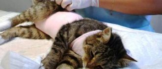
The procedure for manual bladder emptying is recommended to a cat by a veterinarian during an in-person examination if urinary retention is proven.
Reasons for prescribing bladder massage:
- Hypertonicity of the smooth muscles of the bladder itself or the urethral sphincter due to a neurological deficit.
- Sphincter spasm due to cystitis.
- Urethritis.
- Urolithiasis disease.
- Anomalies in the development of the urinary system.
- Urethral obstruction caused by gonadal hyperplasia in uncastrated cats.
- Obstruction of the urethra caused by compression from the outside by tumors, hematomas, purulent-inflammatory processes, scars, etc.
Manipulation is contraindicated when:
- Injuries with rupture of the wall of the bladder or urethra.
- Disintegration of malignant neoplasms of the urinary system.
- Acute urinary retention for more than two days.
- Severe bleeding from the urethra.
The bladder in all animals is considered a special organ, which surprises with its elasticity and is second only to the uterus in its general stretching function.
Every person should know how to palpate a cat’s bladder, since situations arise when, in order to urgently determine the pet’s condition at home, it is necessary to palpate this organ.
How to check an organ?
A person can independently palpate a cat’s bladder at home. When it is hard and enlarged, it signals the development of serious diseases. Sometimes it is difficult to feel the internal organ, especially if it is not complete. In this case, follow these recommendations:
- The cat is placed on a flat surface so that it rests on all its paws. If the pet's body is in a different position, it is impossible to palpate the bladder.
- With one hand they support the cat under the peritoneum, and with the other they try to secure it as firmly as possible by the skin on the back, while trying not to hurt the pet.
- The person’s hand is positioned so that the thumbs can feel the croup, and the rest can touch the stomach.
- When the bladder is overfilled with urine, it may be mistaken for a tumor or a lump that has an elastic consistency. If you press on it, the cat may meow or scratch due to pain. In such a case, emergency catheterization may be required to remove accumulated urine.
Structure of the bladder
The bag is designed quite unusually. This can be explained not only by its elasticity, but also by its connection with the urethra and ureters. The internal part of the organ is lined with mucous membrane, which saves the body from various bacteria entering it through the bladder. The structure of the organ is as follows:
- Outer shell. It is formed from connective tissue, under which there is a layer of smooth muscle, which performs the timely expulsion of the contents of the organ and the urge to urinate, which begins to appear even before the bladder is completely full.
- Mucous tissues. This is a complex membrane formed from special elastic fibers, which provides proper support to the walls of the bladder, even when completely filled. It also supports the mucous membrane of the inner surface of the organ, preventing it from moving.
- The mucous membrane is located in the cavity of the organ. Thanks to this tissue, the bladder can collect after urination and stretch to a huge size when overfilled. The secret that the cells of this layer of the bladder produce helps suppress the development of dangerous bacteria in the cat’s body, but only if he has excellent immunity.
Like any organ, the bladder has its own diseases and has nerve endings, which is why the animal experiences pain when it is ill. You should not think that since the bladder, in general terms, is considered a temporary bag for collecting fluid before excreting it from the body, then it cannot get sick, like other organs that are distinguished by the complexity of their functioning.
ATTENTION! When an animal has a tendency to develop diseases of the genitourinary system, it is unacceptable to leave this process and do nothing, since the reasons for this can be completely different, including life-threatening ones.
Answers to questions from an online veterinarian. Free consultations
Should the doctor replace it with something? We are afraid that the cat will not live to see the end of the course....Thank you
↓↓0↑↑ Elena (-9 / 3) July 27, 2014 23:43 »»
Hello, Elena. Very similar to an overdose, which leads to a strong increase in the tone of the smooth muscles of the internal organs. It is urgent to discuss with your veterinarian the possibility of taking another anticholinesterase drug or reducing the dosage of proserin. It must be said that adequate dosage is of decisive importance.
Insufficient - has no effect, and high can lead to serious consequences. If you suspect that your cat has any pain, you should not give it Proserin until the pain syndrome is relieved (in the acute phase of the disease, the drug is strictly contraindicated). By the way, the drug injections themselves are very painful.
Why is it necessary to eliminate the cause of the disease?
One of the symptoms of bladder or kidney disease is fluid retention. Mostly cats suffer from such phenomena. For example, urolithiasis mainly occurs in cats, since the urethra in cats is much wider than in cats. Neutered pets suffer from these ailments more than others.
They do not receive all the hormones, and without quality nutrition and vitamins they become weaker. Be sure to limit neutered animals from fish products, as they can provoke an exacerbation.
ATTENTION! If your cat has kidney stones, give him the medicine "CatErvin". Thanks to the pleasant smell, animals will happily absorb these drops. According to the recommendation, the product should be given to the cat for prevention once every 3-4 months. It has diuretic properties, destroys small stones and sand itself.
To rid your pet of kidney stones, buy food for neutered cats that is specially marked. Infectious and cold pathologies can also cause fluid retention. Cats, especially those accustomed to a warm home, should not become overcooled.
To avoid such difficulties, you need high-quality care, proper nutrition and preventive measures. When you see that your pet cannot urinate, try to understand how long it has been since he urinated. Healthy animals walk three times a day, kittens up to four times. Cats can “accumulate” liquid in themselves, so concentrated urine is considered normal.
If a cat cannot defecate for several days, this is a very dangerous problem, since the body can be poisoned by urea. Manifestations of poisoning: lethargy, apathetic mood, lack of appetite.
How can you tell if your cat's bladder is full? This can be done quickly. Place the animal on its paws, clasp it with both hands so that your thumbs are located on the cat's rump, and use the remaining fingers to feel and palpate the belly. If your cat is constantly running to the toilet and squeezing out little things all the time, this may be enough to get rid of the poisoning.
Therapeutic manipulations to combat cystitis
Even if the owner has at least a little knowledge about medicine and is confident in himself, you should not treat your pet at home. It is better to spend a couple of hours and money on professionals so that the cat feels great and lives for a very long time. After all, the treatment of each ailment has individual characteristics, which take into account various points.
To get rid of some types of disease, you just need to change the food and give your pet plenty of herbal tinctures, washing out the inflammatory process from the body. In more difficult situations, treatment with antiseptics, antispasmodics and sulfonamides will be required, and in the presence of dangerous flora, antibiotics and antibacterial agents are added.
IMPORTANT! In case of acute pathology, prompt and high-quality treatment for cats involves washing the bag. First aid for problems with going to the toilet is to install a catheter to empty the organ. If symptoms include an increase in body temperature, you need to put in IVs to eliminate the discharge.
Useful materials:
- Cutaneous horn General description of the disease Cutaneous horn on the forehead or face (ICD 10 code - L57.0) -...
- Cloudy eye in a cat Common causes of cloudy eyes in a cat The most common causes of cloudy eyes are glaucoma, cataracts or keratitis.…
- Itching and odorless discharge Main causesBefore considering the factors that provoke the appearance of discharge that has a sour odor, it is necessary to immediately note...
- Normal temperature in animals Normal temperature in different types of animals Veterinary services Day hospital for animals Veterinary certificates Vaccination…
Prevention
To prevent the development of the disease, pay attention to your pet’s diet, which should not only be varied and of high quality, but also completely balanced in terms of the content of nutrients. Feed your pet with professional prepared food and high-quality meat products
Supplement your natural diet with vitamin and mineral complexes.
Avoid hypothermia and stressful situations that weaken the animal’s body. Strengthen your pet's immune system using homeopathy remedies and complex vitamins. Systematically carry out preventive deworming and vaccinations.
If the animal's condition worsens, do not delay your visit to the veterinary clinic. Remember that many diseases can only be treated in the early stages.