Hypertrophic cardiomyopathy is a common form of cardiac pathology, accompanied by thickening and hypertrophic changes in the muscle layer of the organ. The disease is characterized by a deterioration in myocardial nutrition and a reduction in blood volume. The disease can be primary in nature, or develop as a result of concomitant ailments.
According to veterinary statistics, hypertrophic cardiomyopathy is diagnosed in 45% of furry pets with symptoms of heart failure.
Interesting facts from the medical history
Hypertrophic cardiomyopathy is a relatively new disease in veterinary practice. Intensive research into cardiac pathology began in the early 2000s in the United States. A large population of Maine Coon and Ragdoll cats was scientifically analyzed for carriage of a mutation leading to heart disease.
American scientists have concluded that a mutation in the gene responsible for the myosin-binding protein is the main cause of the genetic predisposition of Maine Coons and Ragdoll cats to hypertrophic cardiomyopathy.
Based on the research conducted, genetic test systems were created. However, their widespread use was not justified, since even with the selection of mutation-negative sires, cases of heart disease were encountered in the offspring.
By 2010, German scientists had completed large-scale studies of a large population of Maine Coons and Ragdolls in order to identify carriers of the mutation. It turned out that the genetic tests proposed by American scientists and widely used throughout the world are reliable only for cat populations in the United States.
We recommend reading about heart disease in cats. You will learn about congenital and acquired heart diseases, symptoms that should alert the owner, diagnosis and treatment. And here is more information about the causes and treatment of endometritis in cats.
Prevention
Sterilization and castration of sick animals is the only way to prevent the spread of the disease.
About buying a cat of a breed that is at risk:
- you should make sure that there is no hereditary burden of the disease;
- timely vaccination prevents the development of dangerous infections that can provoke myocardial inflammation;
- regular screening in a veterinary clinic every six months in order to identify hypertrophic cardiomyopathy at an early stage;
- Cardiac ultrasound once a year for young cats;
- regular, once every six months, deworming;
- proper nutrition with taurine, L-carnitine and fatty acids.
By adhering to preventive rules, you can protect your pet from a serious disease - hypertrophic cardiomyopathy.
Causes of development of hypertrophic cardiomyopathy
Studying the causes leading to the development of heart disease in furry pets,
allowed us to make an unambiguous conclusion about the genetic predisposition of certain breeds to hypertrophic cardiomyopathy. It is reliably known that more than 10 genes are involved in the development of the disease.
Most veterinary specialists are inclined to believe that the main cause of the development of the disease in domestic cats is gene mutations. Defects in the transmission of genetic information leading to hypertrophic cardiomyopathy most often occur in breeds such as Maine Coon, Ragdoll, Persian, Sphynx, and Abyssinian cats.
Scientific research shows that if a defective gene is present in each pair of chromosomes (homozygous animal), the risk of heart disease increases significantly compared to a heterozygous cat (if in a pair of chromosomes one is normal and the other is defective).
Among such popular breeds as the British Shorthair, Siamese, Russian Blue, Siberian, there is no direct genetic relationship between mutational changes and the development of cardiac pathology. However, these breeds are often susceptible to secondary forms of the disease.
In addition to the genetic cause that influences the development of cardiomyopathy, veterinary specialists identify the following factors contributing to the disease:
- Congenital pathologies of the myocardium in the form of thickening of the walls of the organ and an increase in its size - “bull” heart.
- Endocrine diseases: overactive thyroid gland, acromegaly. Increased production of thyroid hormones by the thyroid gland leads to tachycardia and worsens the trophism of the heart muscle. Increased production of growth hormone (acromegaly) leads to thickening of the walls of the heart.
- Imbalance of the diet in terms of taurine. The amino acid reduces the load on the heart, has an anti-ischemic effect, regulates the contraction of myocardial muscle fibers and protects cell membranes from damage. Taurine deficiency leads to disruption of the functional state of the heart muscle.
- Constantly high blood pressure in a pet leads to wear and tear on the heart muscle.
- Malignant neoplasms, in particular lymphoma, contribute to changes in the structure of the myocardium.
- Chronic intoxication of various etiologies. Poisoning with household pesticides, overdose of medications, and waste products of helminths have an adverse effect on the muscle fibers of the heart muscle, leading to ventricular hypertrophy.
- Pulmonary diseases, such as pulmonary edema.
Let's talk about the reasons
Of course, we have already mentioned that they did not seem to be identified... In fact, this is not entirely true. Veterinarians already know some things. For example, that many types of cardiomyopathy are secondary to other systemic diseases:
- Hyperthyroidism (thyroid gland working too hard).
- Arterial hypertension, that is, high blood pressure).
- Acromegaly (excessive production of growth hormone). Against this background, by the way, hypertrophic cardiomyopathy occurs especially often in cats.
What happens to a cat’s heart during pathology?
Disturbances in the functioning of the heart muscle begin after certain morphological changes occur in the organ. With the development of hypertrophic cardiomyopathy, the left ventricle and interventricular septum are primarily affected by pathological destruction.
A defective gene results in the body being unable to produce a sufficient amount of a specific protein - myosin, which is the basis of the myocardium. The body begins to compensate for the lack of muscle fibers with connective tissue. The myocardial wall thickens. The organ seems to be scarring.
Thickening of the myocardial wall leads to a decrease in the volume of the left ventricle, and often the left atrium. In addition, connective tissue reduces the elasticity and extensibility of the heart. The pumping function of the organ weakens.
Thickening of the myocardium leads to the fact that blood stagnates in the atria and the functioning of the atrioventricular valve is disrupted. Aortic obstruction occurs and circulatory deficiency occurs.
The development of hypertrophic cardiomyopathy affects all parts of the heart and affects the blood circulation of the body as a whole. This is explained by the fact that a spasm of the peripheral bloodstream occurs, and the pulmonary vessels become overfilled with blood. In a sick animal, blood clots form due to slow blood flow in the distended chambers of the heart.
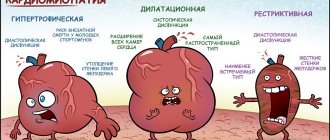
Types of disease
This disease is divided into four separate categories. Division criteria are the characteristics of those changes that occur in the tissue of the heart muscle:
- Hypertrophic cardiomyopathy (HCM). This is the most common form of heart disease. It is characterized by thickening of its walls and an increase in total volume. As a result, the immediate volume of blood is reduced, nutrition and oxygen supply to the organ deteriorate, since this hypertrophy is pathological and was not initially intended by the body.
- Dilated cardiomyopathy (DCM). The volume of the organ increases, but the thickness of the muscle tissue itself remains the same. The consistency of the heart resembles a rag. Naturally, it can no longer contract normally, and therefore the development of oxygen starvation of the entire body is very likely.
- Restrictive cardiomyopathy (RCM). Characterized by the development of fibrosis of the heart muscle. Simply put, the organ becomes rigid and loses its elasticity. As in the previous case, all tissues and organs in the cat’s body cease to receive the proper amount of oxygen and nutrients. Life expectancy is short; an animal with such disorders rarely lives more than a couple of years.
- Intermediate cardiomyopathy (ICM). “Officially” this variety does not exist, but in practice it is distinguished. This diagnosis is made in cases where the heart of a sick animal may show signs of two or three types of cardiomyopathies (dilatation and fibrosis, for example).
Types of hypertrophic cardiomyopathy
In veterinary practice, it is customary to distinguish between primary and secondary cardiomyopathy. The primary form of the disease, which has a genetic predisposition, usually manifests itself before the animal is 5 years of age. The secondary form is most typical for older animals and is more often observed in cats over 7 years of age. This type of pathology develops as a result of diabetes mellitus, kidney disease, and endocrine system.
According to the nature of the course, primary cardiomyopathy can be obstructive and non-obstructive. In the first case, the mitral valve is involved in the pathological process. In the non-obstructive form, there are no changes in the bicuspid valve.
Symptoms of cardiomyopathy in cats
Heart disease is most often observed in males. Cats are less susceptible to myocardial disease. As for age, the pathology can affect both a young animal and an older animal. There is no clear connection between the disease and the age of the pet.
The disease can occur in obvious and hidden forms in terms of the manifestation of clinical signs. If there is obvious pathology in the animal, the owner may observe the following symptoms:
- Lethargy, apathetic state of the pet. The cat stops actively participating in games, tries not to make unnecessary movements, lies and sleeps a lot. The animal may experience a low temperature - hypothermia.
- Heavy breathing, shortness of breath. During active physical activity, the animal experiences difficulty in inhaling due to the slowing of blood flow in the pulmonary veins. The owner can observe how the cat begins to breathe rapidly, sticking out its tongue. In this case, breathing movements are performed not by the chest, but by the stomach.
- Attacks of suffocation, loss of consciousness, fainting. Severe shortness of breath often ends with such symptoms due to oxygen starvation of the brain. The pulse is thread-like in nature.
- Due to lack of oxygen, the mucous membranes become bluish (cyanosis).
- A reflex cough is observed due to the pressure of the enlarged heart on the trachea. The animal takes a characteristic pose: leaning on all its limbs, stretches its neck and head forward. The front paws are widely spaced for better ventilation of the lungs.
- Hydrothorax and ascites. As a result of exudate, edema forms in the chest and abdominal cavity.
- Paralysis of a cat's hind legs develops in advanced cases of the disease, when blood clots close the lumen of large blood vessels in the pelvic area.
- Young animals gain muscle mass poorly and lag behind breed standards and their peers in development.
In many cases, hypertrophic cardiomyopathy occurs insidiously, without obvious clinical signs and ends in death. Sudden death is often the only symptom that an animal has heart problems.
Diagnosis of hypertrophic cardiomyopathy
The difficulty of identifying cardiac pathology in a pet is due to the hidden nature of the disease and the long-term absence of a clinical picture. A veterinarian may suspect problems with the myocardium during a clinical examination and listening to heart murmurs. Auscultation of the chest helps to identify systolic murmurs, cardiac arrhythmias, the so-called ri.
Having detected heart murmurs and rhythm disturbances, a veterinarian will usually order a chest x-ray, electrocardiography, and cardiac echocardiography.
X-ray examination can detect not only enlargement of the left ventricle and atrium, but also detect pleural effusion. An ECG of the heart reveals disturbances in its functioning in 70% of patients with feline hypertrophic cardiomyopathy.
X-ray (lateral and AP) of a cat with HCM
The most informative method of diagnosis and differentiation from other diseases in cardiomyopathy is ultrasound examination of the organ. The method allows you to evaluate the thickness of the heart wall and the diameter of the aortic opening. Using cardiac ultrasound, a veterinarian can assess the size and shape of the atria, blood flow in the chambers of the heart, and detect blood clots.
For information about what echocardiography shows in cats with HCM, watch this video:
ICD
Feline hypertrophic cardiomyopathy
Recently, there has been a sharp increase in owners' requests for cats in extremely serious condition (weakness, shortness of breath, paralysis of the pelvic limbs), pulmonary edema. Cases of death of animals from pulmonary edema after planned surgical procedures (castration and sterilization) have also become more frequent.
What is the reason? The answer, as a rule, in these cases is HYPERTROPHIC CARDIOMYOPATHY.
Hypertrophic cardiomyopathy (HCM) is a disease characterized by hypertrophy (thickening) of the wall of the left and/or occasionally the right ventricle. Hypertrophy is often asymmetrical, predominantly affecting the interventricular septum. Irregular, chaotic arrangement of muscle fibers in the myocardium is characteristic. zheludok
HCM is the most common cause of heart failure, arterial thromboembolism, and sudden death in cats.
Hypertrophic cardiomyopathy can be primary or secondary.
Primary HCM is a disease that is inherited. This disease occurs more often and is caused by mutations in genes encoding the synthesis of myocardial contractile proteins.
There are breeds that are predisposed to developing HCM. These are Maine Coons, Ragdolls, Sphynxes, British and American Shorthairs, Scottish Folds, Norwegian Forest cats and some others . That is, kittens inherit this disease from their parents, and by the age of 1-3 years they may develop signs of heart failure. However, this is not a guarantee that your outbred Murka cannot have this pathology, what if her grandmother with a British or Persian cat sinned?
In secondary HCM, changes in the myocardium (heart muscle) develop under the influence of other diseases (for example, hyperthyroidism). In such animals, signs of heart failure may develop either at a very old age, or may not have time to develop at all.
A distinctive feature of this disease is the significant difficulty of early diagnosis. In a cat with HCM, the disease may first manifest as pulmonary edema and/or death. That is, the signs will not develop for a long time and gradually, will not be noticed by the owner, but severe manifestations of the disease will immediately and sharply develop.
Often, a cat begins to show signs of heart failure (mainly shortness of breath - rapid breathing and/or breathing with an open mouth) after stress, which is either transporting an animal or visiting a veterinary clinic for some reason not initially related to heart disease. Only a small percentage of owners of cats diagnosed with HCM can recall noticing that the cat was breathing heavily after exercise (provoked by the owner or another animal playing). At the same time, the insidiousness of this pathology lies in the fact that during examination, auscultation and even on a chest x-ray, in the absence of complaints, more than half of the animals with HCM may not have any abnormalities.
The mechanism of development of this pathology is that as the heart muscle thickens, the volume of the left ventricle decreases, because of this, the volume of blood pumped through it decreases. Because of this, in turn, the pressure in the left atrium increases, it increases, the pressure in the vessels of the lungs increases, and then, in later stages, pulmonary edema and/or hydrothorax (accumulation of free fluid in the pleural cavity) develops.
The only way to know for sure whether a cat has HCM or not is with echocardiography (ultrasound of the heart). Examination, auscultation, x-ray, ECG are additional studies and only allow you to suspect something is wrong.
One of the frequent and extremely severe complications of HCM, which can appear against the background of the absolute apparent well-being of a cat, is thromboembolism (blockage of a vessel with a blood clot formed in the dilated left atrium). Most often, blockage occurs at the level of the femoral arteries, in this case the first symptom will be sudden paralysis of the pelvic limbs and severe pain - the cat screams and drags its hind legs. In such cases, the count is in hours, if not minutes. An extremely small percentage of patients recover; most often these are animals with mild symptoms. A recovered animal has a high probability of relapse (repetition of the situation) in the coming months. Of course, the sooner the patient arrives at the veterinary clinic, the greater the chance of restoring blood flow.
Considering all of the above, timely diagnosis is extremely important. The sooner the doctor begins treatment, the longer the patient can live and the lower the risk of developing adverse complications.
Echocardiography should be performed on a cat that has no health complaints if:
a) a cat of a breed at risk;
b) you have noticed that the cat is inactive or after exercise breathes with an open mouth;
c) if one of the first two signs is combined with the fact that your pet requires general anesthesia.
As mentioned above, the disease may not manifest itself in any way; the doctor during the examination before anesthesia does not reveal any changes, while in a cat with HCM, general anesthesia can lead to serious complications in the form of pulmonary edema and death in the coming hours or days after the operation. This applies primarily to young animals who come to the clinic for castration. As a rule, these patients are about a year old, and the vast majority of those who were diagnosed with HCM showed no signs of the disease. The reason for conducting echocardiography in this case was either the doctor’s wariness regarding the presence of HCM, or the owners’ increased anxiety about the upcoming anesthesia. The detection of HCM in these animals is not an absolute contraindication to general anesthesia, but it is a higher degree of anesthetic risk, it is a different approach to general anesthesia, it is the need for longer and more careful postoperative observation, the possibility of owners in the coming days after surgery in the event of complications developing contact the clinic urgently.
We wish you and your pets health!
Deputy Chief Veterinary Doctor
Andreeva Ekaterina Alexandrovna.
Treatment of hypertrophic cardiomyopathy in cats
Therapy for this pathology is aimed primarily at reducing congestion, regulating heart rate, preventing pulmonary edema and preventing the formation of blood clots.
If hydrothorax is detected in a sick cat, a puncture of the chest is performed in a specialized clinic in order to pump out the pleural effusion. Furosemide is used parenterally to reduce congestion and eliminate edema. The dosage and frequency of use is determined by a veterinarian based on an echocardiographic examination of the diseased organ.
Beta blockers have a good effect in the treatment of hypertrophic cardiomyopathy in cats. The drugs reduce heart rate and suppress tachyarrhythmias. Beta blockers reduce myocardial oxygen demand and fibrosis in the organ.
In the treatment of the disease, calcium channel blockers are used, for example, Diltiazem, Delacor, Cardizem. The drugs reduce the heart rate and have a positive effect on the relaxation of the heart muscle.
In addition to complex therapy, maintenance and nutrition play an important role in the treatment of sick animals. A sick cat should be protected from stressful situations and given rest. The diet should be balanced primarily in terms of taurine content. On the recommendation of a veterinarian, the animal can be given the amino acid orally.
Taurine for cats
Prognosis for a cat when the disease is detected
The forecast depends on the following factors:
- timely detection of pathology;
- manifestations of clinical signs;
- severity of symptoms;
- likelihood of pulmonary edema;
- presence of thromboembolism.
Veterinary practice shows that cats with moderate enlargement of the left ventricle and atrium often live to an advanced age. In the presence of severe heart failure and congestion, the prognosis is cautious. Cats with significant hypertrophy of the heart muscle live 1 - 3 years. The prognosis is even more cautious, even unfavorable, with the development of thromboembolism.
We recommend reading about cirrhosis in cats. You will learn about the causes of primary and secondary cirrhosis in a pet, symptoms, diagnosis, treatment, prognosis. And here is more information about the best methods of treating urolithiasis in cats.
Other drugs
Also during treatment, auxiliary agents are used that increase the effect of the “main” drugs. Pimobendan is very good. Promotes the expansion of blood vessels (increased nutrition of cardiac tissue), increases the sensitivity of the muscle fibers of the organ (improved contractility). Its use is indicated in cases of congestive heart failure (dilated cardiomyopathy).
Diuretics (furosemide). They are extremely important, as their use helps prevent the development of pulmonary edema and other congestion in the body. The dosage can be flexibly “adjusted” to the characteristics of each animal, which significantly increases the effectiveness of treatment.
Unfortunately, the true effectiveness of many drugs in treating heart disease in cats is unknown, as more clinical trials are needed. Different drugs have different effects on cats. But statistics on the use of these drugs are gradually accumulating, so that in the coming years we can expect the emergence of more effective treatment regimens.