Nature of Mastocytoma Symptoms Diagnosis Treatment and Prognosis
Mastocytoma is a malignant tumor of mast cells that can appear anywhere in the body. Typically, internal organs or skin are affected in cats.
Skin tumors usually appear around the head and neck. There may be several or one tumor, dark or pinkish in color.
Also, such a tumor often occurs on the spleen, liver and gastrointestinal tract. Mastocytomas in the bone marrow can lead to systemic (body-wide) damage to the body. This form is sometimes called mastoleukemia.
What is mastocytoma?
Mastocytoma is a neoplasm consisting of neoplastic mast cells. The mast cells themselves originate from the hematopoietic tissue of the red bone marrow. Normally, they develop from half-stem precursors and migrate to organs and tissues, where they perform their function. When the genetic information in the nucleus of these cells is disrupted, a tumor develops. This oncological disease is in first place in terms of frequency of occurrence of all skin tumors in dogs, and in second place in cats.
What symptoms indicate pathology?
As the disease progresses, growths appear on the pet’s skin that have a dense consistency and a dark or pink tint. At this point, the animal’s fur begins to fall out and an ulcer appears. The skin on the scalp is often affected. If several tumors are observed at once, this condition mainly indicates that a well-differentiated histiocytoma has also affected the internal organs. Such symptoms pose the greatest danger to the cat’s life. Siamese cats that are less than 4 years old sometimes develop tumors on the skin; they resolve on their own within 2-3 months.
Bloating in a pet develops when internal organs are affected by formations.
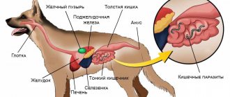
If we talk about those situations where mastocytosis is localized on internal organs, then most often it affects the spleen. Moreover, this type of pathology is dangerous because metastases can spread to the liver, respiratory organs, gastrointestinal tract, lymph nodes and bone marrow. If we talk about the defeat of the latter, then systemic damage to the body, which in veterinary medicine is called mastoleukemia, cannot be ruled out. When internal organs are damaged, the following symptoms develop:
- severe weight loss;
- vomit;
- diarrhea;
- bloating;
- lethargy;
- breathing problems.
How dangerous is this disease?
The tumor itself is dangerous due to its biological behavior. Inside the cells there are such basic biological substances as histamine and heparin, which are normally involved in allergic and inflammatory reactions. When tumor cells are damaged as a result of impacts, accidental friction, inspection, or a biopsy, these substances are released into the surrounding tissue, as a result of which a complex of symptoms may develop, which is called Darier syndrome, including redness, swelling, itching, and soreness. If we consider the systemic effects of the tumor on the body, we can highlight the development of an erosive-ulcerative process mainly in the stomach and small intestine, and the risk of anaphylactic shock. All this is the result of a long and constant release of histamine and heparin by the tumor. We do not see these symptoms in all patients, but when making a diagnosis of mastocytoma, we must always be on guard.
What are the limitations of cytology?
Limitations in cytology mainly relate to the diagnosis of neoplasms. The volume of cells taken for cytology is small; there may be cases when tumor cells simply do not get there or it is not possible to get a complete picture or determine the specific type of tumor.
Since cytology is minimally invasive, there are few contraindications, but if the animal has bleeding disorders and the doctor needs to perform a biopsy of a liver or kidney tumor, it is recommended that the procedure be performed in the operating room under the supervision of a surgeon in order to stop the bleeding if necessary. In the vast majority of cases, a thin needle will not cause bleeding.
Video about cytology in animals - interview with the head of the diagnostic laboratory:
How to recognize mastocytoma in an animal?
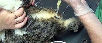
mastocytomas are great imitators. They can be in the form of ulcers, swellings, plaques, loose subcutaneous masses, which can lead to a false conclusion. However, making a diagnosis in most cases is not difficult. Using a fine-needle biopsy, cellular material is collected, from which a smear is then made and stained according to Pappenheim.
Obtaining material for cytological examination from an intradermal formation using fine-needle aspiration biopsy
About 90% of mastocytomas have a characteristic appearance under light microscopy. Otherwise, it is necessary to stain the biopsy material with toluidine blue, which makes it possible to differentiate granules of biologically active substances in the cytoplasm. However, tumor mastocytes without an intracellular granular component, the so-called histiocytic type of mastocytoma, are uncommon in cats. In this case, immunocytochemistry or immunohistochemistry is necessary. Also, to fully understand the biological behavior of a neoplasm in a particular animal, analysis for C-Kit and Ki-67 . These two indicators are leading for assessing the sensitivity of the tumor to chemotherapy and for prognosis of the course of the disease. Cytological analysis makes it possible to make a diagnosis quickly and cheaply, and this is its advantage. But the degree of malignancy is determined only on the basis of a pathohistological examination or biopsy material.
Cytology
Histology
In dogs, the disease always occurs in isolation, that is, each lesion is a separate tumor that grows independently of the others. In cats , one or more mastocytes are often the result of a systemic disease. But in any case, the animal undergoes a comprehensive examination.
Clinical picture of mastocytes
Clinical picture of mastocytes
Diagnostics
Required research:
- Fine needle biopsy - a needle is inserted into the tumor and a sample is taken for analysis.
- Complete clinical blood test, blood biochemistry and urine analysis. These tests help determine the animal's overall health and, in some cases, indicate whether tumors have spread to internal organs.
- X-ray. X-rays of the abdomen and/or chest help determine tumors and their spread within the body.
- Ultrasonography. May provide more detailed visualization of the tumor and help detect metastases.
- Biopsy - removal of the tumor and its subsequent examination.
How to diagnose mastocytoma?
To determine the stage of the disease, as in the vast majority, we, in addition to morphological methods, use visual research tools. Since mast cell tumors tend to metastasize to the liver and spleen, plain ultrasound examination is the first step. If more active tumor growth is suspected, an X-ray examination of the chest cavity is performed in 2-3 projections. Clinical and biochemical blood and urine tests are also routinely performed. In case of abnormalities in the cellular composition of the blood, red bone marrow aspiration is sometimes resorted to. One of the key points in staging the disease is assessing the condition of the sentinel lymph node, which serves the area where the tumor appeared. For this purpose, X-ray contrast agents are used, after injection of which the “staining” of the lymphatic system is monitored on radiography.
What are the advantages of cytology?
Cytology is a fairly fast diagnostic method (unlike, for example, histology). It allows you to quickly obtain information important for prescribing treatment.
In most cases, to take cell samples, there is no need to sedate the animal or use any special fixation methods—a swab or taking blood from the ear is practically painless. A fine-needle biopsy may be performed under sedation to keep the patient still. Such a study is most often carried out under visual control - using an ultrasound sensor or endoscope; in some cases, the area of sampling is localized using X-rays.
In some cases, without cytology, it is impossible to correctly prescribe further treatment.
How are neoplasms treated?
Surgery is the method of choice in the treatment of mastocytes. Removal of the tumor may be therapeutic in the initial stages of the disease, after which the animal recovers; or palliative (i.e. auxiliary, in the last stages) mainly to reduce the volume of the tumor mass before chemotherapy. The operation must be carried out in compliance with all principles of surgical oncology . Removable sutures are applied and removed after 14 days. But healing may take longer due to the biology of mastocytomas.
Unfortunately, animals at risk are often seen in an advanced state, when the tumor is no longer resectable, i.e. does not make it possible to remove the pathological focus, and the animal is inoperable. Therefore, chemotherapy, especially in this case, is the only life-saving treatment option. The most effective drugs are tyrosine kinase inhibitors (toceranib, imatinib, masivet), which act specifically on tumor cells that have a mutation in the C-Kit gene and a high ability to divide cells based on Ki-67 indicators (the more pronounced this marker, the more actively the tumor grows ). Their disadvantage is their high cost, especially when used for life. These drugs belong to the so-called targeted therapy . Treatment protocols also use lomustine (CCNU), prednisolone, vinblastine, vincristine, and platinum drugs in combinations. This or that approach to therapy is determined by the veterinarian individually for each specific case.
If the dynamics of therapy and recovery are positive, monitoring by a veterinary oncologist is required once every 4 months with blood tests, urine tests, ultrasound and x-rays.
Treatment and prognosis
Depending on whether the tumors are located only on the skin or other organs are involved, treatment may be as follows:
- Surgery. Removal of the tumor is the main treatment method.
- Chemotherapy. May be recommended in case of metastases. The effectiveness of this method has not been confirmed.
- Irradiation. If the tumor cannot be completely removed by surgery, radiotherapy can destroy any remaining cancer cells.
- Other options. Your veterinarian may recommend additional treatments to alleviate secondary effects of the disease.
The prognosis depends on many factors. If the tumor is located only on the skin, has not metastasized and can be completely removed through surgery, then the prognosis is good. For internal tumors or tumors with metastases, the prognosis may not be as optimistic. Your veterinarian may recommend that you consult with an oncologist to consider additional treatment options.
Early diagnosis and treatment significantly improves the prognosis. Check your cat's skin regularly and contact your veterinarian at the first sign of illness.
Symptoms of ascites in cats
The most characteristic sign of ascites in cats is an increase in abdominal volume. With a large amount of liquid, it becomes tight and large.
A specific sign of ascites in this case is the following test: the cat is placed on its hind legs, after which the shape of the abdomen takes on the appearance of a pear as a result of fluid flowing down. After the cat returns to its natural position after some time, the abdomen again becomes evenly enlarged on both sides.
However, with a small accumulation of ascites fluid, as well as in cats with long and fluffy coats, the increase in volume will be hardly noticeable.
When palpating the abdominal wall, the cat experiences discomfort, and in the presence of peritonitis, also pain.
Sick animals show a decrease in activity, they get tired faster after exercise, and lie down more. Appetite decreases, digestive disorders are often observed: diarrhea or constipation, possible vomiting and intestinal flatulence.
As the pressure in the abdominal cavity increases, the pressure on the diaphragm also increases. The animal breathes rapidly, shortness of breath appears, and it happens that the cat begins to breathe through its mouth, like a dog.
In some cases, exhaustion and fever are possible.