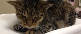
In the daily work of a veterinarian, fractures of various origins in cats occupy one of the leading places with owners’ requests to veterinary institutions. The number of fractures in cats at different times of the year varies significantly; for example, in the summer, danger can lurk for a cat, both at home and in an outdoor environment. At home, fractures in cats occur as a result of falls from an open window or balcony, as well as as a result of the inability to get out of the window frame in the tilted position. On the street, a cat can face even more danger - these are injuries received as a result of fights with other animals, various falls or fractures as a result of road accidents.
What symptoms may indicate that a cat has a fracture?
If a cat has a broken paw (front or back) - and veterinarians have to treat such fractures most often, you will see the following signs of a fracture:
- the animal does not lean on the affected limb at all
- the cat is taking care of its sore paw, an attempt to touch it results in a flash of pain, your pet may try to bite you
- severe swelling of the soft tissue appears at the site of the fracture - the diseased paw is twice as thick as a healthy one, a large hematoma may appear
- the configuration of the diseased paw is changed, it seems asymmetrical in relation to the healthy one
- a bone fragment can be seen through the skin
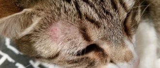
With specific fractures in cats, you can notice other external signs - for example, if the cat has a broken jaw, it cannot eat, trying to open its mouth causes pain, the muzzle may lose symmetry. If a cat has a fracture of the spine or pelvis, she cannot rest on her hind legs, they drag, and control over bowel movements and urine output is often lost.
Diagnostic methods
A fracture of the humerus or pelvis in a cat does not heal on its own. With slight damage, the bone structures sometimes grow together, but in this case the risks of complications and disability of the animal are high. To prevent this from happening and to carry out treatment correctly, it is recommended to quickly contact a veterinary clinic for help. To make a diagnosis and assess the severity of a pelvic fracture, the following diagnostic procedures are performed:
- Ultrasound of the pelvis;
- x-rays;
- urine and blood analysis;
- orthopedic examination;
- computed tomography of the pelvis;
- neurological examination.
What can you do yourself to help your cat?
The main task of first aid for a fracture in a cat is to understand whether there is bleeding and stop it. As a rule, severe bleeding accompanies open fractures, fractures caused by bites and gunshot fractures. To stop bleeding, it is better to use a pressure bandage, which is effective in 90% of cases. We recommend a pack of sterile gauze pads to stop bleeding. If they are not nearby, then you can use a handkerchief, mitten, piece of fabric, or feminine pad. Sterility is not so important now, the main thing is to stop the bleeding, which is life-threatening for the cat. A swab made of napkins or other fabric should be placed directly over the source of bleeding and bandaged quite tightly - with gauze or an elastic bandage, or a piece of fabric. Now - hurry up to the veterinary clinic. Our doctors recommend not wasting time calling a doctor to your home in such a situation, because full care for a fracture can only be provided to a cat in a clinic setting.
If there is no bleeding, but you see that the cat most likely has a broken front or back leg and it is “dangling around a lot,” you can immobilize it in the position it is in. Don’t try to “set” the fracture yourself! This is very painful. If handled improperly, the sharp edges of the bone can damage blood vessels and nerves, causing pain and additional injury to the cat. You just need to ensure that the cat's paw remains motionless while transporting the animal to the clinic. If you are in doubt or afraid, it is better not to do anything and take your cat to a veterinary clinic as quickly as possible.
First aid
If a fracture of bone structures is suspected, the animal must first be provided with complete rest. The injured cat must be moved to a flat surface. A wide board, a plastic panel from a car, thick cardboard or plywood are suitable for this purpose. If you don’t have such an object at hand, you can wrap the cat in a towel or blanket.
Transporting a cat with a pelvic fracture
To ensure immobilization, the pet should be secured to a hard surface using ropes or belts. In field conditions, a belt, harness, or cord are suitable. The cat must be secured on the board in the forearm area. An injured animal should be transported in the back seat of a car. It is important that damaged bones are kept at rest.
Despite the severe pain, veterinarians do not recommend giving your pet any painkillers. In the case of an open fracture, the owner must take measures to stop the bleeding, cover the wound with a clean cloth and transport the animal to a specialized facility as quickly as possible.
How will the doctor act?
First, the doctor will numb your animal. He then examines the cat and its broken leg. This is of particular importance for those patients who were injured by a car or fell from a height, because in addition to the problems that are obvious to the owner - a fracture - the cat may have a chest or abdominal injury. These injuries to a cat may not be noticeable, but require significantly more urgent and complex treatment than a fracture. After the examination, your veterinarian will take an X-ray of your cat's broken leg to evaluate the fracture and plan treatment, as well as further diagnose other injuries, if any. Sometimes sedation is required for x-rays.
As a rule, 99% of fractures of the front and hind legs in cats require treatment with surgery - osteosynthesis. This operation is performed as planned, often 3-5 days after the fracture. This period is explained by the physiological characteristics of the body: during an injury, massive hemorrhage occurs in the area of the fracture, and then this blood and parts of the destroyed tissue that fall into it become so-called “osteogenic elements” - substances that stimulate the bone to heal. If surgery is performed immediately after a fracture, the entire contents of the hematoma will spill out, and healing will be slower and more difficult. Another difficulty for manipulating bone fragments is created by swelling of the soft tissues, which subsides precisely by 3-5 days after the fracture. The exception is open fractures - due to the open gate for infection, they require urgent (within 24 hours) surgery.
Before surgery, the doctor will fix the broken paw with a bandage.
Of course, fractures of the jaw, pelvis, and spine require a special approach in cats—we’ll talk about them a little later.
What to do and how to treat?
Treatment without surgery
For minor ailments, a tourniquet or plaster is applied to the site of the lesion, which fixes the bones in the desired position.
If the signs of a pelvic fracture do not appear acutely, and the integrity of the bones is not severely damaged, then conservative therapy can be used. In rare cases, plaster or a narrow bandage is used to treat pathology to help fix the bone structures in one position. The retainer must be worn for a month or longer, and the cat must remain motionless. Therapy is supplemented by taking the drugs presented in the table:
| Medicine | Name |
| Corticosteroids | "Prednisolone" |
| "Dexamethasone" | |
| "Methylprednisolone" | |
| Strong analgesics | "Fentanyl" |
| "Tramadol" | |
| Painkillers | "Spazgan" |
| "Pentalgin" | |
| "Baralgin" |
After a fracture, the cat experiences stress, so the veterinarian prescribes sedatives such as Fitex and Fospasim.
Surgical intervention
Fractures in cats are treated surgically, and the type of manipulation is selected by a veterinarian based on the results of examinations. More often, osteosynthesis is performed, during which fragments are removed and damaged bone structures are fixed using a plate, rounded on both sides, with screws or steel knitting needles. If the injury is severe, then an operation such as arthroplasty or endoprosthetics is performed. The sooner surgery is performed after a pelvic fracture, the higher the chances of the cat regaining motor activity and avoiding complications.
Why is osteosynthesis necessary when you can simply apply a cast?
Plaster casting is not used to treat fractures in cats for many reasons. To begin with, it is very difficult, even impossible, to force a cat to take care of a broken paw with a cast. And, even more so, provide the animal with bed rest or hang its leg in traction. Cats try to get rid of the cast as quickly as possible, bite it, gnaw it, try to remove it on furniture, causing additional harm to themselves. The second reason is that since cats and dogs almost never break their legs by “slipping on ice” or “unsuccessfully jumping,” they almost never have fractures of the “crack” type or simple fractures without displacement. For this reason, fractures in cats often require complex reduction of fragments and comparison of fragments. Finally, a number of studies have shown that for the fastest and most complete healing of a fracture, several factors are required - the most complete comparison of fragments, their most rigid fixation, preservation of blood supply and early support on the limb. All these requirements cannot be met if fractures of paws in cats are treated with the application of plaster, therefore, throughout the world, osteosynthesis is preferred in both veterinary and human orthopedics, in which recovery occurs faster and with better quality.
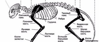
Anatomical features
The left and right halves of the pelvic bone are not at all a single monolith, but several parts at once, firmly fused together during the growth and maturation of the animal’s body. Each half is made up of the ilium, ischium, and pubis. It is for this reason that the impact of critically strong mechanical factors causes not one, but several fractures at once. But that's not even the worst thing. The pelvis forms the acetabulum of the hip joint, which connects to the spinal column through the sacroiliac joints.
In case of pelvic fractures, these parts are also damaged, and this is not at all good: rupture of the round ligament and deformation of the femoral head are treated only through a difficult and expensive operation, often involving the installation of synthetic implants.
Treating such injuries at home is unrealistic!
Each case of pelvic fracture is considered individually. The overall severity of the injury, the age of the cat, the experience of the surgeon, and the financial situation of the animal owner are assessed. All this is necessary to determine the most appropriate treatment strategy. Improper care for a sick pet, an incorrectly performed operation or other negative factors - all of these can have severe consequences in the future. This includes incomplete bone fusion, the formation of false joints, and even cases of complete non-fusion.
This leads to changes in the diameter of the pelvic cavity, osteomyelitis (inflammation of the bone tissue), arthritis of the hip joint, or even loss of a limb. Finally, bone fragments from the pelvic cavity can (under very unfortunate circumstances) damage the diaphragm and pass into the lungs. And these are severe pneumonia and other pathologies of the respiratory system.
What operations are there?
Osteosynthesis is performed using metal structures with which parts of the bone are fixed to each other. Metal parts can be placed inside the bone (pins, wires), pass through the bone (screws, screws, wire sutures) or attached to its surface (plates). In addition, there are methods for fixing fractures in which the wires pass through the bone fragments, and the main structure that ensures the strength of their connection is located outside the limb (Ilizarov apparatus and other external fixators).
Osteosynthesis is performed under general anesthesia. The doctor selects a fixation structure depending on the type of fracture and accompanying soft tissue injuries. In veterinary medicine, we specialize in complex fractures (comminuted, crushed, multifocal, gunshot). We have all the necessary equipment and experience to treat them, including using external fixators and the Ilizarov apparatus. Many cats, who in other clinics were offered amputation of their paws due to complex fractures, retained their limbs thanks to the joint efforts of our doctors and their owners.
Symptoms: how to determine if there is a problem
The fact that something is wrong with a cat’s tail will be visible to the naked eye. A crease or unnatural bend will be visible at the site of injury. Also, often when injuries occur, the innervation is disrupted, and the animal is unable to move the injured part.
The main signs of possible problems with a cat's tail are:
- visually noticeable fracture or disruption of integrity;
- pain when trying to touch the animal;
- traces of a bite, injury or infection are visible on the tail;
- a clearly noticeable inflammatory process and an increase in temperature, which can be detected even with a simple touch;
- the damaged part of the tail swells and swells;
- the tail hangs unnaturally;
- an open injury in which the bones are gaping;
- ensuing paralysis;
- problems with motor function of the legs are noticeable;
- problems with the toilet (either urinary and fecal incontinence appears, or the cat cannot defecate on its own).
An examination by a specialist will help you understand whether there is a fracture or whether it is a simple bruise.
How is the postoperative period going?
As a rule, after osteosynthesis, the cat remains in the hospital for 6 hours to 24 hours under the supervision of doctors. Then, suture treatment and painkillers are usually prescribed for 3-5 days. We recommend restricting movement for 4-6 weeks (cats can be placed in a large cage). The cat usually begins to lean on its paw 3-5 days after surgery.
After 3-4 weeks, you will need to visit the veterinary clinic with your cat again to take a control x-ray to assess the speed of fusion. On average, the rate of fusion, depending on the complexity of the fracture, varies from 2 to 8 months. After fusion, with the exception of rare cases (intra-articular fractures, fractures of the pelvic bones, very complex fractures in which the fixing elements firmly grow into the general mass of callus), metal structures are removed.
Likely consequences
If a fracture of the pelvic bones in a cat is not treated correctly, then there is a high risk of complications, such as:
The disease requires a careful approach to treatment, as there is a possibility of complications such as lameness.
- improper fusion of bone structures;
- lameness;
- narrowing of the pelvic canal;
- difficult bowel movements and chronic constipation;
- loss of sensation due to damage to nerve endings.
What are the characteristics of different types of fractures?
Previously, we looked at the actions of cat owners and veterinarians in the event of a “paw fracture” - that is, a fracture of the long tubular bones of the limbs. In a cat, these are the following types of fractures with their symptoms and treatment: hip fractures - that is, the femur, as well as the neck and head of the femur, tibia fractures - the tibia and fibula, humerus fractures - the humerus and forearm fractures - the radius and ulna. . These types of fractures are encountered most often in the practice of a veterinary traumatologist. Let us now move on to the nuances characteristic of other types of fractures.
What types are there?
The symptoms of a broken paw may vary for each cat, depending on the type of disorder. The table shows the classification of pelvic injuries and a brief description of each type:
| View | Peculiarities |
| Fracture with separation of the pelvic bones | Violation of the integrity of the pubic, ischial and/or ilium bones |
| Deviation in the sacroiliac joint | |
| Fracture of the spine and inner wing of the bone | |
| Trauma Without Disconnection | Damage to the internal and external tubercle of the ilium |
| Acetabular fracture | |
| Mixed | Combination of several injuries at once |
A hip fracture can lead to complete or partial immobilization of the animal.
A femur fracture in a cat can also be unilateral or bilateral, with the latter often leading to complete immobilization of the pet or the pet dragging its two hind legs behind it. Considering the degree of injury, the following types of deviation are distinguished:
- crack;
- break;
- a complete fracture in the area of the cat’s pelvic bone structures.
Fractured toes in a cat
Owners usually refer to fractures of all the “small” bones of the hand and foot in cats as a “finger fracture.” Thus, these are fractures of the bones of the wrist and tarsus, and fractures of the metacarpus and metatarsus, and fractures of the small bones that make up the fingers. Cats often get these types of fractures after falling out of a window; they can happen if the cat's paw is stepped on. Sometimes such fractures are the result of a car injury.
External signs of these fractures are either a complete inability to step on the paw, or very severe lameness; Toe fractures in cats are not often open and are rarely accompanied by severe swelling. However, there is usually a significant pain reaction.
Surgical treatment (osteosynthesis) is necessary in case of fractures of the wrist and tarsus bones, less often used for fractures of the metacarpal and metatarsal bones (more often if all bones are broken), and is extremely rarely used for fractures of the finger bones - as a rule, fixation with a bandage and restriction of motor activity is sufficient for 1-1.5 months.
In any case, you need to consult a veterinarian and x-ray the limb.
How to care for a cat with a fracture
A complete and speedy recovery is only possible with proper care for an injured pet. First of all, the owner should take care of limiting the cat’s physical activity. For this purpose, cellular content can be used in the first week of conservative treatment or the postoperative period.
With injuries to the pelvic girdle, the animal may develop problems with urination and bowel movements. The owner should closely monitor the cat's excretory functions and, if necessary, provide assistance.
To prevent constipation, it is recommended to feed your pet wet food, excluding dry mixtures. On the recommendation of a veterinarian, you can use Vaseline oil or Duphalac in the absence of stool. It is not always possible to fully massage the abdominal area due to pain.
It is important to prevent the animal from jumping and causing repeated injury. Under no circumstances should the cat be allowed outside until it has fully recovered. The pet should be fed with special food recommended by a veterinarian. If necessary, a course of vitamin and mineral preparations is prescribed.
During the rehabilitation period, the pet must undergo a control x-ray examination to determine the correct fusion of the bones. The date of its implementation will be determined by the attending veterinarian.
Spinal fracture in cats
This type of fracture is one of the most difficult in veterinary practice. What are the symptoms of a spinal fracture in cats? A spinal fracture in a cat is the result of a serious injury - a car injury, a fall from a height, serious bites and bruises. As a rule, these fractures occur in the thoracic or lumbar spine, then the cat cannot rest on its hind legs (paralysis of the hind legs), and sometimes urine flows from it. If a cat has damaged the spine in the sacral region, the ability to walk is retained, but there are difficulties with urination/defecation. If a cat breaks its spine in the neck area, it can be completely paralyzed - both its front and hind legs. These injuries are extremely painful and your cat may bite or scratch you when you touch them. Cats with such injuries must be moved extremely carefully; a rigid stretcher is preferable. But if there is no hard one nearby, don’t waste time looking. Bring your cat to the veterinary clinic as soon as possible. In such cases, the clock counts, so don’t hesitate. We categorically do not recommend calling a doctor to your home in such cases - he will only be able to anesthetize the animal, in the case of a spinal fracture, this will only be a waste of time.
The fact is that when a cat has a spinal fracture, the biggest problem is damage to the spinal cord from fragments. This is the cause of paralysis. Nerve fibers can be torn - and then, unfortunately, the situation is irreversible. Or fragments and splinters may simply compress the spinal cord. In this situation, the sooner help is provided to the animal, the greater the chance of saving it and restoring normal support.
Treatment of a spinal fracture in a cat, cat or kitten is always surgery. During this process, the doctor will examine the spinal cord and determine whether it is intact and whether there is hope for restoration of all functions. Then he will remove small fragments and fix the damaged vertebrae in their normal position - usually knitting needles and screws are used for this. After such an operation, the cat usually remains under observation in the hospital from a day to a week, depending on its condition. If the spinal cord is not severed, recovery usually occurs 3-4 weeks after surgery, and the first signs of improvement appear the very next day after surgery.
Damage to the sacral and caudal vertebrae in cats
Displacement of the sacral and caudal vertebrae (SCD) in cats has been identified as a separate clinical syndrome, which is very different from sacral bone fractures in dogs and cats (Smeakand Olmstead, 1985). Separation of the CCP occurs when the tail is separated under the influence of an external force, and fractures of the sacral bone are the result of a blow with a blunt object. Displacement of the CCP can occur when the tail is driven over by a car wheel or pinched by a heavy door. Denervation of the tail, pelvic organs, and pelvic limbs results from rupture of the cauda equina and avulsion of the nerve roots from the end of the spinal cord. The anatomy of the injury is the same as in dogs.
Clinical symptoms depend on the extent of nerve damage and the presence of other orthopedic and soft tissue injuries. Hyperesthesia of the base of the tail is common. Some cats may experience paraparesis, which resolves within a few weeks of injury. Most cats with CCP displacement experience analgesia and impaired tail motor function. This may be the only neurological sign in some cats (Group 1). Other cats have residual urine volume and tail denervation with normal anal muscle tone, perineal sensation, and the ability to maintain a normal posture when urinating (group 2). Cats with decreased anal muscle tone and perineal sensation, and an inability to maintain a normal posture when urinating, may have damage to one or both of the pudendal and pelvic nerves or a rupture of the spinal cord (Group 3). Cats with a lack of anal muscle tone and perineal sensitivity have complete urinary and fecal incontinence (group 4). For groups 1 and 2 the prognosis will be very favorable. In all cats, tail function will be restored, and in most of the 2nd group, normal urination will also be restored. With appropriate medical treatment, cats in group 3 will have a relatively favorable prognosis. Approximately 75% of these cats recover completely. Cats in Group 4 will have a guarded prognosis, but approximately half recover fully. An acceptable outcome is recovery of most neurological function within the first month after injury, and if it does not occur by this point, it is unlikely to occur at all. Surgical decompression of the nerve roots in cats with CCP separation has controversial results but is generally not recommended because the damage is primarily rupture rather than compressive. Tail amputation should not be immediately performed after initial diagnosis. Indications for tail amputation are ischemic necrosis, frequent contamination with feces and urine, and persistent pain.
Every cat owner tries to protect it from all kinds of diseases and injuries, but, unfortunately, this is not always possible. Among the many health problems encountered in a furry pet, injuries and even tail fractures occur quite often.
Fractured ribs in a cat
The ribs make up the main part of the chest's frame, and broken ribs can cause your cat to have serious breathing problems. In addition, broken ribs can cause pneumothorax (a dangerous accumulation of air in the chest) or lead to lung injury with bleeding. Rib fractures in cats usually occur as a result of a car injury, a fall from a window, or fights with large dogs. The main symptoms of rib fractures in cats are: wounds in the chest area, chest asymmetry, shortness of breath, breathing with an open mouth. As a rule, all serious changes - pneumothorax, bleeding into the chest - are invisible in the first stages, therefore, with any chest injury - especially if bite marks are visible between the ribs - it is necessary to bring the cat to the veterinary clinic as soon as possible.
After examination, anesthesia and x-rays, the veterinarian will evaluate the injury to the ribs and structures of the chest, the presence of blood and air in it. As a rule, single rib fractures (if there are no other injuries) do not require surgery - a special bandage is applied to the chest and pain relief is performed. In the case of multiple rib fractures and/or injury to the lungs and pleura, the intervention of a trauma surgeon is necessary to stop the bleeding, install drainage and reconstruct the broken ribs. If help is provided in time, a cat can be saved even with severe chest injuries. After such an operation, the cat will have to spend some time in the clinic’s hospital and wear a special bandage for about a month.
Patient Assessment
Fractures of the sacrum and disruption of the sacral and caudal vertebrae in dogs and cats are almost always the result of trauma sustained in a collision with a motor vehicle. Pelvic fractures, which are the most common orthopedic disorder, are also the most common result of a motor vehicle accident (Kolata et al., 1974). One study noted that 20% of cats with a pelvic fracture had an associated sacral fracture. This is why careful assessment of injured animals is so important: 70% of such animals may have associated chest injuries (pneumothorax, hydrothorax, pulmonary contusion, rib fractures and traumatic myocarditis). When examining a patient, it is necessary to assess the integrity of the urinary system, perform abdominal palpation and radiography. Animals should always urinate several hours after hydration prescribed to treat an injury. Of 100 dogs with pelvic fractures following blunt force trauma, 39 had associated urinary system injury (ureteral and bladder rupture, urethral fissures, bladder mucosal irregularities, bladder displacement and hernias, hydroureter and hydronephrosis) (Selcer; 1982). At least three dogs with urinary dysfunction had sacral bone fractures, and more animals had sacroiliac joint dislocations.
An initial assessment of the integrity of the pelvic bones requires a rectal examination. Assess the tone of the anal muscles. A complete neurological assessment of the patient should then be performed to predict and assess the extent of the damage. To do this, examine myotactile reflexes, perineal sensitivity, bladder tone, voluntary mobility and sensitivity of the tail, and palpate the bladder. The ability to initiate and complete voiding is also assessed and residual urine volume is measured. Abnormal rectal palpation, pelvic neurological deficits, and inability to walk increase suspicion of pelvic and sacral fractures.
Dogs and cats with sacral fractures and tail denervation almost always have pelvic organ denervation. Animals with a normal perineal reflex and anal tone but a large residual urine volume are likely to have an intact pudendal nerve but impaired pelvic nerves. This occurs because the pelvic nerves are considered more fragile than the pudendal nerve fibers. The bladder of such patients is very easy to isolate. The inability to completely empty after the start of urination may be a consequence of impaired reflexes of the muscular layer of the bladder. These animals have a higher chance of recovery than animals with impaired perineal sensitivity and anal muscle tone. Dogs and cats with a lack of anal muscle tone, a large and easily excreted bladder, and perineal hypoalgesia will have severely damaged pudendal and pelvic nerves that are unlikely to recover.
In some animals, bladder function cannot be directly assessed due to other injuries to the urinary system, such as rupture of the bladder and urethra. However, there is a high degree of overlap between urinary continence and fecal continence because these functions depend on the pudendal and pelvic nerves. Therefore, fecal incontinence is associated with urinary incontinence.
Some dogs and cats with sacral fractures have sciatic nerve deficits, which are characterized by deficits in conscious proprioception, flexion of the pelvic limbs, weakening of the tibialis, gastrocnemius, and withdrawal reflex (Kuntze et al., 1995). It is possible that this deficit results from damage to the sacral components of the sciatic nerve or damage to the peripheral sacral nerves in ipsilateral pelvic fractures, which often accompany sacral and caudal vertebral disorders.
Pelvic fracture in cats
A cat can get this unpleasant type of fracture, mainly from a fall from a height or a car injury. The pelvic bones form a structure through which the pelvic organs (bladder, uterus, colon) are protected from the external environment. Also, with the help of the pelvic bones, the cat’s hind legs are “attached” to the spine. Therefore, if a cat breaks her pelvis, her support on one or both hind legs is usually impaired. There may also be blood in the urine and stool. This type of fracture requires a veterinary traumatologist to additionally check the integrity of the internal organs so as not to miss ruptures of the bladder, ureter, uterus and intestines. Sometimes these injuries require separate surgery and are more urgent than treating the cat's pelvic fracture itself. If these problems are excluded, the usual treatment for a pelvic fracture in a cat is osteosynthesis using plates, wires and wire sutures. Rehabilitation after surgery usually lasts from 2 weeks to 2 months, depending on the severity of the fracture and associated injuries. Approximately 50% of all pelvic fractures in cats are treated with this method. Those that are not associated with severe displacement and do not affect the hip joint area can be treated without surgery. Such cats are placed in a cage for 1.5-2 months and given painkillers and hygiene.
Reasons for deviation: what could they be?
According to statistics, the main source of a cat's hind leg fracture is a fall from a height. At risk are pets that live at home and occasionally like to go outside and climb trees.
Damage to a cat's lower leg also occurs as a result of careless handling on the part of the owners, who often show aggression. There are also other reasons due to which closed or open fractures in the pelvic area occur in domestic animals:
- A cat gets hit by a car. Not only is damage to the pelvic bone structures possible, but the radius bones, internal organs, and other injuries are also observed. Collisions with vehicles lead to serious damage and sometimes even death.
- Impacts of different types and severity. A femoral neck fracture in a cat can occur as a result of a fight with other members of the cat family.
- Diseases. With some pathological processes occurring in the cat's body, metabolism is disrupted and the strength of bone tissue is reduced, which is why cats run the risk of breaking the jaw, pelvis and other bones.
Tail fracture in cats
This is one of the simplest fractures in veterinary practice. Typically, a cat's tail fracture occurs when the cat's tail is pinched or stepped on. Symptoms of a broken tail in a cat can include immobility (this is why sometimes cats’ tail “hangs” and does not move), curvature, and pain. In the case of an open fracture of the tail, the cat may develop lacerations, and if the owner does not ask himself in time how to treat this injury, they will fester. Except for extremely rare cases when a cat's tail fracture occurs very close to the body (at the base of the tail, when the fragments are sometimes displaced so that the main artery or nerve supplying the tail is torn), these fractures are very easy to treat. If the tail injury is serious and the nutrition of the tail below the fracture is disrupted, the tail, unfortunately, has to be amputated just above the fracture site. If a cat's tail is broken without significant displacement, simply external fixation for about 4 weeks is enough for a complete recovery.
Diagnosis of a cat's condition
After collecting an anamnesis, the veterinarian will examine the injured cat and palpate the area of injury. If there are associated injuries, necessary assistance will be provided. After stabilization of the animal's condition, an x-ray examination will be prescribed. Diagnosis is carried out after preliminary sedation.
To establish the type, location, and severity of a fracture, lateral and ventrodorsal x-rays are used in veterinary practice. Examination of the animal in two mutually perpendicular projections allows one to obtain data on the proximal part of the femur, assess damage to the lumbar vertebrae, and also examine the caudal region of the abdominal cavity. In some cases, contrast X-ray diagnostics using iodine preparations is used.
Fracture of the pelvis in a kitten along the acetabulum and ilium
Modern veterinary clinics also have the ability to conduct a computed tomography scan of the pelvic area. The method allows you to obtain layer-by-layer photographs of all bone structures and internal organs, as well as assess their condition. In some cases, your veterinarian may order a magnetic resonance imaging scan.
The animal must undergo a neurological examination to identify damage to the sciatic nerve and nerve endings of the lumbosacral plexus.
In case of concomitant injuries, the furry patient undergoes a clinical blood and urine test and an ultrasound examination of the internal organs.
Jaw fracture in cats
Jaw fractures in cats are very rare in veterinary practice, but if such a fracture does occur, it needs close attention. As a rule, these fractures are open; through the wound of the mucous membrane, numerous bacteria from the oral cavity can penetrate into the thickness of the jaw. Treatment for jaw fractures in cats requires immediate treatment. Typically, such fractures occur when falling from a window. An additional risk factor is old age and poor oral health (gingivitis, periodontitis). With these diseases, the jaw bone becomes weak and can easily break with minimal impact. Symptoms of a broken jaw in cats: severe pain, inability to close or open the mouth, inability to eat, loss of facial symmetry, bleeding from the mouth. What to do if your cat has a broken jaw? Take him to the veterinary clinic immediately. To treat fractures of the lower jaw in cats, surgery is necessary - osteosynthesis with a plate, knitting needles or wire sutures, depending on the location of the fracture. Within a day after the operation, the cat will be able to eat soft food and will quickly recover.
You can contact our veterinary center regarding any type of fracture in your cat. To do this, you do not need to make an appointment with a surgeon or traumatologist. Bring your cat to any general practitioner every day from 10.00 to 22.00. He will conduct an examination, provide pain relief, evaluate associated problems, take x-rays, and record the fracture until the time of surgery.
If you have already been examined and want to undergo osteosynthesis in our clinic, you can sign up for the operation and ask all your questions by calling us at the Veterinary by phone.
Symptoms and diagnosis
If the owner suspects damage to the tail of his pet, it is necessary to immediately find out how serious the problem is and whether it is really a pearl. Knowing the main symptoms of a tail fracture, the owner will be able to respond adequately:
- The organ at the site of possible injury will be bent. Depending on the severity of the damage, the depth of the bend will depend. If the pearl is indeed present, then a kink in the tail will be evident.
- The tissue around the affected area will be swollen.
- Presence of a bleeding wound.
- Any touch to the injured part will cause severe pain to the animal.
- Increased temperature of the damaged organ and the whole body.
After an injury, the owner should keep an eye on the cat, because a fracture of the tail is not always accompanied by a clear bend. It may appear smooth, but the fact that the injury is serious will be indicated by the pet’s behavior and various signs.
The cat's gait will definitely change: it becomes uneven and staggering. The tail will drag along the ground in an unnatural manner. With a very severe injury, even failure of the hind legs is possible. The cat may behave aggressively because the injury causes him severe pain.
The animal may have involuntary urination, and sometimes there is uncontrolled excretion of feces. But such symptoms do not always indicate that the cat has a broken tail. This is possible if the spine is damaged.
If it is confirmed that the pet has a tail fracture, he needs urgent hospitalization at the veterinary clinic. Doctors will use x-rays to diagnose and prescribe the necessary treatment. In difficult cases, if an animal’s tail injury has led to paralysis, an MRI, ultrasound, and blood test may be prescribed. Also, if the clinic has such a service, electromyography is prescribed. This is a method by which they determine how well nerve impulses travel through the muscles and peripheral nerves, that is, they determine the severity of the injury.
Cats take various manipulations calmly, but severe pain and the presence of strangers can provoke panic in the animal. It will be difficult to carry out a full diagnosis. In such cases, the veterinarian may decide that general anesthesia is necessary. In this way, he will make his work easier and the condition of the cat, who is experiencing torment.
Types of fractures
In veterinary practice, it is customary to distinguish the following types of destruction of the spinal column:
- Compression fracture of the dorsal vertebral body
Compression fracture of the dorsal vertebral body. Damage most often occurs due to the vertical direction of the mechanical impact, as well as when the animal’s back is sharply bent. This damages the vertebral body, intervertebral discs and ligaments. - Distraction fractures. Destruction is characterized by the destruction of the intervertebral disc due to excessive stretching of the spinal column. Subluxation and dislocation of bone structures are also observed.
- Rotational damage. A fracture is characterized by the destruction of all anatomical elements of the animal’s spine with obvious neurological symptoms.
The most severe are distraction and rotational fractures, characterized by damage to the spinal cord, a pronounced neurological picture and an unfavorable prognosis for the animal.
Veterinary clinic of Dr. Shubin
Description, causes and significant pathophysiology.
Sacroiliac dislocation is a disruption of the connection between the wings of the sacrum and the ilium.
There are several generally accepted names for this pathology, namely: iliosacral fracture, iliosacral dislocation, iliosacral fracture-dislocation and similar ones; in this article these terms will be used as synonyms. The main cause of a sacroiliac fracture is trauma; most often, this fracture develops during a traffic accident and a fall from a height.
The sacroiliac joint for its displacement requires either bilateral separation with the pelvic ring preserved, or additional fractures of the pelvic bones in three places and separation of one of the halves of the pelvis. Displacement of the wings of the ilium mainly occurs cranially and dorsally; significant complications of this displacement are medial deviation of the ilium and narrowing of the pelvic canal.
It is always important to remember that the cause of a sacroiliac fracture is trauma, which often involves associated injuries. The area of the iliosacral joint contains the femoral and sciatic nerves, which can be easily damaged; this fact requires a thorough neurological examination of the patient before deciding to fix the fracture. Neurological deficit is also characteristic of transverse fractures of the sacrum and involvement of the spinal canal.
Clinical signs and diagnosis
An iliosacral fracture is characterized by a decrease in the ability to bear weight on the affected side, however, with damage to the opposite long bones or pelvic fractures, the animal may exhibit partial support function on the side of the fracture.
When palpating a conscious animal, sacroiliac instability is rarely detected; when the patient is sedated or anesthetized, the doctor can determine pathological mobility of the ilium. If the fracture zone is significantly displaced, the conscious animal may show signs of severe pain.
The main means of determining a sacroiliac fracture is a radiographic examination; for its correct implementation, in most cases, sedation of the animal or general anesthesia is indicated, this relieves pain and allows taking pictures in the proper positions. To assess the fracture, two orthogonal projections are used - ventrodorsal and lateral; the ventrodorsal projection has the greatest diagnostic significance. The basis for diagnosis is a clear violation of the joint configuration. Also, in the ventrodorsal projection, concomitant fractures of the sacrum and pelvis and the degree of narrowing of the pelvic canal are assessed. In doubtful cases, a computed tomography (CT) scan may be required.
Treatment
Conservative treatment
The mainstay of treatment for an iliosacral fracture is surgical fixation, but conservative therapy (pain control and rest) may be indicated in some cases. It should be remembered that most patients with hip asymmetry will eventually regain normal ambulation. Most patients undergoing conservative treatment of an iliosacral fracture experience restoration of normal function within 12 weeks.
Below are the main indications for conservative treatment:
• Minimal SI joint displacement and mild discomfort.
• Sacroiliac fracture in small patients. In these patients, minimal load promotes rapid healing of the sacroiliac fracture, while surgical stabilization increases the risk of implant displacement into the spinal canal with the development of subsequent neurological problems.
• Subacute sacroiliac fracture is characterized by a sufficient period from the moment of injury (more than 1 week), this fracture is difficult to reduce without significant dissection of scar tissue. Surgery in such cases may increase morbidity and risk of neurological damage, and the decision to perform it is usually made to reduce the risk of pelvic canal stenosis.
• Financial restrictions on the part of animal owners.
The essence of conservative treatment is to limit mobility for 3 weeks after the injury, after which there is a gradual restoration of mobility (eg walking on a leash), the optimal use of physical rehabilitation. In dogs during the resting period, modification of the bedding with frequent changes in the animal's position is important to prevent the development of bedsores. Also, during the rest period, the discharge of feces and urine is monitored, and if problems arise, they are corrected.
The main contraindication to conservative treatment of an iliosacral fracture is narrowing of the pelvic canal.
Surgery
Indications for surgical treatment of a sacroiliac fracture/dislocation are early restoration of motor function and prevention of narrowing of the pelvic canal. The main method of treatment is open reduction of the fracture with the placement of fixing screws. Sometimes transiliac insertion of a rod is indicated to reduce the forces acting on the screw and prevent medial displacement of the ilium.
Before initiating surgical stabilization of a sacroiliac fracture, it is important to be aware of the possibility of associated injuries and adequate stabilization of the injured animal.
Surgical approach
Significant Surgical Anatomy
The sacroiliac joint consists of two components: the lunate synovial joint and the fibrocartilaginous synchondrosis. The stability of the joint is ensured by the dorsal and ventral ligaments coming from the fibrocartilaginous synchondrosis.
Patient position and preparation
In case of a unilateral sacroiliac fracture, the position of the animal on its side is permissible, the affected limb is located on top, for the convenience of the surgeon, the pelvis can be located at an angle to the table (45 degrees). With a bilateral sacroiliac fracture, the animal is located on the chest.
The surgical field is prepared as a surgical one, 10 cm from the iliac crest cranially, caudally, dorsally and ventrally.
Description of the procedure
A. The skin incision begins cephalad on the dorsal iliac crest and continues caudally parallel to the midline to the level of the hip joint. The subcutaneous tissue, gluteal fascia, and fat are incised along the same line until the cranial and caudal dorsal iliac crest are exposed.
A. Figure source: Piermattei's Atlas of Surgical Approaches to the Bones and Joints of the Dog and Cat (2014)
B. To expose the lateral (gluteal) surface of the iliac wing, an incision is made along the periosteal origin of the gluteus medius muscle on the lateral border of the ilium adjacent to the cranial dorsal ridge and terminating behind the caudal dorsal ridge. To expose the sacrum, a second incision is made at the periosteal origin of the spinosacral muscle, at the medial border of the ilium. This incision is extended caudally, and in some cases it may be necessary to cut part of the fibers of the superficial gluteal muscle.
B. Figure source: Piermattei's Atlas of Surgical Approaches to the Bones and Joints of the Dog and Cat (2014)
C. The gluteus medius muscle in young animals is elevated subperisotally, in adult animals it is simply scraped off from its insertion site. The elevation of the gluteus medius extends caudally to the caudal dorsal iliac crest. With further caudal extension of the incision, damage to the cranial gluteal artery, vein and nerve may occur. A similar elevation of the lumbosacral muscle on the medial side of the ilium provides limited exposure of the dorsal surface of the sacrum. Muscular elevation of the sacrum should be limited to the area lateral to the intermediate ridge to avoid damage to the dorsal nerve roots exiting through the dorsal foramen of the sacrum.
The ilium is displaced ventrally using bone holding forceps, and fibrous debris in the fracture area is removed to achieve a clean appearance of the sacral alar surface.
Closing
The superficial fascia, lumbosacral muscle and gluteus medius muscle are returned to their place through a series of sutures crossing the wing of the ilium. Caudal to this, the fascia of the superficial gluteal muscle is also sutured. This is followed by layered closure of the gluteal fascia, gluteal fat, subcutaneous fascia and skin.
Precautions
This approach should not extend caudally beyond the caudal dorsal iliac crest. The cranial gluteal nerve arises as branches of the lumbosacral trunk, caudoventral to the sacroiliac joint. Then, the cranial gluteal nerve is supplemented by the caudal gluteal artery and vein, passes dorsally through the body of the ilium and emerges in the fascicle of the deep gluteal muscle.
Stabilization
Once the joint is exposed, a blunt Hochmann retractor is placed between the ilium and the ventral bony shelf of the sacrum and the ilium is retracted ventrally. This maneuver exposes the lunate articular cartilage and the fibrocartilaginous articular surface of the cartilage. For proper placement of the screw in the sacrum, direct visualization of its lateral surface is preferable.
The opening in the sacrum is located 2 mm cranially and 2 mm proximally to the center of the lunate-shaped articular cartilage. The depth of retraction in the sacrum is calculated so that the tip of the screw extends at least 60% of the distance across the body of the sacrum.
Determining the proper location of the hole in the ilium is done by palpating the articular convexity on the medial surface of the wing of the bone. A screw is passed through the iliac foramen until the tip appears on its medial surface, then the ilium is retracted caudally to align with the articular surface of the sacroiliac joint. Under visual control, the tip of the screw is guided to the prepared opening of the sacrum, the screw is tightened with a screwdriver.
If necessary (according to the physician's preference), another screw can be added, it is located dorsal and cranial to the first screw. The second screw should be shorter to avoid entering the spinal canal.
Also, according to the physician’s preference, the structure can be strengthened by transiliac insertion of a Steinmann wire (rod) over the dorsal surface of L7.
Drawing. Scheme of fixation of an iliosacral fracture with a screw and transiliac rod. Source: Small animal surgery, fifth edition, 2021
Postoperatively, a radiographic examination is performed to determine the reduction of the joint and the location of the implant. Immediately after the operation, the animal’s movements are somewhat limited (for dogs, only walking on a leash). A repeat radiographic examination is performed after 6 weeks to monitor fracture healing and the width of the pelvic canal. Postoperatively, short-term use of NSAIDs may be indicated. Implants (screws) are removed only if complications develop.
Complications
The main complication of an iliosacral fracture is a narrowing of the pelvic canal, which can be accompanied by constipation and difficulty in childbirth in females. Also, misalignment of the screw in the sacral body can cause severe neurological deficits, including fecal and urinary incontinence. A rare complication is migration of the implant; this phenomenon usually develops when the length of the screw is short or when its position in the body of the sacrum is violated.
Forecasts
Once the iliosacral fracture has been adequately stabilized, the prognosis for returning ambulation is good. Return of normal motor activity after surgical treatment should be expected after 6 weeks. With conservative treatment, the recovery time is longer.
Factors influencing the outcome of surgical treatment are the accuracy of the placement of the implant and its length. The optimal length of the screw is considered to be 60% of the width of the sacrum, while the development of postoperative weakening of the implant is unlikely.
Shubin Valery Anatolyevich, veterinarian, Balakovo
Symptoms of a bone fracture
A surgical disease in the form of a violation of the integrity of bone structures is characterized primarily by pain. A sick animal exhibits the following clinical signs:
- Impaired support function. With a unilateral fracture, the cat does not step on one of the hind limbs and holds it suspended. Fracture of the pubic bones is characterized by mixed lameness or lameness of the supporting limb. If the integrity of the ischium is violated, a sick animal experiences lameness of the hanging paw. With bilateral damage, the pet is unable to stand or walk. Motor function is completely absent.
- The animal is worried, screams, meows, can show aggression, and bite. This behavior indicates severe pain. The most pronounced pain syndrome is in the sacroiliac discrepancy due to damage to the nerve endings in the lumbosacral region of the spine.
- When examining a cat, asymmetry in the pelvic girdle is often noted. This phenomenon is especially often observed during maklok fractures. When the tuberosity of the ischium is fractured, a depression is visually observed in this place, which does not correspond to the anatomical structure. When the pubic bone is damaged, the hind paw is often abducted to the side.
- Active as well as passive movements of the limb in the pelvic area are accompanied by a crunching sound (crepitus) caused by fragments of damaged bone structures.
- Visually and when palpating the pelvic area, swelling and swelling are noted. Sometimes the swelling spreads to the anal area.
- The animal refuses food and water, does not lick itself or groom itself.
- There are difficulties with urination and bowel movements.
- Neurological damage is characterized by decreased sensitivity, paresis and paralysis of the hind limbs.
Injuries in the pelvic girdle are often accompanied by damage to the bladder, rupture of the rectum, injuries to the genital organs and other associated pathologies. In addition, a fracture of the pelvic bones can be open.
Is it possible to treat on your own?
Many pet owners who have encountered the problem described remember their confusion in the first minutes when they discovered that the cat’s tail was broken and were at a loss what to do. You need to realize that if we are talking about a fracture, then you cannot do without the help of a specialist. It is the veterinarian who will make the correct diagnosis and prescribe the necessary treatment.
At home, you can try to treat a dislocation yourself (if you are confident in the diagnosis) or a fracture not complicated by displacement. In this case, a tight bandage of a special fixing bandage is applied to the damaged area, which will protect it from unnecessary movements. The bandage is applied for 3 weeks.
But in this case, you need to be prepared for some problematic issues that arise during treatment: it is rarely possible to apply a bandage tightly enough, a sick cat can scream and struggle, and remove the fixing bandage on its own (in this case, experts recommend putting a special collar on the animal).
How to distinguish a fracture from a tail bruise
In practice, tail bruises are extremely rare. The tail vertebrae are quite delicate and fragile, and therefore they break easily under strong mechanical stress. The organ itself is thin and flexible, and it is not easy to bruise it.
The tail is not a very “voluminous” organ; in case of fractures, it is easy to notice its strong curvature. Often a broken ponytail is shaped more like a poker. This does not happen with bruises.
When palpating, it is easy to feel areas where the vertebrae are crushed or severely diverged. During the examination, the animal becomes very nervous and screams in pain. With bruises, such severe pain usually does not occur.
Rehabilitation of a cat after a fracture
The recovery program for a pet after surgery depends on the severity of the injury and the degree of manifestation of the neurological disorder. The complex of rehabilitation measures includes physiological procedures (massage, darsanvalization), the use of drugs that improve tissue regeneration, and vitamin therapy.
The duration of the rehabilitation period ranges from several months to a year.
We recommend reading about a broken jaw in a cat. You will learn about the types of jaw injuries, the causes and symptoms of this fracture, diagnosis and treatment options. And here is more information about dislocation in a cat.