Owners, when bringing a sick cat to a veterinary clinic, are almost constantly faced with the fact that veterinary specialists, in addition to a clinical examination of the sick cat, conduct a blood sample test from the sick animal in order to make an accurate diagnosis of the disease. Modern effective treatment of a sick animal cannot be based only on examining the sick animal.
If not so long ago no one seriously took the possibility of conducting a study to assess the condition of an animal using a clinical and biochemical blood test in the laboratory, today every self-respecting veterinary specialist, and even more so a modern veterinary clinic, considers it the norm to do a blood test on their patients.
A blood test is one of the most informative ways to examine pets. By conducting a blood test, a veterinarian has the opportunity not only to confirm or refute the clinical diagnosis, but also to identify hidden pathological processes in the animal’s body (subclinical pathology) that have not yet produced characteristic symptoms.
The main types of research are general clinical and biochemical blood tests. Using the CBC, you can establish the basic composition of the blood. Biochemical research allows a veterinary specialist to more accurately judge the functional state of the animal. Before performing an operation or other intervention, a blood test is a mandatory norm.
How to properly prepare an animal for blood donation
- Before taking blood for analysis, the cat is not fed for 10-12 hours, and there must be free access to water.
- We limit the animal’s physical activity (refusal of games, etc.).
- Taking any medications is prohibited. If your cat regularly takes any medications as prescribed by your veterinarian, consult about stopping them.
- Before taking blood, therapeutic procedures, ultrasound, x-rays, and massage are unacceptable.
The procedure for taking blood for analysis
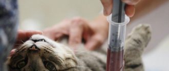
Blood is taken from the vein of the animal. To do this, the cat is placed on its side, and if the cat resists, it is secured in a special veterinary bag. Blood is taken from the front paw by shaving off a small area of fur. The injection site is treated with a disinfectant solution, a needle from a disposable syringe is inserted into the vein, blood in the amount of 2 ml is drawn into a test tube with heparin or sodium citrate.
What blood tests are performed in veterinary clinics?
In modern veterinary clinics, two laboratory blood tests are performed:
- General or clinical.
- Biochemical.
General blood test for a cat
A general blood test based on the number and condition of formed blood elements shows the health status of the cat’s body. When performing a general blood test, parasites such as dirofilaria (dirofilariasis), hemobartenella can be detected in a cat’s blood.
What indicators does the clinic’s veterinary specialist receive when conducting a general blood test:
- Hematocrit
- Hemoglobin.
- Average content and concentration of hemoglobin in an erythrocyte.
- Color indicator.
- ESR (erythrocyte sedimentation rate).
- Red blood cells.
- Leukocytes.
- Neutrophils.
- Lymphocytes.
- Eosinophils.
- Monocytes.
- Platelets.
- Basophils.
- Myelocytes.
Biochemical blood test for a cat
Biochemical blood tests allow veterinary specialists to identify subclinical (hidden) cat diseases. A biochemical blood test allows you to determine the functioning of the body’s enzymatic system and provide information about damage to a particular organ in a cat.
A biochemical blood test for a cat includes enzymatic, electrolyte, fat and substrate indicators.
Basic biochemical parameters:
- Glucose.
- Protein and albumin.
- Cholesterol.
- Bilirubin is direct and total.
- Alanine aminotransferase (ALT).
- Aspartate aminotransferase (AST).
- Lactate dehydrogenase.
- Gamma glutamyl transferase.
- Alkaline phosphatase.
- a –Amylase.
- Urea.
- Creatinine.
- Calcium.
- Magnesium.
- Creatine phosphokinase.
- Triglycerides.
- Phosphorus, inorganic.
- Electrolytes (potassium, calcium, sodium, iron, chlorine, phosphorus).
What is aspartate aminotransferase in the blood and what are the norms of this enzyme
Aspartate aminotransferase (AST) is commonly called an endogenous enzyme belonging to the group of transferases, a subgroup of transaminases (aminotransferases), which is widely used in medical practice for the purpose of laboratory diagnosis of various damage to the liver and cardiac muscle (myocardium) (heart muscle).
In this article we will look at what aspartate aminotransferase in the blood is, what the standards for this enzyme are and other information.
The AST enzyme is synthesized intracellularly, and normally only a small part of it enters the lymph and blood. In the presence of damage to the liver (cholangitis, hepatitis, primary and metastatic liver cancer) and heart (myocardial infarction), as a result of the process of cell destruction (cytolysis), AST enters the systemic bloodstream, which can be detected using laboratory methods.
An increase in aspartate aminotransferase AST, which exceeds an increased level of alanine aminotransferase (ALT), is most characteristic of damage to the heart muscle; in cases where the alanine aminotransferase level exceeds the AST level, this, as a rule, indicates processes of destruction of liver cells.
Such an unpronounceable phrase as aspartate aminotransferase in the blood means a special enzyme that takes an active part in the normal interaction of almost all amino acids and metabolic processes.
It is important! A large amount of AST is found in the tissues of the heart muscle, in the nervous tissue, and also in liver cells. It is thanks to these properties that almost all diseases that are in one way or another associated with these organs require testing for the level of AST in the blood. Aspartate aminotransferase is considered one of the types of transaminases that transport aspartic acid through molecules.
It is worth noting the fact that the coenzyme analogue of AST is the well-known vitamin B6.
Indicators of blood tests obtained and their characteristics
Each indicator of a blood test shows the functioning of individual organs or entire systems, while the veterinarian takes into account not only each data separately, but also the relationship to each other.
Hematocrit is a conditional indicator showing the ratio of all formed elements of blood to its volume, i.e. determines the thickness of blood. Shows how much blood can carry oxygen.
Hemoglobin is a protein contained in red blood cells that ensures the movement of oxygen and carbon dioxide throughout the animal’s body.
The average concentration of hemoglobin in an erythrocyte shows in percentage terms how much erythrocytes are saturated with hemoglobin.
The color (color) blood index shows how much hemoglobin is contained in red blood cells in relation to the normal value.
ESR is an indicator that determines the presence of an inflammatory process in the body.
Erythrocytes are red blood cells that take part in tissue gas exchange and maintaining acid-base balance. It’s bad when test results go beyond the norm, not only in the direction of decrease, but also of increase.
Leukocytes (white blood cells) - indicate the state of the animal’s immune system. Leukocytes include lymphocytes, neutrophils, monocytes, basophils and eosinophils. For a veterinarian, the relationship between these cells is of diagnostic importance.
- Neutrophils are responsible for destroying bacteria in the blood.
- lymphocytes - speak of a general indicator of immunity.
- monocytes - perform the function of destroying foreign substances in the blood,
- caught in the blood.
- eosinophils - perform the function of fighting allergens.
- basophils - together with other leukocytes, help the body recognize and identify foreign particles that have entered the blood.
Blood chemistry
Glucose is an informative indicator indicating the functioning of a complex enzymatic system in the body, including individual organs (liver, pancreas, kidneys). Glucose metabolism in the body involves 8 different hormones and 4 complex enzymatic processes. A disorder is considered to be either high or low blood sugar levels in a cat.
Total protein in the blood reflects the correctness of amino acid metabolism in the body. Shows the total amount of all protein fractions - globulins and albumins. Proteins in an animal’s body take part in almost all life processes of the body. For specialists, both their increased and decreased amounts are important.
Albumin is the most basic blood protein produced by the liver. Albumin in a cat’s body performs a large number of functions (transporting nutrients, maintaining reserve reserves of amino acids for the body, maintaining osmotic blood pressure, etc.).
Cholesterol is a structural component that ensures the strength of cellular structures and takes part in the synthesis of many vital hormones. Veterinary experts judge lipid metabolism in a cat’s body based on cholesterol levels.
Bilirubin is a bile pigment found in the body in two forms - direct and indirect. Indirect bilirubin is formed in the blood as a result of the breakdown of red blood cells, and bound (direct) bilirubin is converted from indirect bilirubin in the liver. Bilirubin shows the functioning of the hepabiliary system (biliary and hepatic). Refers to “color” indicators i.e. when its content in the body is increased, the tissues turn yellow (jaundice).
Alanine aminotransferase (ALT, ALaT) and aspartate aminotransferase (AST, ACaT) are enzymes produced by liver cells, heart cells, red blood cells and skeletal muscles. It is an indicator of the functions of these organs or departments.
Lactate dehydrogenase (LDH) is an enzyme involved in the final step in the breakdown of glucose. Veterinary LDH specialists monitor the functioning of the cardiac and hepatic systems, and also judge the risks of tumor formation.
ɤ-glutamyltransferase (Gamma-GT) – in combination with other liver enzymes, provides insight into the functioning of the hepabilary system, pancreas and thyroid glands.
Alkaline phosphatase is determined to monitor liver function.
ɑ-Amylase – produced by the pancreas and parotid salivary gland. Their work is judged by its level, but always in conjunction with other indicators.
Urea is the result of protein processing, which is excreted by the kidneys. Some remains circulating in the blood. Using this indicator, you can check your kidney function.
Creatinine is a muscle byproduct excreted from the body by the renal system. The level fluctuates depending on the condition of the urinary excretory system.
Potassium, calcium, phosphorus and magnesium are always assessed in complex and in relation to each other.
Calcium is involved in the conduction of nerve impulses, especially through the heart muscle. By its level, you can determine problems in the functioning of the heart, muscle contractility and blood clotting.
Creatine phosphokinase is an enzyme that is found in large quantities in the skeletal muscle group. By its presence in the blood, one can judge the work of the heart muscle, as well as internal muscle injuries.
Triglycerides in the blood characterize the functioning of the cardiovascular system, as well as energy metabolism. Usually analyzed in conjunction with cholesterol levels.
Electrolytes are responsible for membrane electrical properties. Thanks to the electrical potential difference, cells pick up and execute commands from the brain. In pathologies, cells are literally “thrown out” from the nerve impulse conduction system.
Biochemical blood test
Material to be tested: serum, less commonly plasma.
Taking: On an empty stomach, always before diagnostic or therapeutic procedures. The blood is taken into a dry, clean (disposable) tube (a tube with a white or red cap).
Factors influencing results:
- with prolonged compression of the vessel, they increase when studying the concentrations of proteins, lipids, bilirubin, calcium, potassium, enzyme activity,
- at room temperature after 10 minutes there is a tendency for glucose concentration to decrease,
- high concentrations of bilirubin, lipemia and turbidity of samples overestimate cholesterol values,
- bilirubin of all fractions is reduced by 30-50% if serum or plasma is exposed to direct daylight for 1-2 hours,
- physical activity, fasting, obesity, food intake, trauma, surgery, intramuscular injections cause an increase in a number of enzymes (AST, ALT, LDH, CPK),
It should be taken into account that in young animals the activity of LDH, alkaline phosphatase, and amylase is higher than in adults.
Normal values are specific to each laboratory.
Aspartate aminotransferase (AST, ASAT)
Aspartate aminotransferase (ACT, ASAT) is an intracellular enzyme involved in amino acid metabolism. It is found in high concentrations in the liver, heart, skeletal muscles, brain, and red blood cells. Released when tissue is damaged.
Norm: for cats - 9 - 29 units.
Increased:
- Necrosis of liver cells of any etiology, necrosis of the heart muscle, necrosis or injury of skeletal muscles, fatty liver, damage to brain tissue, kidneys;
- acute and chronic hepatitis:
- use of anticoagulants, vitamin C.
Downgraded:
- Has no diagnostic value (rarely with a lack of pyridoxine (Vitamin B6).
Alanine aminotransferase (ALT, AlAT)
Alanine aminotransferase (ALT, AlAT) is an intracellular enzyme involved in the metabolism of amino acids. It is found in high concentrations in the liver, kidneys, muscles - the heart and skeletal muscles. Released when tissue is damaged, especially when the liver is damaged.
Norm: for cats - 19 - 79 units.
Increased:
- Cell necrosis, acute and chronic hepatitis, cholangitis, fatty liver, liver tumors:
- use of anticoagulants.
Downgraded:
- Has no diagnostic value.
Creatine phosphokinase (CPK, CK)
Norm: for cats - 150 - 798 units. In young animals during the growth period, CPK activity increases 2-3 times.
Increased:
- Myocardial infarction (2-24 hours; CPK-MB is highly specific), myocarditis;
- Injuries, surgeries, muscular dystrophies, heavy physical activity;
- convulsions, infections, embolisms;
- damage to brain tissue, cerebral hemorrhage;
- anesthesia;
- poisoning (including sleeping pills);
- Slight increase in congestive heart failure, tachycardia, arthritis.
Downgraded:
- Has no diagnostic value.
Gamma glutamyl transferase (GGT)
Gamma-glutamyltransferase (GGT) is present in the liver, kidneys, and pancreas. The test is extremely sensitive for liver diseases
Norm: for cats - 1 - 10 units.
Increased:
- Hepatitis, cholestasis, tumors and cirrhosis of the liver, pancreas;
- post-infarction period.
Downgraded:
- Has no diagnostic value.
Lactate dehydrogenase (LDH)
Norm: for adult cats - 55 - 155 units. In young animals during the growth period, LDH activity increases 2-3 times.
Increased:
- Damage to myocardial tissue (2-7 days after the development of myocardial infarction):
- leukemia;
- necrotic processes;
- tumors:
- hepatitis;
- pancreatitis;
- jades:
- muscular dystrophy:
- damage to skeletal muscles;
- hemolytic anemia, circulatory failure, leptospirosis;
- infectious peritonitis of cats.
Norms of blood tests in cats
- General (clinical) blood test
- Blood chemistry
| The name of indicators | Units | Norm |
| Ø hematocrit | % (l/l) | 26-48 (0,26-0,48) |
| Ø hemoglobin | g/l | 80-150 |
| Ø average hemoglobin concentration in an erythrocyte | % | 31-36 |
| Ø average amount of hemoglobin in a red blood cell | pg | 14-19 |
| Ø color indicator; | 0,65-0,9 | |
| Ø ESR | mm/hour | 0-13 |
| Ø red blood cells | million/µl | 5-10 |
| Ø leukocytes | thousand/µl | 5,5-18,5 |
| Ø segmented neutrophils | % | 35-75 |
| Ø band neutrophils | % | 0-3 |
| Ø lymphocytes | % | 25-55 |
| Ø monocytes | % | 1-4 |
| Ø eosinophils | % | 0-4 |
| Ø platelets | million/l | 300-630 |
| Ø basophils | % | — |
| Ø myelocytes | % | — |
Norms of blood tests in cats
General (clinical) blood test
Blood chemistry
| The name of indicators | Units | Norm |
| Ø glucose | mmol/l | 3,2-6,4 |
| Ø protein | g/l | 54-77 |
| Ø albumin | g/l | 23-37 |
| Ø cholesterol | mmol/l | 1,3-3,7 |
| Ø direct bilirubin | µmol/l | 0-5,5 |
| Ø total bilirubin | µmol/l | 3-12 |
| Ø alanine aminotransferase (ALT) | U/l | 17 (19) -79 |
| Ø aspartate aminotransferase (AST) | U/l | 9-29 |
| Ø lactate dehydrogenase | U/l | 55-155 |
| Ø ɤ-glutamyltransferase | U/l | 5-50 |
| Ø alkaline phosphatase | U/l | 39-55 |
| Ø ɑ-Amylase | U/l | 780-1720 |
| Ø urea | mmol/l | 2-8 |
| Ø creatinine | mmol/l | 70-165 |
| Ø calcium | mmol/l | 2-2,7 |
| Ø magnesium | mmol/l | 0,72-1,2 |
| Ø creatine phosphokinase | U/l | 150-798 |
| Ø triglycerides | mmol/l | 0,38-1,1 |
| Ø inorganic phosphorus | mmol/l | 0,7-1,8 |
| Ø Electrolytes | ||
| Ø potassium (K + ) | mmol/l | 3,8-5,4 |
| Ø calcium | mmol/l | 2-2,7 |
| Ø sodium (Na + ) | mmol/l | 143-165 |
| Ø iron | mmol/l | 20-30 |
| Ø chlorine | mmol/l | 107-123 |
| Ø phosphorus | mmol/l | 1,1-2,3 |
Blood tests in cats (transcript)
All deviations in indicators are considered in complex and in relation to one data to another within the same results from the study of one blood sample. Only an experienced veterinarian should interpret blood tests (results).
General (clinical) blood test
| The name of indicators | Promotion | Decline |
| 1. Hematocrit |
|
|
| 2. Hemoglobin |
|
|
| 3. ESR |
|
|
| 4. Red blood cells |
|
|
| 5. Leukocytes |
|
|
| 6. Segmented neutrophils (mature) |
|
|
| 7. Band neutrophils (immature) |
| |
| 8. Lymphocytes |
|
|
| 9. Monocytes |
|
|
| 10. Eosinophils |
| |
| 11. Platelets |
|
- incoagulability of blood. |
| 12. Basophils | hemoblastoses | Normally absent |
| 13. Myelocytes |
| Normally none. |
Blood chemistry
In the article I will provide a breakdown of the biochemical blood test in cats. I will describe normal indicators, tell you what deviations from the norm indicate, give a comparative table, and what this may be connected with.
Why might aspartate aminotransferase be elevated?
If we talk about the causes of elevated aspartate aminotransferase, this occurs in the following cases:
- For hepatitis;
- liver necrosis;
- cirrhosis;
- alcoholism;
- liver cancer and metastases;
- myocardial infarction;
- hereditary and autoimmune diseases of the muscular system (Duchenne muscular dystrophy);
- mononucleosis;
- hepatosis;
- cholestasis.
It is necessary to understand that there are a number of different pathological conditions, as a result of which an increase in the activity of this enzyme is also observed. These conditions include:
- Injuries;
- Burns;
- Poisoning with poisonous mushrooms;
- Heatstroke.
It is important! If we talk about a decrease in aspartate aminotransferase, then this condition is characteristic of vitamin B6 deficiency and the presence of extensive liver damage (cirrhosis, necrosis).
Reference values (the norm of aspartate aminotransferase in blood serum is 10−30 IU/l.
The norm is considered to be a fairly low content of this enzyme, but if damage occurs in the tissues, the level of aspartate aminotransferase in the blood begins to gradually increase, thus it is released from the damaged cells.
The level of AST in the blood directly depends on the extent of tissue damage.
The level of AST in the blood can sometimes exceed normal limits by even five times, and such indicators persist for about a week.
Increased activity of this representative of transaminases is a clear indicator of the extremely serious condition of the patient, for whom there is a risk of an unfavorable outcome. An increase in AST activity may be a consequence of necrotic phenomena in the liver and myocardial infarction.
Decoding the biochemical blood test in cats
A biochemical blood test allows you to evaluate the functioning of the internal organs of cats and dogs.
Enzymatic activity is assessed by: ALT (alanine aminotransferase), AST (aspartate aminotransferase), amylase and alkaline phosphatase.
The following indicators are considered normal:
| Index | Units | Norm |
| Bilirubin | µmol/l | 3-12 |
| Total protein | g/l | 54-77 |
| Creatinine | µmol/l | 70-165 |
| Urea | µmol/l | 5,4-12,1 |
| Glucose | mmol/l | 3,3-6,3 |
| Amylase | mg/(s*l) | 8-32 |
| Cholesterol | mmol/l | 1,6-3,7 |
| AST | U/l | 9,2-39,5 |
| ALT | U/l | 0-95 |
| Alkaline phosphatase | U/l | 39-55 |
| Phosphorus | mmol/l | 1,1-2,3 |
| Calcium | mmol/l | 2-2,7 |
Dangerous deviations in indicators in a cat
Deviations from the norm (increased or decreased) indicate that a malfunction has occurred in the body. Monitoring allows you to timely identify the development of the disease and begin treatment.
Bilirubin
Bilirubin is part of bile.
An overestimated value indicates the development of liver diseases (hepatosis, hepatitis), as well as blockage of the bile ducts.
Scheme of bilirubin formation in the blood
A decrease in bilirubin levels is observed with anemia and bone marrow lesions.
Total protein
An increase is observed with dehydration due to vomiting and diarrhea. A decrease in protein levels is typical for intestinal diseases, chronic liver diseases (cirrhosis or hepatitis), renal failure, and fasting.
Creatinine
An increase in blood creatinine levels may indicate the development of hyperthyroidism or kidney failure. A decrease in this value is observed during protein starvation.
Urea
An increase in urea indicates impaired kidney function and blockage of the urinary tract. Also, an excess of this value is observed when feeding your pet food rich in animal protein.
Uric acid crystals under a microscope
A decrease in urea indicates intestinal dysfunction, liver pathologies or protein deficiency in the diet.
Glucose
The reasons for increased blood glucose are as follows:
- Cushing's syndrome;
- diabetes;
- release of adrenaline into the blood due to increased physical activity or severe stress;
- chronic kidney or liver diseases;
- pancreatitis;
- pancreatic tumors.
A decrease in value is observed with an overdose of insulin, prolonged fasting, poisoning with poisons or alcohol.
Blood glucose
Low glucose levels are also characteristic of pancreatic diseases.
Amylase
An increase in the rate is observed in the following diseases: pancreatitis, diabetes mellitus, peritonitis, volvulus, renal failure.
A decrease in the indicator may be the result of taking anticoagulants, poisoning, or necrosis of pancreatic tissue. The analysis determines total amylase and pancreatic amylase. The norm is 500-1200 units/l.
Cholesterol
An increase in cholesterol levels is typical for pancreatitis, diabetes mellitus, hypothyroidism and kidney disease.
AST and ALT
An increase in these indicators indicates the destruction of liver cells, which was caused by cirrhosis, hepatitis or other diseases. Also, an increase in AST and ALT can be a consequence of injury or heart failure.
Alkaline phosphatase
Increased alkaline phosphatase can occur in pregnant animals and in pets that eat fatty foods.
A decrease in the level of alkaline phosphatase is observed with anemia, vitamin C deficiency, and long-term use of corticosteroids.
Alkaline phosphatase is a whole complex of enzymes that is found almost throughout the body in small quantities.
Phosphorus
An increase in phosphorus is characteristic of leukemia and bone tumors. Also, a high value is observed in renal failure, hypervitaminosis of vitamin D, and disorders of the endocrine system.
Long-term diarrhea also leads to a decrease in the indicator.
Calcium
An increase in calcium is typical for:
- dehydration;
- destruction of bone tissue due to cancer;
- excess vitamin D.
Calcium deficiency is observed with pancreatitis, lack of vitamin D, taking anticonvulsants, and chronic renal failure.
№AN8ALT, ALT (alanine aminotransferase)
Alanine aminotransferase (ALT) is an enzyme belonging to the group of transaminases. ALT is localized predominantly in the cytosol of cells and catalyzes the reversible transamination reaction, that is, the transfer of an amino group from alanine to α-ketoglutaric acid with the formation of pyruvate and glutamic acid.
The reaction occurs in the presence of a coenzyme - a derivative of vitamin B6. Excess or deficiency of vitamin B6 proportionally affects the activity of transaminases. In dogs and cats, the highest concentrations of ALT are found in hepatocytes (especially near the portal area), so serum ALT determinations are typically included in biochemical profiles for these animals. This enzyme is also present, but in lower concentrations, in the kidneys, pancreas, intestines, skeletal and cardiac muscle tissue and red blood cells. In some mammals (for example, rabbits), the ALT content in the liver and heart muscle is almost the same. Because ALT predominates in liver tissue in dogs and cats, its serum activity is a more specific indicator of liver damage than AST. The level of serum ALT activity is proportional to the number of damaged cells, but is not always related to the severity of tissue damage. Serum ALT activity may be higher after sublethal injury to a large number of hepatocytes than after necrosis of a few liver cells. Cholestasis and biliary obstruction may increase serum ALT activity due to the toxic effects of bile salts on hepatocytes. Simultaneous examination of the level of alkaline phosphatase and GGT in the blood serum will identify a possible cholestatic component in hepatic pathology. If ALT activity increases, testing bile acids or plasma ammonia levels will assess liver function. An increase in ALT activity is also observed in severe lesions of muscle tissue (especially in dogs).
Although ALT activity in skeletal and cardiac muscle is significantly lower than the activity of this enzyme in the liver - 5 and 25%, respectively, damage to these muscles can cause a significant increase in ALT levels due to greater muscle mass. With severe muscle injuries, AST and CPK levels also increase, but the increase in ALT is less pronounced than the increase in AST. Testing the activity of a more specific enzyme found in muscle, such as CPK, can help determine whether muscle tissue is the source of ALT leakage. An increase in ALT activity is also observed due to acute intestinal enteropathies, intravascular hemolysis (especially in cats), during hypoxia, as a result of toxic effects on hepatocytes, and in a number of other cases.
Serum ALT levels are not usually considered significant unless they are 2 to 3 times the upper limit of normal. The serum half-life of ALT ranges from 17 to 60 hours in dogs and 3.5 hours in cats. Following severe acute hepatocellular liver injury (eg, exposure to toxic substances), enzyme levels increase significantly after 12 hours and peak within 1 to 2 days.
When exposure to damaging factors ceases, the rate of normalization of serum ALT activity does not always correspond to what is expected, taking into account the half-life of the enzyme in the blood serum, and is 1–3 weeks. This slow decrease can be explained by an increase in ALT activity during reparative regeneration of liver tissue.
PRE-ANALYTICS
To obtain more accurate results, animals must be on a fasted diet for at least 12 hours before the study. The sample is stable for 1 week at storage temperature +2°С…+8°С; remains stable for more than 1 week when frozen -17C...-23C. Severe hemolysis and lipemia can cause a moderate increase in the ALT value in the test sample.
INTERPRETATION
The study results contain information for physicians only. The diagnosis is made based on a comprehensive assessment of various indicators and additional information.
Units of measurement of the VET UNION laboratory
: Units/l.
Reference values:
Dogs: 0 – 6 months. — 10–32 U/l; 6–12 months — 10–45 U/l; over one year - 10–65 U/l. Cats: 0–6 months — 10–50 U/l; 6–12 months — 10–75 U/l; over one year - 10–85 U/l. Ferrets: 78–149 U/L - albinos; 82–289 U/l - dark. Horses: 4–20 U/l. Leopard:10–146 U/l. Tiger: 21–109 U/l. Chinchilla: 10–35 U/l. Hamster: 22–128 U/l. MRS: 60–84 U/l. KRS:17–37 U/l. Rabbit: 14–80 U/l. Rat: 20–92 U/l. Guinea pig: 10–25 U/l. Mouse - 26 -77 U/l.
In neonatal infants fed colostrum, the ALT level in the first three days of life can reach 337 U/l. The results of the study are influenced by substances that change the concentration of phosphopyridoxal (vitamin B6). Due to vitamin B6 deficiency, ALT activity levels remain low even in cases of hepatocellular lesions. When acetaminophen (paracetamol) is prescribed, an increase in serum ALT activity is sometimes observed as a result of the action of the resulting reactive hepatotoxic metabolites. An increase in serum ALT activity may occur in dogs with hyperadrenocorticism or when corticosteroid hormones are prescribed (for steroid hepatopathy). The increase in ALT levels is usually slight (2–5 times) and is detected a few days after the start of therapy. After cessation of therapy, serum ALT activity slowly decreases. Such changes are individual and depend on the dose and duration of treatment. Some drugs have a hepatotoxic effect and therefore cause an increase in serum ALT levels (for example, sulfonamides, sodium thiacetarsamide, erythromycin, rifampicin). Anticonvulsants (eg, phenobarbital, primidone, phenytoin) also cause a slight increase in serum ALT in dogs.
Level up:
Hepatitis. Hypoxia (anemia, cardiovascular diseases). Neoplasia of the hepatobiliary system. Steroid hepatopathy. Effect of hepatotoxic drugs or toxins. Liver lipidosis. Pancreatitis. Traumatic lesions of the liver tissue. Parasitization of the liver fluke. Copper storage disease. Damage or extensive necrosis of muscle tissue, myositis. Muscle fatigue.
Downgrade:
Liver atrophy (congenital portosystemic shunts). Decrease in vitamin B6 levels.