Are dogs smarter than cats?
One of the arguments in favor of the opinion that dogs are smarter than cats is the ability to train the animal. Like humans, dogs use different areas of the brain to process different features of human speech. The left hemisphere of a dog's brain processes the meaning of words, while the right hemisphere is used to analyze the intonation with which they are spoken. Scientists were able to find out that dogs can recognize new words, clearly associating them with some action or object.
At the same time, cats also demonstrate similar abilities. As the work of Japanese scientists shows, mustachioed purrs are also capable of remembering the voice, words and even intonation of their owners. The results of Japanese colleagues were recently confirmed by American scientists.
Research also shows that cats are able to remember what happens to them and recall relevant information when necessary. For example, during the experiment, scientists tried to determine whether cats could remember after 15 minutes which bowl they had already eaten from and which bowl was still full. It turns out that cats are actually capable of accessing short-term memory to figure out what is where.
Human thinking is connected with cooking
The most beautiful face: the world admires the appearance of a model from Israel today (photo)
Mazda SUVs will be rear-wheel drive by 2022 with Skyactiv-X engine
A pregnant wife spent a week wearing what her husband chose for her: what she had to wear
Human cognitive mechanisms take place in a relatively small cortex because ancient Homo sapiens learned to cook. Cooking allows people to extract more calories from their food with less energy spent on digestion. This is a topic that Herculano-Hoesel has written about previously in her book The Human Advantage: How Our Brains Became Remarkable.
According to Herculano-Houzel, bears and lions could benefit from human practices if they could master the finer art of gastronomy.
Sense organs
Through the senses, the animal receives basic information about the world around it. As you know, cats have very sharp eyesight and hearing. They can see even in the dark and are able to hear sounds that humans cannot hear.
A description of the anatomical structure of the organs of vision and hearing is important not only in order to get to know your pet better, but also to recognize the presence of pathological changes and know how to help your pet.
Eyes
Visible part of the eye:
- upper eyelid;
- lower eyelid;
- third eyelid;
- Iris;
- sclera;
- pupil.
Cats have relatively large eyes. Cats have stereoscopic vision. This means that they can perceive size, shape and judge the distance to a certain object. Also, cats can see the world around them not only in front of them, but also from the side. Their eyes are capable of capturing images within a range of 205 degrees around them.
Cats' eyes glow in the dark due to the ability of this organ to accumulate rays that enter the eyes during daylight hours. They cannot see in complete and utter darkness. But even a minimal amount of light entering the room allows them to clearly distinguish objects due to the reflection of light from the surface of objects.
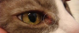
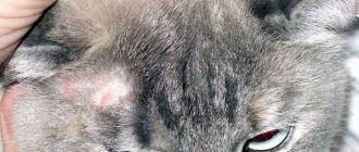
© shutterstock One of the features of cats' eyes is the presence of a third eyelid. This membrane serves as protection against foreign objects entering the cornea. Usually the third eyelid is not visible. It can be noticed in those moments when the animal has just woken up. If it is visually noticeable all the time, or even covers part of the eye, this is a signal about the presence of some pathology in the body.
Ears
A cat's ears are made up of these parts::
- ear canal;
- eardrum;
- middle ear bone;
- vestibular apparatus;
- snail;
- auditory nerve.
Cats have the ability to perceive sounds over a wide range. The physiology of a cat and the structure of its ear allows it to hear high-frequency sounds that are inaccessible to human hearing. A cat can hear about 100 different sounds, while for a person this number is limited to fifty.
There are about 30 muscles around and on the ears that are responsible for movement in this area. Attentive owners notice the cat's ability to move its ears in different directions.
Cat owners should pay extra attention to the structural features of the ear. Your pet should have its ears checked and cleaned regularly. Due to the rather complex structure of the ear, one can often miss the presence of various inflammatory processes and the presence of ear mites.
Neuron Density
Herculano-Houzel told Live Science that neurons are cells that use a lot of energy to support the body. According to her, their number in the brain is also the best reflection of the brain's capabilities. But the size of the brain is not an indicator of how many nerve cells it contains.
“If you simply compare animal species based on brain size, you get some pretty strange results. For example, cows and chimpanzees have the same size brains, Herculano-Hoesel said. “On the other hand, when you compare the number of neurons, the assessment of the level of cognitive development is based on criteria such as behavior and intelligence. For example, the human brain contains a large number of nerve cells - up to 16 billion in the cortex, which is responsible for our thinking."
A woman designed her bathroom in Japanese style. Now I want to repeat
This narrow house seems inconspicuous only from the outside, once you look inside
Bacteria in the gut influence the immune response to Covid
How to determine the level of intelligence?
These examples certainly demonstrate the high intellectual capabilities of each species. But are these capabilities related to the size of the animals' brains? Previously, scientists really tried to draw such a parallel. However, the results of recent studies refute this assumption.
After studying the brains of several animals, including dogs and cats, researchers came to the conclusion that the level of intelligence of animals does not depend on the size of the “gray matter”. A more interesting indicator is the number of cortical neurons contained in the cerebral cortex - special cells that process, store and transmit information using electrical and chemical signals.
According to researchers, the total number of neurons in an animal's brain, especially in the cortex, determines the wealth of its intellectual abilities and the ability to predict events based on past experience. Scientists have found that the cerebral cortex of dogs contains 530 million, and cats have half as much - 250 million.
Can we conclude from this that dogs are smarter than cats? The researchers themselves say that dogs are biologically more adapted to complex mental processes than cats. But at the same time, they explain that this does not mean that cats are stupid.
The difficulty in objectively assessing the intelligence of these species is that cats and dogs are too different and behave differently. Each type is better in some ways and worse in others.
What does science say?
Another sign of cat intelligence is a refusal to participate in research.
David Grimm writes in the online publication Slate that two leading animal researchers with whom he discussed feline intelligence had great difficulty working with their subjects because the cats simply did not participate in the experiments and did not follow directions. Leading animal scientist Dr. Adam Miklosi even had to go to the cats' home because in his laboratory they categorically did not make contact. However, the more scientists learn about cats, the more they want to try to subdue them. You just need to get them to follow commands, but it is obvious that this is very difficult.
Nervous system
The central nervous system is represented by the brain, spinal cord and brain stem. It receives and transmits signals and commands to the peripheral nervous system.
The brain is the main organ of the central nervous system of cats. The normal size of a cat's brain is 5 centimeters in length. Domesticated breeds have a smaller brain volume than wild breeds. Otherwise, the physiology of domestic cats changes slightly compared to wild cats.
© shutterstock
The peripheral nervous system includes the entire system of nerves in the animal's body - nerves in the skull and spinal cord, plexuses of nerve fibers and nerve endings . This system is responsible for motor activity, reflexes, and pain.
The autonomic nervous system ensures the autonomous functioning of all internal organs. It is also responsible for the cat’s innate reflexes associated with hunting, food production, protection, reproduction, and orientation in terrain and space.
Hello student
midbrain
The midbrain, mesencephalon, being the rostral continuation of the rhombencephalon, has a fundamentally similar structure: it contains a space that separates the bottom from the roof. The roof or vault, tectum mesencephali, in contrast to the rhombencephalon, is a compact structure. In domestic mammals, the roof is built in the form of a plate with four dorsal protrusions and is called the plate of the quadrigemina, lamina quadrigemina with rostral and caudal hills, colliculi rostrales et colliculi caudales.
The rostral colliculi, colliculi rostrales, have a layered structure, as in the comparable optic lobe, lobus opticum of the bird brain. The rostral hills are connected to the lateral geniculate body, corpus geniculatum laterale, through the leg of the rostral hill, brachium colliculi rostralis, and then to the optic tract and contain the corresponding reflex centers.
The caudal colliculi, colliculi caudales, in carnivores are larger than the rostral colliculi, the nervous substance is divided into nuclei, and they conduct acoustic reflexes. They are included in the auditory pathway and connect on both sides through the leg of the caudal hill, brachium colliculi caudalis, with the medial geniculate body, corpus geniculatum mediale.
The midbrain cavity, cerebral aqueduct, aquaeductus mesencephali is a gap that has different lateral extensions in different animal species. The cavity contains on the lateral side a continuation of the sulcus limitans from the rhombencephalon, which makes it possible to divide into lateral plates (tectum, from the sensory pathways) and main plates (tegmentum, the center of the efferent nuclei).
The covering of the legs, the cap, tegmentum mesencephali, is a direct continuation of the corresponding parts of the rhombencephalon. A particularly important feature is the presence of legs in the tegmentum, as in the rhombencephalon, formatio reticularis, which, like important motor centers, includes autonomic functions and the sensitive system of excitation of the body's waking state.
One pathway approaches the three original areas of the midbrain, which is adjacent to the ventral side and provides a functional connection between them: this is the motor pathway from the cortex cerebri, which in humans, in the form of the tractus corticospinalis, represents a direct connection from the cerebral cortex to the motor neurons of the spinal cord. Also, carnivores have a well-formed so-called pyramidal system, which is represented in the midbrain region by the cerebral peduncles, crura cerebri.
In domestic mammals, motor activity is controlled mainly by subcortical centers. In the midbrain, in the area of the reticular formation, there is a center that includes very large neurons and, due to the presence of iron-containing pigments, has a red color, nucleus ruber. An important pathway to the spinal cord, tractus rubrospinalis, emerges from this nucleus. The reticular formation is delimited on the ventral side from the crura cerebri by the black substance, substantia nigra. The latter is named so due to the presence of melanin-containing neurons and carries out motor activity. The reticular formation continues into the diencephalon, where it is located in the motor centers of the subthalamus.
Above the crus cerebri there is a dorsoventrally transverse peduncular tract, tractus cruralis transversus, in the form of a narrow white stripe in the tegmentum mesencephali.
In accordance with the longitudinal division of the zones, also in the midbrain in the main plate (ventral to the aquaeductus mesencephali) there are somatomotor nuclei paramedian: in the area colliculi rostraeles motor nuclei, nuclei motorius (and parasympaticus) n. oculomotorii, and in the area of the caudal colliculus nucleus motorius n. trochlearis.
As an example of the afferent nature of the tectum mesencephali, one should cite the sensitive nucleus n. trigeminus. Proprioceptive fibers from the masticatory muscles end here, which, as in the spinal ganglia, have the form of pseudounipolar nerve cells.
INTERMEDIUM BRAIN
The diencephalon, diencephalon, develops from the forebrain and connects, on the one hand, with the midbrain, on the other, with the telencephalon. The cavities of these parts of the brain narrow, as in the telencephalon, into a fissure. Also worth mentioning is the wide connection between the diencephalon and the telencephalon.
The diencephalon generally belongs to the brainstem and is covered by the telencephalon. The roof of the diencephalon remains very thin for a long distance and consists only of a single-layer ependyma with a connective tissue base. Loops of vessels depart from it, which are also covered with ependyma and are located in the third ventricle in the form of a plexus chorioideus. Laterally, the thin roof of the third ventricle reaches the tuberosity band, taenia thalami, rostrally passing into taenia chorioidea. It runs caudolateral to the border in the form of a border strip, stria terminalis telencephalon. In the triangle constructed in this way, the surface of the thalamus lies freely.
During development, the diencephalon connects dorsolaterally and dorsorostrally with the telencephalon, ventrally forms the border plate, lamina terminalis, of the rostral end of the third ventricle, while its base connects with the optic chiasm, chiasma opticum, gray tubercle, tuber cinereum, pituitary gland, hypophysis and mastoid body, corpora mamillaria. The optic tract, tractus opticum, covered with the telencephalon, arcuately caudodorsal, also departs from the chiasma opticum and passes with its lateral root, radix lateralis, in the region of the dorsocaudal part of the thalamus (metathalamus) into the lateral geniculate body, corpus geniculatum laterale. The axons of the optic ganglion cells end in this area. The lateral geniculate body is connected to the anterior colliculus of the quadrigeminal pedicle by the legs of the rostral colliculus, brachium colliculi, medial root, rostralis. The radix medialis of the optic tract conducts fibers directly into the colliculi rostrales, or area praetectalis of the midbrain (optical reflex center and pupillary center). From the optic tract in the region of the midbrain (brachium colliculi rostralis), a bundle of fibers is separated, especially pronounced in carnivores, which runs along the lateral surface (crus cerebri) in the form of the tractus cruralis transversus, and at the oculomotor root enters the tegmentum inesencephali. There is no more detailed information on the functioning of the accessory optic root.
Ventral to the lateral geniculate body in dogs there is a relatively large medial geniculate body, corpus geniculatum mediate, which is connected to the caudal hills of the midbrain by the legs of the caudal hill, brachium colliculi caudalis, and is a center for switching impulses in the auditory pathway.
With a loose division of the diencephalon, it is divided into real paired “tubercles”, the thalamus, thalamus, a thin region above it, the epithalamus, and the base, the hypothalamus, hypothalamus.
Both thalami form the main part of the diencephalon. They emerge from the region of connection with the telencephalon. As their volume increases, they meet at the midline and grow together into a rounded area. The third ventricle is formed in a ring around this area. The connection area, which has nuclei and fiber connections, is called interthalamic fusion adhaesio interthalarnica
The thalamus is a complex nuclear region where switching occurs for afferent and efferent pathways. All sensory pathways, with the exception of the olfactory pathway, pass through the thalamus before sensory stimulation becomes conscious in the cerebral cortex, cortex cerebri. In addition, there are connections to the propulsion system.
Rice. 7. Cat's brain. Dorsal view
B' sulcus ectosylvius rostralis. B” sulcus cctosylvius caudalis; With sulcus suprasylvius; E sulcus marginalis; G sulcus coronalis: N sulcus ansatus; J sulcus cruciatus
a» gyrus intersylvius; b gyrus ectosylvius; c gyrus ectomarginalis rostralis, c' gyrus ectomarginalis medius, c'” gyrus cctomarginalis caudalis; d gyrus marginalis; e gyrus praecruciatus, e' gyrus postcruciatus; e + e' gyrus sigmoidcus
1 bulbus olfactorius, 2 fissura longitudinalis cerebri; 3 cerebellum, vermis, 3′ hemisphaerium; 4 fasciculus gracilis; 5 fasciculus сuneatus
Rice. 8. Cat's brain. View from the lateral side on the left
A fissura lateralis cerebri (fissura sylvia); B' sulcus ectosylvius rostralis, B” sulcus ectosylvius caudalis; With sulcus suprasylvius; E sulcus marginalis; G sulcus coronalis; H sulcus ansatus; J sulcus cruciatus; L sulcus rhinal is lateralis; M sulcus praesylvius; U sulcus diagonalis
a' gyrus sylvius rostralis, a" gyrus sylvius caudalis, a"' gyrus intersylvius; b gyrus ectosylvius; c gyrus ectomarginalis rostralis, c' gyrus ectomarginalis medius, c'» gyrus ectomarginalis caudalis; d gyrus marginalis; e gyrus praecruciatus, e' gyrus postcruciatus; e-e' gyrus signioideus; g gurus compositus rostralis, g' gyrus compositus caudalis, h gyrus olfactorius lateralis
1 bulbus olfactorius; 2 tractus olfactorius lateralis; 3 lobus piriformis, 4 n. opticus; 5 pons; 6 cerebellum, vermis, 6′ hemisphaerium; 7 plexus chorioideus ventriculi quarti; 8 n. trigeminus, 9 n facialis; 10 n. vestibulocochlearis; 11 corpus trapezoideum; 12 v. abducens; 13 no. glossopharvngeus, 14 n. vagus; 15 no. accessorius, 15′ radices spinales n. accessorii; 16 no. hypoglossus; 17 tractus spinalis n. trigemini
The hypothalamus contains the center for controlling the autonomic nervous system and the endocrine system. This is especially clearly expressed in the connection between the hypothalamus and the pituitary gland.
The epithalamus, epithalamus, includes the pineal gland, glandula pinealis (epiphysis), the dorsocaudal protrusion of the diencephalon, functionally the endocrine gland, the center of neurovegetative regulation. The gland, for its part, is controlled by the retinal exposure phase and controls the biological clock rhythms (biological clock). The epiphysis is connected by two thin ligaments - the frenulum, habenulae, with the diencephalon. The epithalamus also includes a ligament along the taenia thalami, stria medullaris thalami, as well as the commissura caudalis lying at the transition to the midbrain.
Rice. 9. Cat's brain. Ventral view
A fissura lateralis cerebri (fissura sylvia): B' sulcus ectosylvius rostralis, B» sulcus ectosylvius caudalis; G sulcus coronalis; L sulcus rhinalis lateralis; M sulcus praesylvius; U sulcus diagonalis
a'gyrus sylvius rostralis, a"gyrus sylvius caudalis, a"'gyrus intersylvius; b gyrus ectosylvius; f gyrus proreus; g gyrus compositus rostralis, g' gyrus compositus caudalis, h gyrus olfactorius lateralis, h' gyrus olfactorius medialis 1 bulbus olfactorius; 2 tractus olfactorius lateralis; 3 lobus piriformis; 4 n. opticus, 4′ chiasma opticum, 4″ tractus opticus; 5 infundibulum; 6 corpus mamillare; 7 crus cerebri; 8 n. oculomotorius; 9 no. trochlearis; 10 pons; 11 corpus trapezoideum; 12 no. trigeminus; 13 no. facialis; 14 no. vestibulocochlearis; 15 no. abduccins; 16 pyramids; 17 aedulla spinalis.
Rice. 10. Cat's brain. View from the medial side (sagittal section)
The roof of the third ventricle, recessus suprapinealis, as well as plexus chorioidei are not marked
J sulcus cruciatus; N sulcus corporis callosi, P sulcus hippocampi; Q sulcus splicnialis; Sulcus suprasplenialis; T sulcus genualis
d gyrus marginalis; e gyrus praecruciatus, e' gyrus postcruciatus; e + e' gyrus sigmoideus; h' gyrus olfactorius medialis; i gyrus splicnialis; k gyrus einguli; I gyrus parahippocampalis; m tuberculum gyri dentati: n gyrus callosus; o gyrus genualis; p gyrus paraterminalis; q gvrus occipitalis
1 bulbus olfactorius; 2 corpus callosum, 2′ genu; 3 septum pellucidum; 4 lamina terminalis; 5 comniissura rostralis; 6 glandula pinealis with rccessus pinealis ventriculi tertii; 7 adhaesio interthalamica; 8 n. opticus, 8′ chiasma opticum; 9 hypophyses; 10 corpus mamillare; 11 commissure caudalis: 12 tectum mesencephali, colliculus rostralis, 12′ colliculus caudalis with rccessus colliculi caudalis; 13 aquaeductus mesencephali; 14 tegmentum mesencephali; 15 pons; 16 corpus trapezoidum; 17 cerebellum, 18 velum mcdullare rostrale, 18′ velum mcdullare caudale; 19 recessus tecti ventriculi quarti; 20 recessus caudalis ventriculi quarti, 21 canalis centralis spinal cord
III ventriculus tertius; IV ventriculus quartus - foramen intervcntriculare
The tegmentum of the midbrain peduncles (with formatio reticularis and substantio nigra) penetrates between the thalamus and hypothalamus. This area, together with the pallidum, which is closely connected with the ganglia of the telencephalon (putamen), is designated as the subthalamus. It is part of the central motor system.
External factors that provoke the appearance of nervous disorders
Any changes that cause bright, negative emotions in a cat can cause disruptions in the functioning of the nervous system and the formation of various diseases. In most cases, abnormalities in the functions of the central nervous system in a pet appear when:
- moving to another place of residence;
- traveling in public transport, car;
- visiting veterinary hospitals;
- participation in exhibition events;
- change of owner;
- changing the interior, etc.
Of course, it is not always possible to completely avoid such situations. It should be noted that these factors do not cause neurological diseases for all animals. Owners need to be attentive to their pets, thoroughly study their characteristics and emotional reactions to various stimuli. This will reduce the risk of disorders and keep the cat healthy.
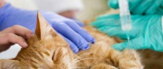
Often, disorders of the nervous system are the result of infectious, viral diseases. Therefore, it is necessary to pay special attention to prevention, timely vaccination, and minimize the risk of contact with sick animals.
If any serious ailments occur, it is necessary to contact professionals for diagnosis and treatment. You cannot independently determine the type of disease, choose procedures, medications. Many medications have side effects and contraindications. A qualified doctor will select the necessary funds and determine the optimal dosage, taking into account the condition, age of the pet, and the stage of development of the disease. The recommendations of a specialist should be strictly followed. An untreated disease can become chronic and cause complications in the form of neuralgic pathologies, which are extremely difficult to get rid of.
Hepatitis in cats
Most often, cats experience not just hepatitis, but cholangiohepatitis, that is, inflammation of the liver involving the bile ducts and gallbladder.
There are many reasons for the development of cholangiohepatitis: long-term loss of appetite as a result of stress or another disease, impaired absorption of proteins in the gastrointestinal tract, obesity, etc.
In addition, due to the unusual anatomical connection between the common bile duct and the main pancreatic duct in cats, bacteria or digestive enzymes from the duodenum or pancreas are refluxed, which leads to the development of bacterial or sterile hepatitis or cholangiohepatitis.
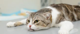
Clinical signs in most adult cats are the same and not specific: lethargy, drowsiness, lack of appetite, progressive weight loss. Less common are chronic vomiting, diarrhea, fever, abdominal effusion, and signs of damage to the central nervous system. Hepatic encephalopathy in cats most often manifests itself as depression and drooling.
Specific signs of liver inflammation: jaundice, enlarged liver. Similar clinical signs may result from non-inflammatory liver diseases such as lipidosis, bile duct obstruction, chronic viral infections such as feline viral peritonitis, and cancer.
The diagnosis is made based on a combination of anamnestic data, blood tests, including infections, and ultrasound results. In many cases, a final diagnosis is possible only after histological examination of a biopsy of liver tissue.
In the treatment of hepatitis, antibiotic therapy and choleretic drugs play an important role. anti-inflammatory drugs, as well as nonspecific treatment aimed at eliminating dehydration, removing toxic substances, etc.
Leave feedback. Hepatitis in cats
How are cats different from dogs?
For example, cats have very well developed sensorimotor intelligence of a predator. They are far superior to dogs in this regard. The amazing ability of cats to “play with gravity” is not limited to the ability to balance, jump and land on all fours. Even domestic cats constantly train (playing, chasing toys and your feet), which indicates preserved hunting skills. Cats can hunt prey that is several times larger and more dangerous than them. This, for example, was recently confirmed in Australia, where cats have become a real epidemic.
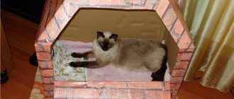
Unlike dogs, cats are very attentive and careful. Finding themselves in an unfamiliar environment, the first thing they do is carefully examine and sniff everything. And only then, having made sure that there is no danger, will they find a warm and cozy place for themselves. A cat will never run up to an unknown object or person. Dogs do not have this behavior, which can sometimes cost them their lives, and certainly does not indicate high mental abilities.
In turn, dogs have retained the so-called collective intelligence. They are social animals. In the wild, wolves, considered the ancestors of dogs, always hunt in a pack, where each has its own role. Domesticated dogs easily adapt among people and are very active in communicating with people. It is for this reason that dogs are believed to understand people much better than cats. Cats are introverts. They are accustomed to a solitary lifestyle.
At the same time, cats are excellent manipulators. Where a dog seeks the approval of its owner, a cat does not ask - it demands. And at the same time she is ready to use any trick to get what she wants. Even at 2 am.
Cats perceive information very selectively
They pay attention only to those things that seem interesting to them, but at the same time they show complete indifference to everything that does not interest them. Dogs are more curious in this regard
Does all of the above mean that one of these species is smarter? No. This just goes to show that they are very different and each is smart in their own way.
And is it even important to know “who is smarter” if we are talking not just about a beloved pet, but about a full-fledged member of the family?
Organs of the circulatory system
The process of blood circulation, like the internal structure of a cat, is practically no different from a similar process in other mammals. It is provided by two circles of blood circulation. The first is the transport of blood from the heart to the capillaries through the arteries. The second is the transportation of venous blood to the heart and lungs.
The pulse in cats should be measured on the inside of the thigh, where the femoral artery is located. A healthy adult at rest has a heart rate of up to 130 beats per minute.
Similar to humans, cats' blood can have different groups: A, B, AB. Group AB, as in humans, is the rarest. Most often cats have group A.
Cats' blood clots much faster than humans'..
Respiratory system
Breathing provides the body with oxygen and also gets rid of excess water.
The cat's respiratory system is similar to most mammals.
Respiratory organs include:
- Nose. The inhaled air enters the nasal cavity, where mucus is contained, which traps small dust particles, hairs and bacteria and prevents them from penetrating into the lungs.
- The nasopharynx is the part of the pharynx through which air continues to flow further along the respiratory tract.
- The larynx is a cartilaginous tube that seals the airways when swallowed.
- The trachea is a cartilaginous tube that carries air into and out of the lungs. It runs from the larynx to the heart, where it divides into 2 bronchi.
- The lungs are the key organ of the respiratory process. The lungs are divided into left and right lobes. The left lung still has an additional lobe, so its size is slightly larger. The lungs contain blood and respiratory vessels. The trachea is divided into bronchi in the lungs, the bronchi are divided into bronchioles, and these, in turn, are divided into alveoli - small air bubbles that transmit oxygen to the blood vessels. Parenchyma is a collection of various vessels in the lungs.
- The diaphragm is a muscle that allows the lungs to expand.
The very process of a cat's breathing can be described as follows: under the action of the pectoral muscles and diaphragm, the lungs expand and pull air through the nasal cavity into the respiratory tract until it reaches the alveoli, which come into contact with the blood vessels and saturate them with oxygen, while at the same time removing carbon dioxide from them.
Organs of the digestive system
The digestive tract of cats is provided with such organs:
- Mouth . Consists of lips, cheeks, tongue, gums, palate (soft and hard), teeth, tonsils, pharynx and salivary glands.
- Throat . Serves to connect the nasal cavity with the lungs, the oral cavity with the esophagus. Covered with mucous membrane and has strong muscles.
- Esophagus . Serves to transport food from the mouth through the pharynx to the stomach. Consists of skeletal muscles, the contraction of which helps move food.
- Stomach . Has one camera. Located in the abdominal cavity (front). Food enters the stomach, is stored in it and processed into chyme, which then enters the small intestine.
- Intestines . The total length of a cat's intestines is about 2 meters. The intestines are 3 times longer than the cat’s entire body.
- Small intestine . It is about 1.5 meters long. The main process of digestion of proteins and carbohydrates occurs in the small intestine.
- Colon . In the colon, the final breakdown and absorption of useful substances occurs, as well as the removal of residues in the form of feces.
- Pancreas . The ducts of the small intestine exit into it. Over the course of one day, it secretes several liters of a special secretion, which helps break down substances supplied with food.
- Gallbladder and liver . Filters blood that comes from the stomach and intestines. The liver produces bile, which is necessary for processing fats.
Digestive and excretory systems
The digestive system regulates the process of eating, absorbing nutrients and eliminating undigested residues.
The organs of the digestive system are involved in the process of digesting food
The digestion cycle is carried out per day. This process involves:
- oral cavity;
- pharynx;
- esophagus;
- stomach - the pH environment in the stomach is more acidic than in humans, which allows it to digest rough food and fight bacteria in the food;
- small intestine, in cats it is short and does not allow carbohydrates to be digested well;
- colon;
- liver;
- kidneys
The process of digestion begins in the mouth as soon as food enters it. The salivary gland softens hard food, facilitating its passage into the stomach and esophagus.
The process of digesting food begins in the mouth
Under the influence of saliva, food begins to break down in the mouth. The complete food processing process takes place in 4 stages:
- The fundus of the stomach contracts, pushing the contents toward the pylorus.
- The contents of the stomach enter the duodenum in the following order: liquid, carbohydrates, proteins, fats.
- Food passes through the small intestine where nutrients are absorbed.
- Food debris enters the large intestine, and feces are formed and excreted.
Keep your brain fit
Regular exercise helps keep your brain in shape by increasing capillaries. And this, in turn, ensures maximum supply of oxygen and glucose to the body. The most effective exercises are those that last at least 30 minutes a day.
The most effective method of developing the brain is considered to be engaging in new, hitherto unfamiliar activities, as well as communicating with people who are superior in intelligence to the interlocutor. The more educated a person is, the less likely it is for brain diseases to occur, because mental activity causes the production of additional tissue that replaces the damaged one.
The cat's brain in the evolution of the cat family
In terms of its dimensions, a cat is a miniature creature, the exception, of course, is the Maine Coon breed. However, it is wrong to think that if an animal is smaller than a person, it means it is stupider and less intellectually developed than a person. Scientists have proven that the mass of this biological computer does not affect intelligence at all. In addition, its structure has existed without any significant changes for tens of millions of years. Even the first representatives of the cat family on Earth had exactly the same structure of this organ. They were replaced by the ancestors of lions, tigers, panthers, etc. And then these representatives themselves. It is worth noting that the filling of a lion’s skull in its structure is an exact copy of the filling of the skull of any domestic or street cat. The differences are only due to the habitat and body mass. The greater the body weight, the greater the functions of the trunk that need to be controlled, the more space in the skull for “gray matter”
Structure of the National Assembly and provoking factors
The basis is created by neuron cells. When neurons come into contact, reflex circuits are formed through which nerve impulses pass.
- CNS (brain, spinal cord).
- Peripheral (cranial and spinal nerves connecting the central trunk with receptors of tissues and organs).
Factors that can provoke a nervous system disorder include anything that changes the animal’s usual life.
- change of owner or place of residence;
- riding public (personal) transport;
- visit to the veterinary clinic;
- abrupt transition from one food to another;
- weaning from the mother cat;
- holidays (guests, fireworks, music).
A special place is given to the physical ailments of the cat, in which it experiences pain and discomfort. This also includes veterinary manipulations associated with “improving” appearance, or with preventive methods (castration, sterilization, declawing, etc.).
Emotional stress causes the development of deep neuroses. Although this is not a prerequisite, cats, just like people, have individual characteristics. Some react inadequately to moving the tray, while others are not bothered by loud music in the room, washing, or long trips in the car.
The breeder’s task is to know how sensitive their pet is and to prevent unnecessary worries!
General information
The essence of one of the very first experiments on studying the memory of cats was as follows. To confirm the presence of short-term memory in cats, bowls of food were shown in front of hungry animals. After the demonstration, the food was placed under cover so that the cats could see where their food had been taken. After half an hour, the animals returned to the room and almost all of them ran up to those places of shelter where the previously hidden portion of food stood.
The second part of the experiment tested long-term memory. The bowls were also demonstratively hidden, but the cats were allowed into the room only after 24 hours. The result was not so good in terms of how many cats remembered everything. Some of them ran towards the stern, still confidently. Some of the experimental subjects sniffed the shelter and looked for food. And some of the cats showed no interest in the search at all.
The conclusion from this study is simple - cats have short-term memory in the vast majority, but long-term memory is not observed in all individuals of the species. Therefore, you can judge what kind of memory a cat has over time only by looking at each individual cat.
INTERESTING: in cats, their ability to remember decreases with age in the same way as in a person or dog. The mechanisms of both long-term and short-term memory suffer.
Stress factors
Loneliness and lack of attention from the owner can negatively affect a cat's health. Many people are accustomed to thinking that stressors can only be psychological in nature, but in fact there are several types:
- Physical - the impact of temperature on the cat (burn, frostbite, sudden change from cold to hot and vice versa), humidity, sunstroke, electric shock, painful injuries with extensive damage to the body and heavy bleeding.
- Chemical - this group includes any toxic or irritating substances that can (household chemicals, medications, etc.). By the way, even tobacco smoke blown into a cat’s nose for the sake of a joke, or the smell of alcohol can cause stress.
- Biological – prolonged fasting, insufficient supply of nutrients and infections of any etiology (viruses, fungi, bacteria).
- Mental stressors are probably the largest and most common group of stressors in cats, which would be worth dwelling on in a little more detail.
Cat brain
Does your cat react when you call its name?
Can a cat distinguish your voice from a stranger's voice?
Can a cat find a toy hidden behind a large object, such as a closet?
If the answer to all these questions is YES
, this means your cat is quite smart.
At least, this is reported by feline behaviorist
Kristin Vitale at the Human-Animal Interaction Laboratory at the University of Oregon
Today Felis silvestris catus
(the official name of the cat) is one of the smartest animals on Earth. According to the specialist, this may be due to the flexibility of cats in terms of behavior.
Marvelous
, but to date, relatively little research has focused on cat cognition or internal mental processes, especially compared to the significant amount of research on the psyche of dogs.
Respiratory system
The anatomy of a cat is not much different from other mammals. This also applies to the respiratory system. It includes the following organs :
- bronchi;
- larynx;
- lungs.
- nose;
- nasopharynx;
- trachea;
© shutterstock The breathing process begins from the nose and nasopharynx.
The nose has 2 nasal cavities inside, in which, when inhaled, the process of recognizing odors, heating the air and purifying it from impurities, dust, and debris occurs. The cavities are separated by a septum of hyaline cartilage. The larynx is located between the trachea and pharynx, and is located above the hyoid bone. Main functions of the larynx :
- air conduction;
- preventing food from entering the respiratory system;
- formation of sounds.
The larynx consists of five movable cartilages and a mucous membrane. It also contains the vocal cords, vocal muscle and glottis. This is where all the sounds that a cat makes are produced.
The purring of cats occurs due to the special location and functioning of the organs of the larynx. Purring occurs without effort on the part of the animal, and has the same rhythm as breathing. In this case, the muscles contract at a frequency of more than 1000 times per minute.
The vocal cords of cats differ in their structure from the vocal cords of other animals. Attentive owners may notice that the pet’s “speech” is not limited to meowing. And even ordinary meowing can be different. It is quite simple to study the “language” of your cat, and you can accurately guess what exactly the pet is telling us. For example, dogs can only make about 10 different sounds. And representatives of some cat breeds can express themselves using about 100 sounds that exist in their “lexicon”.
A healthy animal in a calm state takes about 20-25 breaths per minute. Kittens take breaths and come out more often.
Brain and endocrine system
Anatomically, a cat's brain is similar to the brain of any mammal.
The structure of a cat's brain is similar to that of any mammal.
Different parts of the brain are responsible for one or another function in the body:
- the parietal lobe processes information received through the senses;
- the large brain is responsible for consciousness;
- the corpus callosum connects the right and left hemispheres;
- the frontal lobe is responsible for voluntary movements;
- the olfactory bulb is responsible for the perception of smells;
- the hypothalamus secretes hormones and controls the autonomic nervous system;
- the pituitary gland coordinates and controls the work of other glands;
- the spinal cord transmits information from the brain to the body;
- the pineal gland is responsible for sleep and wakefulness;
- the cerebellum controls movements and muscle function;
- the temporal lobe is responsible for behavior and memory;
- The occipital lobe receives visual and tactile signals.
The endocrine system influences the basic functions that occur in the body with the help of hormones. Most hormones are secreted by the pituitary gland and hypothalamus. Also some of them are produced by the thyroid gland, adrenal glands, ovaries in cats and testicles in cats.
The endocrine system influences the body's basic functions
Table: hormonal regulation of cat body functions
| Hormone name | Where is it produced? | Functions |
| Antidiuretic hormone (ADH) | Hypothalamus | Urine concentration |
| Oxytocin | Hypothalamus | Labor and feeding of kittens |
| Corticoliberin | Hypothalamus | Adrenocorticotropic hormone concentration |
| Adrenocorticotropic hormone (ACTH) | Hypothalamus | The adrenal glands produce cortisol when the cat is afraid |
| Thyroid-stimulating hormone (TSH) | Pituitary | Thyroid activity |
| Melanocyte-stimulating hormone (MSH) | Pituitary | Melatonin synthesis in the pineal gland |
| Follicle stimulating hormone (FSH) | Pituitary | Production of sex hormones and eggs in females |
| Luteinizing hormone (LH) | Pituitary | Production of sex hormones and sperm in males |
| Adrenalin | Adrenal glands | Cardiac activity and dilation of blood vessels |
| Progesterone | Ovaries | Preparing the uterus for implantation of embryos, maintaining pregnancy, stimulating the development of mammary glands |
| Testosterone | Testicles, adrenal glands | Development of the cat's reproductive system, development of secondary sexual characteristics |
| Thyroxine | Thyroid | Activation of metabolism, thickening of the uterine walls, increase in heart rate |