One of the most common diseases of the digestive system among cats is cholecystitis. The causative agents of this disease are various microbes, including streptococci, Escherichia coli, etc.
Leading veterinarians at the clinic talk about what this disease is, how it progresses and what the main features of treatment are.
Read in this article
What is this? Causes of the disease Symptoms of cholecystitis in a cat: what to look for? Forms of cholecystitis: acute and chronic First aid for cholecystitis Methods for diagnosing the disease How and with what to treat cholecystitis in cats? What to feed a cat with cholecystitis? Effective recommendations for disease prevention
Causes of the disease
Cholecystitis in cats can be caused by various reasons. The most common is cholelithiasis (GSD). As for provoking factors, these include the pet’s sedentary lifestyle, age and impaired gallbladder tone.
The main causes of cholecystitis in cats:
- Poor nutrition. Cheap mass-market food, expired products and a lack of vitamins (especially vitamin B1) provoke the development of inflammatory processes. Pathogenic microorganisms penetrate the gallbladder and cause inflammation.
- Invasive diseases (helminths). A common cause of helminth infection is feeding your pet raw fish or meat. Parasites clog the bile ducts and penetrate the gallbladder, which leads to disruption of its functions.
- Blockage of the bile ducts. The main reasons that can lead to this condition: cholelithiasis (GSD), pancreatic tumor, etc. All this provokes the development of an acute inflammatory process.
- Infectious diseases . Both bacterial and viral infections can lead to gallbladder inflammation. Among the most dangerous diseases leading to cholecystitis: leptospirosis, viral hepatitis, “feline distemper”, etc.
- Damage or rupture of the gallbladder. It occurs when there is a serious mechanical injury: a cat falling from a great height, strong impacts, collisions, etc. Chronic inflammation can also lead to perforation (breaking through) the walls of the gallbladder.
Prevention
Most often, the main cause of problems with the gastrointestinal tract is improper and poor-quality feeding of the animal. A lack or excess of certain components in food leads to problems that also affect the condition of the gallbladder and kidneys. Feeding raw meat and fish can lead to infection of the animal with various parasites, so it is necessary to carry out heat treatment. Cats should not be fed fish all the time. Such a diet can cause not only cholecystitis, but also urolithiasis. It is advisable to give your cat fish no more than once a week. Dry food should be selected based on its quality and advice from veterinarians. Economy segment feeds have a detrimental effect on the animal’s liver. They contain various chemical impurities that are addictive to cats. Therefore, the animal continues to eat food with appetite, even if it causes him problems with digestion.
If the animal was injured - fell from a great height or was hit by a car - the cat must be urgently shown to a doctor. Occasional vomiting is normal for cats - it helps remove ingested fur from the stomach. If you notice bile in your animal's vomit, you should immediately consult a doctor. Chronic cholecystitis cannot be completely cured without surgery, so it is advisable to deal with the disease before it takes hold in the body.
Thus, problems with the gastrointestinal tract and liver can lead to cholecystitis. An attack in cats is usually accompanied by apathy and loss of appetite, severe vomiting, and fever. If these symptoms appear, you should immediately consult a doctor. Most often, treatment for cholecystitis is medicinal, but in some cases surgery may be necessary, without which the animal will die.
Any liver pathology is bad. More precisely, very bad. The health of a cat directly depends on the condition of this amazing organ. Many people know about this. Unfortunately, pet owners often forget that the liver does more than just filter blood. It is also responsible for the production of bile, which, in turn, seriously affects digestion. For this reason, cholelithiasis that appears in cats should be treated immediately, immediately after the first symptoms are identified.
Symptoms of cholecystitis in a cat: what to look for?
The first thing that indicates cholecystitis in your pet is nausea and vomiting containing bile. A sick cat refuses to eat and therefore rapidly loses weight. Due to the inflammatory process, she will experience increased body temperature and general weakness.
The main symptoms of cholecystitis in cats:
- nausea and vomiting,
- bloating of the abdominal area,
- increase in body temperature,
- lethargy and apathy,
- constipation,
- peeling of the mucous membranes of the oral cavity,
- yellowness of the sclera of the eyes.
In severe forms of cholecystitis, the cat experiences severe pain. She meows loudly, behaves aggressively, does not allow the abdominal area to be touched, etc. Fever may develop (for example, if cholecystitis is caused by an infectious disease).
What symptoms will indicate the disease?
With further progression of the disease, the animal may refuse to eat.
At the initial stages of development, the pathology does not cause any discomfort to the animal. Symptoms of gallstone disease in cats appear after the stone causes serious health problems. As the disease progresses, the following symptoms are noted:
- loss of appetite;
- weakness;
- vomit;
- increase in body temperature.
When eating food, the gallbladder begins to contract and push bile into the intestinal lumen. At this moment, the stones, which have sharp edges, dig into the walls of the organ. As a result, sharp pain occurs. The cat is rolling on the floor and screaming. If there are many stones, but their boundaries are even, there are no symptoms at the initial stages of the disease. As the disease progresses, the cat's mucous membranes turn yellow and the coat becomes rough.
Forms of cholecystitis: acute and chronic
Spicy . Often develops in cats against the background of cholelithiasis (GSD). The gradual accumulation of bile leads to infections and acute inflammation of the walls of the gallbladder. Acute cholecystitis develops rapidly - within a few days or even hours.
Chronic . It occurs as an independent disease, and the entire clinical picture develops gradually. Temporary improvements in the condition are possible. The main danger of chronic cholecystitis is the difficulty of timely diagnosis.
Etiology
The main clinically proven cause of stone formation is a violation of the metabolism of bile acids, phospholipids, bile pigments (biliverdin, bilirubin), cholesterol and inorganic salts.
Cholesterol plays a decisive role in the formation of stones. When cholesterol-retaining factors are disrupted, the ratio of bile acids and phospholipids changes, the colloidal mass thickens and cholesterol crystallizes, which leads to stone formation.
The predisposing factor is anatomical pathology (narrowing of the ducts, atrophy, adhesions, hypertrophy, dyskinesia, tumor growths) of the gallbladder and ducts, which leads to congestion. Without regular outflow, bile thickens, mucus and epithelial cells join the contents, which also contributes to the formation of stones.
Metabolic disorders (anemia, obesity), lack of exercise, unbalanced diet are also a driving factor in the formation of stones in the gall bladder and ducts.
Stones are synthesized in the bladder itself much more often than in the ducts. This is due to stagnation in the bladder cavity.
By their nature, stones can be:
- Cholesterol. Such stones are usually single and light yellow in color. During a long stay in the bladder, calcium salts are added to themselves and become combined;
- Pigmented. They contain bile acids, calcium bilirubinate, cholesterol. These are the stones that are typical for dogs. As a rule, in most cases they are multiple, shiny, black, faceted;
- Combined. The consistency and color of such stones can vary greatly depending on the proportional composition (pigments, calcium carbonate, cholesterol). Such stones are multiple, smooth, and irregular in shape. If there are few stones, then they are large.
Methods for diagnosing the disease
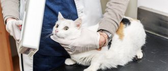
If you suspect that your cat has cholecystitis, contact a veterinary clinic in Moscow immediately. The first thing a specialist will do is conduct a diagnosis. He will not be able to make an accurate diagnosis based on symptoms alone, so additional research will be required.
Appointed:
- general/biochemical blood test,
- ultrasound diagnostics,
- fine needle biopsy,
- scintigraphy (functional imaging) of the gallbladder,
- differential diagnosis.
With cholecystitis, the level of bilirubin (from 7.9 µm/l), alkaline phosphatase and cholesterol increases in a cat. When determining the diagnosis, the veterinarian also takes into account the level of bile acid and endogenous enzyme from the transferase group.
Diagnostics
Data from the anamnesis and physical examination can only suggest the described pathology in the animal, but do not make it possible to make a diagnosis. In addition, the clinical picture does not fully reflect the degree of damage to the gallbladder, and, accordingly, does not allow determining the best method of treatment and prognosis of the disease.
At the first stage of diagnosis, clinical and biochemical (as complete as possible) blood tests are required; urine is an optional test. Laboratory tests can reveal an increase in alkaline phosphatase, hypercholesterolemia, hyperbilirubinemia without signs of hemolytic anemia. Hyperbilirubinemia ultimately leads to bilirubinuria. Increased levels of bile acids, glutamate dehydrogenase and leukocytosis are very characteristic of this pathology and additionally indicate the need for bile testing. An increase in transaminases will be detected only if the liver parenchyma is involved in the inflammatory process.
When choosing a method for diagnosing cholecystitis directly, preference is given to visual studies and predominantly ultrasound diagnostics. X-ray is less sensitive for this pathology and is informative only in the case of calcification of the gallbladder wall or the formation of radiopaque stones (Photos 6 and 7).
Photos 6 and 7.
In this section, we will consider changes in the ultrasound picture of the gallbladder and biliary system observed with cholecystitis, without affecting possible changes in the pancreas, neoplasia of other organs, etc.
- The wall of the gallbladder thickens (thicker than 1 mm in cats and 2-3 mm in dogs), becomes hyperechoic, with uneven edges - a sign of inflammation, edema (portal hypertension, hypoalbuminemia), necrosis, hyperplasia of the gallbladder mucosa, and less commonly, neoplasia (Photo 1) ;
- Along with wall thickening, the appearance of a double-circuit rim (especially in a more acute period) or a diffusely hyperechoic wall, sometimes combined with mineralization (in a chronic process) is often noted (Photos 2 and 3);
- Thickening of the wall and dilatation of the lumen of the common bile duct, increasing its tortuosity. However, it can be quite difficult to differentiate lumen dilatation against the background of obstruction from dilatation against the background of cholestasis in a chronic inflammatory process. In addition, with chronic outflow obstruction, the common bile duct may remain dilated even after the obstruction is removed (this must be taken into account in the postoperative examination);
- The appearance of bile sludge. Physiologically, bile can thicken and transform into bile sludge (bile sludge). It is a mixture of mucus, calcium bilirubinate and cholesterol crystals. Under pathological conditions, its consistency and accumulation can complicate the evacuation of bile into the extrahepatic bile ducts, leading to obstruction of the latter. A characteristic sign of bile sludge is a change in its appearance on the scanogram depending on changes in the position of the animal’s body and the slow achievement of a new horizontal level (the criterion of sludge mobility makes it possible to distinguish it from a biliary mucocele). The general rule is the absence of a distal acoustic shadow. The echogenicity of sludge can vary. Sometimes sludge fills the entire gallbladder, making it difficult to differentiate between liver tissue and the gallbladder. This situation is called "hepatization of the gallbladder" (Photos 4 and 5);
- Biliary mucocele (mucinous hyperplasia of the gallbladder) is characterized by epithelial hyperplasia and papillary growths, excessive accumulation of mucus that stretches the gallbladder. The disease is rare, usually in dogs of small and medium breeds (average age - 9 years). It is one of the reasons for the development of obstruction of the extrahepatic bile ducts and, as a consequence, cholecystitis. As the mucocele forms, a star-shaped contour first appears on the scanogram, then the cross section of the gallbladder acquires a kiwi (fruit) pattern in cross section.
Photos 1 and 2.
Photos 3 and 4.
Photo 5.
If there is any change in the gallbladder or the appearance of bile heterogeneity on ultrasound, it is necessary to perform a fine-needle biopsy to aspirate bile for cytological and bacteriological studies. 22-25 gauge needles can be used for this, and during this procedure it is necessary to remove as much bile as possible to prevent bile from leaking through the puncture hole. The likelihood of such a complication is extremely low; we have not encountered this in our practice, but in the presence of undiagnosed obstruction of the extrahepatic biliary tracts, the risk increases. We also recommend collecting liver parenchyma material for histological examination (the procedure for collecting a biopsy specimen for histological examination is not much more complicated in comparison with a fine-needle liver biopsy, but the result is many times more informative).
One of the modern informative methods is radionuclide scanning of the gallbladder (scintigraphy), which allows you to evaluate the functioning of the gallbladder and determine the location of duct obstruction. Unfortunately, this method is not yet available in our practice.
If biliary peritonitis is suspected, diagnostic laparoscopy or laparotomy is indicated.
How and with what to treat cholecystitis in cats?
Treatment of cholecystitis is determined by a veterinarian individually in each specific case. It takes into account the initial cause of the disease, its stage and severity. Under no circumstances resort to self-medication or folk remedies - this will only harm your cat.
Drug (conservative) treatment . It gives a good result, but only in the initial stages of the disease. The veterinarian prescribes symptomatic medications of various effects:
- antiviral,
- antispasmodic,
- anti-inflammatory,
- painkillers,
- antipyretics, etc.
To eliminate inflammation of the gallbladder, a long course of antibiotic drugs is prescribed. First, the doctor determines the sensitivity of the pathogenic flora to the action of drugs.
Additionally, to speed up and ensure the discharge of bile, plant-based decoctions are used (for example, plantain leaves, dandelions, etc.). In case of severe intoxication of the body, infusion therapy is prescribed.
In parallel with antibiotic drugs, vitamin and mineral complexes are prescribed. Vitamins A, group B (B1, B2, B6 and B12), as well as C are used. If necessary, drugs are prescribed that restore heart function.
Surgical (operative) treatment . Surgical interventions are prescribed if conservative treatment does not produce a successful result. Other indications for surgery include: blockage of the bile ducts, rupture of the gallbladder, frequent relapses of the disease.
Removal of the gallbladder is performed using laparoscopy. If the bladder ruptures or if a complication is detected (for example, an infectious disease), a laparotomy is prescribed. A blood clotting test is performed first.
Pathogenesis
Stones are capable of migration. The movement of small stones from the bile ducts into the gallbladder, and then into the duodenum, is not uncommon. Sometimes they clog the cystic duct, common bile duct, or rise into the hepatic duct. By impeding the flow of bile, the stone acts like a valve, which entails swelling of the bladder walls and collapse of the cavity.
If the stones are streamlined, then no functional disturbances occur in the bladder and liver parenchyma. If the stone is angular, then its enlargement can lead to traumatic injuries and mechanical (obstructive, subhepatic, cholestatic) jaundice. If the obturation is incomplete, then expansion of the overlying parts of the bile ducts with hypertrophy of the walls may occur.
What to feed a cat with cholecystitis?
When treating cholecystitis, your pet must be switched to a special balanced diet. It is recommended to continue the diet until the cat’s condition is completely restored. This takes about 2-4 weeks.
It is necessary to completely exclude fried and fatty foods with a high cholesterol content from the animal’s diet: lamb, sausages, butter, canned fish, condensed milk, etc.
Products that can be included in your cat's diet:
- low-fat meat or fish broth,
- rice or oatmeal porridge,
- rice water,
- boiled vegetables (carrots, potatoes),
- lean minced chicken,
- light cottage cheese.
What to feed or how to organize a diet
A therapeutic diet plays a primary role in the treatment of the disease. Veterinarians advise owners of sick animals to feed special industrial feeds for liver diseases.
Ready-made medicinal mixtures are characterized by a low level of fat, high-energy due to proteins, and rich in antioxidants, vitamins and minerals. Medicinal feeds contain a reduced amount of copper and zinc, which has a beneficial effect on the quality of bile. The composition of the feed has a beneficial effect on liver parenchyma and gall bladder function.
If the animal prefers natural food, veterinarians advise owners to reduce the fat content of the products. The basis of the diet should be lean meat - beef, chicken, turkey. Lactic acid products are useful for a sick cat - low-fat cottage cheese, yogurt.
Rice is the best cereal. The diet should include vegetables - carrots, turnips, zucchini. Particular attention is paid to diet. The cat should receive food often, in small portions.
We recommend reading about peritonitis in cats. You will learn about the types of peritonitis, routes of infection, causes and symptoms in a pet, diagnosis and treatment, and life prognosis. And here is more information about the methods of treating liver cirrhosis.
Effective recommendations for disease prevention
The first and most important rule is do not feed your pet raw meat or fish. Give him all foods only after heat treatment. This will prevent helminth infection and reduce the likelihood of developing cholecystitis.
Preventive recommendations:
- Carry out anthelmintic treatment once a year,
- Vaccinate your cat promptly
- do not allow severe mechanical injuries,
- buy only high-quality food,
- alternate dry and wet food,
- 1-2 times a year, contact a veterinary clinic for preventive examinations.
Cholecystitis is a disease that, without timely and professional treatment, can lead to dangerous consequences for your pet, including death. However, it is possible to prevent this from happening.
The main thing is to entrust the treatment of your beloved pet to experienced veterinarians. In this case, the prognosis is completely favorable!
Dietary rules for the prevention of cholecystitis
To prevent digestive system diseases in pets, including cholecystitis, veterinary specialists give the following advice to owners:
- Make sure your animal's diet is balanced.
- Feed with premium and super-premium industrial feed.
Premium and super-premium cat food
- Avoid eating raw meat and fish. Products must be thermally processed.
- Regularly, once every 3-4 months, carry out preventive treatment against helminths.
- It is mandatory to carry out routine vaccinations.
- Avoid injuring the animal.
Cholecystitis in domestic cats usually develops as a result of infectious, parasitic diseases, as well as in violation of nutritional rules and an unbalanced diet. The chronic form of the disease does not have pronounced symptoms. In acute cholecystitis, the owner observes yellowness of the pet's mucous membranes and skin.
The danger of the disease lies in the risk of organ rupture, peritonitis. Timely diagnosis and correctly prescribed treatment method are the key to a successful prognosis for the furry patient. In order to prevent the disease, high-quality feed that has undergone heat treatment should be used.
How is the treatment carried out?
The veterinarian prescribes No-shpa to the animal in case of small stones.
If, after diagnostic measures, a small amount of gallstones or sand is found in cats, veterinarians prescribe drug therapy. The pet is prescribed antispasmodics and painkillers, for example, “No-Shpa”, “Spazgan”. They allow you to relieve pain caused by the movement of stones. Choleretic drugs (Odeston, Ursosan) and vitamin-mineral complexes are also prescribed. Treatment is not complete without dietary nutrition. During therapy, the pet will need to be provided with rest, as well as drinking clean water in unlimited quantities. Drug treatment is considered a gentle method, however, after completing the course, stones in the gallbladder may remain.
If it was not possible to get rid of the stones or an advanced stage of cholelithiasis is observed, the cat is prescribed surgical intervention. During the operation, the veterinarian removes the stones; in exceptional cases, excision of the gallbladder or certain sections of the liver is required. In this case, after surgery, the animal will need to be provided with dietary nutrition for life. The diet will need to limit fatty foods and complementary foods sold in stores. In the first few weeks, an antibiotic is used, as well as a choleretic drug. Surgery is not used in young or old pets, since surgery in this case can be quite risky.

