Risk group and causes of lymphoma development in cats
Lymphosarcoma in cats can occur at any age. There are several factors that influence the occurrence of this disease.
At risk:
- pets living in a house where they smoke constantly;
- animals with reduced immunity and chronic diseases;
- infected with leukemia viruses (FeLV) and immunodeficiency viruses (FIV);
- Burmese and Siamese cat breeds.
A sick cat suffers from pain for a long time
For your information! Carcinogens that are dangerous to humans negatively affect the health of pets and cause cancer in them.
Causes of lymphoma in cats:
- Feline leukemia virus (FeLV) increases the risk of disease by 62 times. About 20% of cases were infected with this virus;
- immunodeficiency virus (FIV) increases the risk by 6 times;
- constant contact of the animal with carcinogens, especially tobacco smoke, salts of heavy metals, ionizing radiation;
- reduced immunity;
- a number of chronic diseases, including anemia, inflammatory processes, infections (in particular, Helicobacter), etc.;
- genetic factor - belonging to the Siamese or Burmese breed.
Enlarged lymph node
Development mechanism
The body's production of lymphocytes is disrupted. This leads to changes in DNA, causing cells to mutate. The changed cells actively multiply and spread throughout the body. Lymphoma quickly metastasizes. If a cat is sick with both leukemia and lymphoma, then the bone marrow suffers and the process of hematopoiesis is disrupted.
Prevention
Practice shows that cats that are sick and have had leukemia or immunodeficiency are predisposed to lymphosarcoma. A reliable method of preventing these diseases is vaccination.
Lymphoma in cats (lymphosarcoma) is one of the most common malignant tumor (cancerous) diseases of the lymphatic system, diagnosed in veterinary practice in representatives of the Feline family.
With this disease, lymphoid cells penetrate various tissues and sections of the lymphoid tissue system throughout the body. Lymphoma is most often detected in cats aged five to seven years, but there are cases when the cancer affects animals up to two or three years of age. At the same time, in young pets the neoplasm is usually virus-associated, but in adults it is not.
Types and symptoms of the disease
Hemobartonellosis in cats: symptoms and treatment
The disease can develop and proceed in different ways. There are several types of lymphoma in cats. Symptoms and treatment depend on the etiology and course of the disease.
The main symptoms of lymphosarcoma:
- the cat has lost a lot of weight, but its belly has increased;
- she became lethargic, drowsy, refuses to eat;
- with a tumor in the chest, enlarged lymph nodes, an inflamed and swollen throat, difficulty breathing, coughing, and drooling are noticeable;
- with a tumor in the gastrointestinal tract: vomiting, diarrhea, abdominal pain;
- with a tumor of the nervous system: seizures, loss of coordination;
- anemia develops. The number of leukocytes can either increase (leukocytosis) or decrease (leukopenia).
Different forms of the disease have specific symptoms. If they appear, you should immediately contact a veterinarian, conduct a thorough diagnosis, and begin treatment. It is especially urgent to act if the cat suffers from contagious diseases - leukemia or immunodeficiency.
There are 4 forms of feline lymphoma:
- abdominal (alimentary);
- extranodular (MALT lymphoma);
- mediastinal;
- multicentric (multipolar).
Location of lymph nodes in a cat
Abdominal
In abdominal lymphoma, the tumor grows in the abdominal cavity and affects the digestive tract. In most cases (80%), the intestines are affected, but the stomach may be affected (20%).
Symptoms:
- lack of appetite;
- weight loss;
- very loose stools;
- vomit;
- compaction in the intestines;
- enlarged lymph nodes of the liver and mesentery.
Intestinal lymphoma in cats can occur without the influence of the leukemia virus.
Abdominal lymphoma causes loss of appetite and weight loss
Extranodular
The most insidious type of lymphoma is extranodular, extranodal or MALT lymphoma. The tumor consists of lymphoid tissue and mucous membranes of internal organs. Most often, the tumor affects the stomach, kidney, sinuses, and nervous system. This form of the disease is very difficult to diagnose in time because it does not affect the lymph nodes.
With renal lymphoma in cats, kidney failure develops and the kidney becomes enlarged. If the central nervous system is affected, the disease manifests itself in the form of seizures, paresis, paralysis, loss of coordination and other neurological disorders. If the tumor grows in the nasal sinuses, then the main symptoms will be coughing and heavy breathing.
In rare cases, lymphoma affects the skin, and then crusts appear on it for no apparent reason and hair falls out.
Liver lymphoma in cats is characterized by enlargement of its lymph nodes and disruption of the functioning of this organ. A biochemical blood test reveals changes in many indicators. The level of platelets and red blood cells decreases.
Important! Lymphoma of the eyes and heart also occurs, which manifests itself in disruption of the functioning of these organs.
Cancer can affect any organ in your pet.
Mediastinal
The lymph nodes on the neck and chest are enlarged, the cat is breathing heavily, coughing, and suffocating. Often she cannot swallow, and then drooling appears. Fluid accumulates in the pleura and effusion appears. The mucous membranes become bluish in color. A heart murmur is diagnosed. The tumor affects the mediastinum, thymus and nearby lymph nodes. A blood test shows elevated calcium levels. The mediastinal form of lymphoma is almost always accompanied by leukemia.
Lymphoma is the most insidious form
Multicentric
The cause of the multicentric form of the disease is almost always leukemia. The tumor grows in several organs at the same time. Often all major lymph nodes are affected. This type of lymphoma is considered incurable and has a poor prognosis. At the same time, the cat’s health remains normal for a long time. Then she begins to gradually lose weight and loses activity. In later stages, the liver and spleen become enlarged and anemia appears.
Lymphoma under a microscope
Symptoms
Pathological foci can occur everywhere where there are blood vessels containing modified lymphocytes. With almost all types of Lymphoma, the cat loses activity and loses a lot of weight. The tumor most rarely affects the brain, because the blood-brain barrier is an insurmountable obstacle for the virus.
In the latter situation, the cat experiences nervous phenomena accompanied by paralysis. The prognosis is unfavorable. In other cases, the following signs of Lymphosarcoma are observed:
- Difficulty breathing.
- Nutritional sarcoma.
- Multiple lymphoma.
- Paralysis.
- Rhinitis.
- Anemia.
Difficulty breathing
A characteristic sign of mediastinal lymphosarcoma. In place of the thymus gland, an extensive tumor forms, compressing the lungs.
The cat takes a forced position that facilitates breathing movements - it stretches its neck and tries to take a convulsive breath.
Nutritional sarcoma
With this type of Lymphoma, the following pathological symptoms are observed:
- Constipation. Caused by a developing tumor. Promotion of digestive products is difficult. In severe cases, the intestine becomes blocked by a neoplasm or is compressed by a tumor developing in the abdominal cavity. In such a situation, immediate surgical intervention is indicated.
- Vomit. It is a consequence of constipation. The slow movement of chyme through the alimentary channel is accompanied by the development of putrefactive microflora, the excretion products of which cause intoxication. The body tries to fight by increasing peristaltic as well as anti-peristaltic movements.
Vomit
Multiple lymphoma
The pathology is manifested by hypertrophy of the lymph nodes throughout the body. A thorough examination reveals an enlargement of the spleen, as well as the liver and other internal organs.
Paralysis
If the tumor forms near the spine or in close proximity to the spinal cord, the nerves responsible for performing movements of the limbs are blocked. The cat is paralyzed.
Rhinitis
A runny nose, as well as discharge from the nose and eyes, are symptoms of Feline Immunodeficiency. Local lymph nodes become hypertrophied. The pet sneezes and rubs its face with its paws. The discharge may be foul-smelling and multi-colored.
Runny nose with purulent discharge
Anemia
All types of lymphosarcoma occur with signs of anemia. The concentration of leukocytes deviates from the norm in any direction. Symptoms of hypoplastic anemia develop.
Stages of feline lymphoma
Piroplasmosis in cats: symptoms and treatment
From the onset of the tumor process to the death of the cat, there are 5 stages:
- the lymph node enlarges or a tumor appears in an internal organ;
- extranodular neoplasm invades adjacent lymph nodes. A tumor grows in the gastrointestinal tract. On one side of the diaphragm, 2 extranodular neoplasms appear;
- Tumors grow on both sides of the diaphragm, affecting 2 lymph nodes. The third stage includes any neoplasms in the spinal cord or abdominal cavity;
- damage to the spleen or liver;
- damage to the central nervous system or bone marrow. Terminal stage.
Important! The tumor process is inexorably and rapidly progressing, so treatment should be started as soon as possible.
Renal lymphoma in cats. Case example
*history data and research results from third-party clinics are provided by the patient’s owner
The kidney is a fairly common site of metastases, while primary renal tumors are quite rare in dogs and cats. They account for approximately 1.7% and 2.5% of all tumors in dogs and cats, respectively. In cats, the most common kidney tumor is lymphoma. This anatomical location accounts for 5% of all feline lymphomas. [7] Renal lymphoma in cats can be either a primary disease or one of the manifestations of a multicentric form of lymphoma [5]. No sex predisposition has been documented for renal lymphomas in cats; it most often occurs in animals aged 6-7 years. Although FeLV (feline leukemia virus) infection is considered a risk factor for the development of renal lymphomas in cats, available data suggest that only 25-50% of animals with these neoplasms are FeLV positive. There is evidence of an increase in the incidence of feline lymphoma as a result of infection with FIV (feline immunodeficiency virus), but this virus is thought to have only an indirect effect on tumorigenesis. [7]
There is information confirming the possibility of a kidney tumor developing in a recipient after transplantation. According to a study at the University of Pennsylvania School of Veterinary Medicine, 111 cats underwent kidney transplantation from 1998 to 2010, 25 of which subsequently developed a neoplastic process (22.5%). 14 of 25 cats were diagnosed with lymphoma (56%). The mean interval between transplantation and lymphoma diagnosis was 617 days, and the median survival time (MST) after lymphoma diagnosis was 2 days. Tissue from seven of these cats was available for histopathological analysis as specimens collected at necropsy examination (n = 5) or biopsy (n = 2). Five of these cats had multiorgan involvement of various organs, including the liver, spleen, peripheral and mesenteric lymph nodes, small bowel, bladder, heart, mesenteric fat, and peritoneum. Four of the cats with multiorgan disease had renal allograft involvement, two of which also had lymphoma of the native kidney. All lymphomas were classified as diffuse large B-cell lymphomas of intermediate to high grade, which are also the most common subtype of lymphoma in cases of post-transplant lymphoproliferative disorders in humans. [2]
Symptoms of kidney tumors are often nonspecific. The tumor progresses unnoticed, and clinical symptoms are associated with an increase in tumor mass and a decrease in renal function. Anorexia, weight loss and abdominal enlargement are common; macroscopic hematuria, polyuria and polydipsia are less common. [8] Typical clinical manifestations associated with renal tumors also include a palpable renal mass (bilateral enlargement is common in cats with renal lymphomas), abdominal discomfort, swelling of the pelvic limbs due to poor lymphatic drainage, and neurological manifestations (in a large percentage). animals have damage to the central nervous system). Signs of overt renal failure are rare, with the exception of severe bilateral disease, or when the other kidney is damaged as a result of a non-neoplastic process.
Diagnostic testing should include a complete blood count (with white blood cell count), platelet count, serum chemistry panel, and FeLV and FIV tests. Fine-needle aspiration or core biopsy of the bone marrow is indicated to evaluate possible bone marrow involvement and determine the stage of the disease. Bone marrow analysis is especially important in animals with anemia, cellular atypia and leukopenia. In addition, to confirm the diagnosis, a puncture or core biopsy of the kidneys is necessary; it is also possible to take a biopsy during an endoscopic examination or diagnostic laparotomy. In order to determine the immunophenotype, tumor proliferation rate and clonality, histochemical and immunohistochemical methods can also be used.
In many cases, the results of blood tests will be without any abnormalities. Animals with hematuria may show signs of regenerative anemia. The results of the analysis of biochemical indicators of renal function in the blood serum will be mostly normal, with the exception of animals with bilateral severe kidney damage. In rare cases, hypercalcemia may develop as a paraneoplastic syndrome.
Cytological analysis of urine sediment can sometimes detect tumor cells, however, urine analysis usually does not detect them. Proteinuria is often observed, and hematuria is possible. [7]
Cytologically, renal lymphoma aspirates typically contain a homogeneous population of scattered cells with cytomorphological features consistent with large immature lymphocytes. A lot of lysed cells and “blots” in the background are detected. Neoplastic lymphocytes often exhibit moderate to marked pleomorphism, have nuclei containing uniform reticulate chromatin, and have little to moderate amounts of basophilic cytoplasm. Occasionally, nucleoli are visible; rarely, tumor lymphocytes may contain bright pink cytoplasmic granules. [5] Large tumor lymphocytes can usually be easily distinguished from renal tubular epithelial cells. Kidney epithelial cells have more abundant and often vacuolated cytoplasm, round nuclei with indistinct nucleoli, and tend to form small sheets and clusters. [3]. Large cell lymphoma usually does not cause problems with cytological diagnosis, since large malignant lymphoid cells are easily visualized. In this case, the advantages of cytological examination include the ability to quickly establish a diagnosis with low financial costs and minimal trauma for the patient. Some authors note that the lymphoid nature is easier to determine by cytology than by histology. [4]
Small cell lymphoma is rare in the kidney but poses a diagnostic challenge because it cannot be cytologically differentiated from lymphocytic inflammation. In this case, histological examination will be required for differential diagnosis between inflammatory and tumor processes. [3]
Immunohistochemistry with various monoclonal and polyclonal antibodies determines the specific origin and degree of differentiation of the lymphoma. The panel of markers includes leukocyte common antigen (CD45), B cell markers (CD20 and CD79a), T cell markers (CD3 and CD5) and other markers. [4] Immunophenotyping often reveals a B-cell immunophenotype, but a T-cell variant is often possible. [7] In case of low information content of cytological examination, flow cytometry can be used. This non-invasive diagnostic method is a useful tool for recognizing tumor masses. In cases where the morphological or immunophenotypic properties of the lymphoid cell population are ambiguous, PARR (PCR for Antigen Receptor Rearrangements) is used to differentiate lymphomas, lymphoid hyperplasias and inflammations.
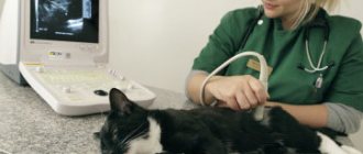
Kidney tumors that occur with nephromegaly can be detected on an abdominal x-ray. [7] Conventional x-rays show abdominal tumor masses, but contrast studies are needed to differentiate them from tumors of other structures - the hepatic lobe, pancreas, and ovaries. [8] Intravenous urography (IVU) or renal angiography can be used to evaluate renal function and parenchymal architecture. In most cases, ultrasound can complement or even provide more detailed results than radiographic methods. Ultrasound of the kidneys allows non-invasive assessment of the architecture of the organ; in addition, using this examination method, accurate and safe percutaneous fine-needle aspiration biopsy (FNAB) or core biopsy of any pathological structures is possible. [7] Ultrasound imaging usually shows bilateral (>80% of cases), unevenly enlarged kidneys with a hypoechoic subcapsular rim, and diffuse tumor spread throughout the cortex. In approximately 1/3 of cases, other abdominal organs are also involved in the process. [6] Ultrasound can also evaluate local tumor invasion into adjacent vascular structures, such as the vena cava. More advanced testing methods include magnetic resonance imaging (MRI) and computed tomography (CT). In addition, scintigraphy can be used to assess renal function and glomerular filtration rate (GFR).
When developing treatment tactics for patients with renal lymphoma, the method of choice is combination chemotherapy, since this disease is usually bilateral and/or generalized. Standard protocols (modifications of CHOP protocols) can be used. The response is often worse than with other forms of lymphoma. Replacement therapy is often required, especially at initial presentation, before a final diagnosis is made. In some cases, with severe azotemia, a reduction in the dose of the cytotoxic drug is required. Doses of the drug can be increased after the initial azotemia has resolved. Particular caution should be exercised when using drugs that require any degree of renal excretion (eg, cyclophosphamide). When using doxorubicin, extensive concomitant infusion therapy is required; the drug should be used with caution due to its severe nephrotoxicity. [1] Nephrectomy can potentially be used in animals with unilateral renal lymphoma. Unfortunately, it is impossible to reliably confirm the fact that only one kidney is affected; Thus, most experts recommend the use of chemotherapy in all cases of renal lymphoma.
When considering the prognosis of the disease, it is worth considering that cats with renal lymphoma respond to chemotherapy worse than other forms of lymphoma, and therefore long-term remission and survival are rarely achieved. [7] From data published in the literature, response to treatment in renal lymphoma occurs in 25% of cases, and the average life expectancy is 4.5 months. [8]
Clinical case
The owner of the cat Mary (an intact, unsterilized female Scottish Fold at the age of 2 years) first consulted a doctor at a third-party veterinary clinic on October 22, 2021, with complaints of a visual increase in the cat’s abdomen in volume. No significant changes in the animal's behavior were observed. After a visual examination, the patient was prescribed an ultrasound examination (hereinafter referred to as ultrasound) of the abdominal cavity, the results of which revealed severe nephromegaly, focal formations in the kidneys up to 15-17 mm, hypoechoic, round, of a homogeneous structure (Fig. 1). Based on the ultrasound findings, a diagnosis of renal lymphoma was suggested. The patient underwent a fine-needle renal biopsy under ultrasound guidance for cytological examination. The result of morphological diagnostics confirmed the preliminary diagnosis of renal lymphoma. Hematological disorders were also noted: moderate normocytic normochromic anemia, increased blood creatinine levels to 619 mmol/l, urea levels to 21.1 mmol/l. An ELISA test for FeLV and FIV showed a negative result. X-ray examination of the chest cavity revealed no signs of metastasis. Unfortunately, no urine analysis was performed. The patient was referred for treatment to an oncologist.
Rice. 1. Ultrasound of the kidneys from October 22, 2016
At the initial appointment with the oncologist on October 25, 2021, palpation revealed nephromegaly, the surface of the kidneys was lumpy. The weight of the animal was 2.0 kg. Monitoring of a general clinical blood test revealed progression of anemia with a moderate regenerative response. Despite the unfavorable prognosis, it was decided to begin treatment with chemotherapy. The patient was offered metronomic chemotherapy with Endoxan at a dosage of 50 mg/m2 body surface orally once every 3 days, Prednisolone at a dosage of 40 mg/m2 body surface in injection form with further transfer to oral administration at a dosage of 1 mg/kg body weight once a day, and also concomitant symptomatic therapy to stabilize the state of red blood. Blood monitoring on October 29, 2021 revealed stabilization of red blood, as well as blood urea and creatinine levels. Further, throughout the entire period of observation of the patient, there were no signs of anemia and increased levels of urea and creatinine in the blood. A control ultrasound of the kidneys on November 18, 2021 (Table 1) revealed significant positive dynamics: the size of the kidneys was within normal limits, moderate heterogeneity of the parenchyma of both kidneys, a focal change on the cranial pole of the right kidney of 16 mm, with a change in the contour of the kidney. Due to the positive dynamics during therapy, a decision was made to continue the prescribed treatment, extending the course to 3 months, with periodic dynamic studies of blood readings and ultrasound of the urinary system. However, the first course of metronomic chemotherapy was completed ahead of schedule - December 18, 2021 - due to a complication in the form of progressive leukopenia (Table 2). Stabilization of leukocyte levels was achieved by injections of the drug Neipomax at a dosage of 5 µg/kg subcutaneously once a day for 3 days. Also during therapy, there was an increase in ALT levels, as well as relative eosinophilia (Table 2). A deterioration in the general condition in the form of decreased activity and heavy shedding was noted once, however, the owners associate these changes with the onset of estrus. The weight after the course of treatment was 2.5 kg.
The first period of remission was 139 days. On May 6, 2021, during the next control ultrasound of the urinary system (Table 1), negative dynamics were revealed: a change in the contour of both kidneys, a focal change in reduced echogenicity on the cranial pole of the right kidney of 7 mm. Weight at the time of treatment was 2.9 kg. The patient was resumed metronomic chemotherapy at the previously indicated dosages. The course lasted 3 months. Control examinations were carried out less frequently during this period due to personal reasons of the patient's owner. During control blood tests during therapy, an increase in ALT was noted (Table 2). Control ultrasound indicates partial regression of the tumor and stabilization of the tumor process (Table 1).
The next period of remission lasted 133 days. On December 16, 2017, during the next control ultrasound of the urinary system (Table 1), negative dynamics were revealed: heterogeneity of the parenchyma of both kidneys (appearance of a hypoechoic rim along the periphery), uneven contours of both kidneys. During the same period, estrus, severe molting, and weight loss of up to 2.6 kg were observed. The patient was resumed metronomic chemotherapy at the previously indicated dosages. The course lasted 3 months. Control examinations were carried out less frequently during this period due to personal reasons of the patient's owner. When conducting control blood tests during therapy, leukopenia was noted (Table 2). Control ultrasound indicates partial regression of the tumor and stabilization of the tumor process (Table 1).
The next period of remission was 129 days. On July 12, 2021, during the next control ultrasound of the urinary system (Table 1), negative dynamics were revealed: heterogeneity of the parenchyma of both kidneys, uneven contours of the kidneys, an increase in the size of the kidneys. Leukopenia was also noted (Table 2), and therefore it was decided to postpone the appointment of Endoxan, limiting the intake of glucocorticoids in the previously indicated dosages. Weight at the time of treatment was 2.8 kg. The course of taking the drug lasted 2 months, against the background of which there was a positive dynamics in the ultrasound picture of the kidneys (Table 1) with partial regression of the tumor and stabilization of the tumor process. The next period of remission at the time of this writing continues and has already lasted more than 240 days.
To summarize, I would like to note a few points:
- The total life expectancy from the moment of diagnosis is already more than 930 days, of which the periods of remission total more than 640 days;
- The patient did not receive high-dose chemotherapy; the treatment period totaled 290 days and consisted of 3 courses of metronomic chemotherapy and 1 course of hormone therapy;
- During chemotherapy, there were no negative changes in the patient’s general condition; The main complications of chemotherapy were leukopenia and periodic increases in ALT levels, but these changes did not affect the general condition of the patient and were easily corrected with symptomatic therapy.
Below are summary data on the results of dynamic examinations (Table 1, Table 2)
Table 1. Results of dynamic ultrasound of the kidneys.
| date | Dynamic Changes |
| 22.10.16 | Severe nephromegaly (up to 60mm), focal formations in the kidneys up to 15-17mm, hypoechoic, round, homogeneous structure |
| 18.11.16 | Dimensions are within normal limits, moderate heterogeneity of the parenchyma of both kidneys, focal change on the cranial pole of the right kidney is 16 mm, with a change in the contour of the kidney (positive dynamics) |
| 25.12.16 | Focal change at the cranial pole of the right kidney 3.5 mm, with a change in the contour of the kidney (positive dynamics) |
| 04.02.17 | Change in the contour of the cranial pole of the right kidney (positive dynamics) |
| 04.03.17 | Change in the contour of the cranial pole of the right kidney (without dynamics) |
| 06.05.17 | Change in the contour of both kidneys, focal change in reduced echogenicity at the cranial pole of the right kidney 7 mm (negative dynamics) |
| 08.06.17; 23.07.17; 02.09.17 | Changes in the contour of both kidneys, heterogeneity of the parenchyma at the cranial pole of the right kidney (dynamics are positive, stabilization) |
| 14.10.17 | Heterogeneity of the parenchyma and uneven contours of both kidneys |
| 16.12.17 | Heterogeneity of the parenchyma of both kidneys (appearance of a hypoechoic rim along the periphery), uneven contours of both kidneys (negative dynamics) |
| 05.01.18 | Heterogeneity of the parenchyma of both kidneys (more pronounced in the right one), unevenness of the contours of the kidneys (more pronounced in the right one), heterogeneity of the parenchyma of the right kidney (positive dynamics) |
| 11.02.18 | Heterogeneity of the parenchyma of both kidneys (more pronounced in the right one), uneven contours of the kidneys (more pronounced in the right one) (positive dynamics) |
| 12.07.18 | Heterogeneity of the parenchyma of both kidneys, uneven contours of the kidneys, increase in kidney size (negative dynamics) |
| 21.07.18 | Slight heterogeneity of the parenchyma of both kidneys, uneven contours of the kidneys, a slight decrease in the size of the kidneys (positive dynamics) |
| 21.09.18 | Slight heterogeneity of the parenchyma of both kidneys, uneven contours of the kidneys (positive dynamics) |
| 15.01.19 | Slight heterogeneity of the parenchyma of both kidneys, uneven contours of the kidneys (no dynamics) |
| 12.04.19 | Slight heterogeneity of the parenchyma of both kidneys, slight unevenness of the contours of the kidneys (positive dynamics) |
Table 2. Results of dynamic changes in selected blood parameters (SCA, b/x), where N is a variant of the norm.
| WBC (х109/l) | EOS (%) | RBC (x1012/l) | HCT (%) | HGB(g/l) | ALT | Creat | Bun | |
| 22.10.16 | 12,75 | N | 5,06 | 22,4 | 79 | N | 21,1 | 619 |
| 25.10.16 | 8,5 | N | 3,10 | 17 | 60 | — | — | — |
| 29.10.16 | 8,25 | N | N | N | N | — | N | N |
| 26.11.16 | 4,0 | N | N | N | N | — | N | N |
| 04.12.16 | 4,0 | N | — | — | — | — | — | — |
| 18.12.16 | 3,75 | N | N | N | N | 104 | N | N |
| 25.12.16 | 2,25 | 21 | N | N | N | — | — | — |
| 08.01.17 | 4,5 | 16 | N | N | N | — | — | — |
| 04.02.17 | 6,25 | N | N | N | N | 89 | N | N |
| 04.03.17 | 9,25 | 10 | N | N | N | 104 | N | N |
| 06.05.17 | 7,0 | 11 | N | N | N | 96 | N | N |
| 19.08.17 | 4,5 | 10 | N | N | N | N | N | N |
| 17.12.18 | 5,0 | N | N | N | N | N | N | N |
| 05.01.18 | 3,75 | N | N | N | N | — | — | — |
| 22.07.18 | 3,75 | N | N | N | N | N | N | N |
| 29.09.18 | 5,6 | N | N | N | N | N | N | N |
| 20.01.19 | 11,3 | 9 | N | N | N | 80 | N | N |
| 13.04.19 | 9,9 | N | N | N | N | N | N | N |
Bibliography.
- Alison Hayes Feline lymphoma. In Practice, 2006; Durham AC, Mariano AD, Holmes ES, Aronson L. Characterization of post transplantation lymphoma in feline renal transplant recipients. J Comp Pathol. 2014 Feb-Apr;
- Leanne N. Twomey Cytodiagnosis of Feline Lymphoma, Jan 2005;
- P. Makovicky, P. Makovicky, L. Vannucci, G. Samasca Histopathological diagnosis of canine and feline lymphoma, September 2015;
- Rose E. Raskin, Denny J. Meyer Canine and Feline Cytology - A Color Atlas and Interpretation. Guide - 2nd Edt, 2010;
- Withrow & MacEwen's Small Animal Clinical Oncology. 5th Edt, 2013;
- J. Dobson, D. Lancelles Oncology of dogs and cats. – Per. from English / Edited by K. Lisitskaya. – M.: Aquarium Publishing House, 2021. – 448 p.;
- R. White Oncological diseases of small domestic animals. – Per. from English Makhiyanova E.B. – M.: “Aquarium Print”, 2021. – 352 p.
Veterinary oncologist, cytologist SVK Svoi Doctor, cytologist IVC MBA Moiseeva Maria Aleksandrovna
Diagnostics
Distemper in cats: symptoms and treatment at home
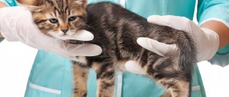
Lymphoma in a cat is diagnosed using several procedures. First, the veterinarian conducts a visual examination of the condition of the skin, fur, and mucous membranes. The doctor checks the lymph nodes, takes blood for clinical and biochemical tests, and weighs the animal. A test is performed to detect leukemia and immunodeficiency viruses. Then they do an ultrasound of the abdominal cavity, during which they carefully look at the condition of the internal organs and note possible pathologies. A chest x-ray is taken to determine the condition of the local lymph nodes and pleura.
Next, a cytological analysis of the affected lymph nodes is required. It quickly diagnoses large cell cancer, allowing treatment to begin immediately. The final stage of diagnosis is a biopsy, histological and immunohistochemical analyzes of tumor tissue. During this operation, the diseased lymph node is completely removed. If spinal cord damage is suspected, a lumbar puncture is performed. Pleural fluid and bone marrow tests are performed.
Lymphoma treatment
The main direction in the treatment of lymphosarcoma is chemotherapy, which involves the administration of cytotoxic drugs to destroy tumors. These drugs have a destructive effect not only on tumor cells, but also on healthy cells of internal organs and body systems, therefore chemotherapy regimens and doses are selected exclusively by a veterinarian. Among the most commonly used drugs:
- Doxorubicin;
- Chlorambucil;
- Cyclophosphamide;
- Vincristine;
- L-asparaginase (in case of leukemia);
- Prednisolone/prednisone (to relieve inflammation and suppress tumor development).
Medicines are given to the animal once a week, and the course lasts 4-6 months. If, at the end of the first course, it is possible to achieve remission, at the veterinarian’s discretion, chemotherapy is stopped or the interval between drug administrations is increased to 2 weeks.
Radiation therapy is not used for lymphoma. If the tumor interferes with the normal functioning of the animal, it is surgically removed, after which chemotherapy and restorative drugs are also prescribed.
How to treat
After all the procedures, it is possible to establish a final diagnosis, assess the severity of the condition, make a prognosis and prescribe adequate treatment.
Unfortunately, owners turn to the veterinary clinic late, so most sick cats are diagnosed with stage 3 lymphoma.
Breeders are often late for visits to the veterinarian
Surgery
If necessary, an operation is performed on the intestine, during which solitary masses and the area with obstruction are removed from it. In addition, in some cases, bone marrow transplantation is performed.
Chemotherapy prescription
The main treatment for lymphoma is chemotherapy. The doctor prescribes a treatment regimen individually for each patient. A sick cat is prescribed a course of treatment with special medications. It is recommended to take the tablets orally, as the cat tolerates this method more easily than droppers and injections. Drugs such as vincristine, prednisolone, cyclophosphamide, chlorambucil, and doxorubicin are used. For leukemia, L-asparaginase is also prescribed.
If the cat does not tolerate chemotherapy well, then more gentle medications are prescribed.
Before prescribing chemotherapy, the number of leukocytes is determined. If there are less than 2500, then treatment is postponed for 7 days, then the analysis is done again.
Note! In many cases, radiotherapy is used. It is effective when only one organ is affected.
Your doctor may prescribe chemotherapy for your cat.
Treatment
The task of healing is to destroy tumor tissue and degenerated cells, in other words, chemotherapy. The main medications are the following:
- Chlorambucil.
- Doxorubicin.
- Vincristine.
- Cyclophosphamide.
- L-Asparaginase. It is preferable to use the drug for a cat that suffered from Leukemia before it developed Lymphosarcoma.
- Prednisolone.
- Radiotherapy. Used mainly for the nasal type of Lymphoma.
All drugs are not harmless to the cat’s body; if complications arise, the veterinarian replaces the drug with a more gentle, although less effective one. In all variants, treatment sessions last several weeks or more. Some treatment regimens require a break between chemotherapy sessions. You need to understand that drugs that destroy tumor cells adversely affect the reproductive organs, parenchyma of the kidneys, liver, and bone marrow.
After the course of treatment, the doctor prescribes the days on which you need to be observed in the clinic. The veterinarian should be prepared for the pet to take general restorative medications for life.
If a cat is diagnosed with Leukemia and develops Lymphoma, to save it it is necessary to undergo harsh chemotherapy, as well as a bone marrow transplant.
What is the survival prognosis for this cat?
How to care for a sick cat
Love and care are the most important things a sick cat needs. It is necessary to provide good care for as long as the cat can live. It is advisable to feed him tasty and nutritious food.
It is necessary to strictly follow the doctor’s prescriptions, regularly take blood tests, and protect your pet from stress. It is important to monitor your cat's well-being. When pain occurs, the animal is prescribed analgesics.
Caring for a sick cat is part of the treatment
Duration of remission and prognosis
Lymphoma in cats is treatable, but not completely cured. Small cell lymphoma in cats is characterized by low malignancy and responds quite well to chemotherapy. Small cell lymphoma most often occurs in the abdominal form. In this case, the prognosis is more favorable.
Without treatment, the cat will live no longer than 8 weeks. If your pet is treated on time, it has a chance to live for 2–18 months. Today the disease is considered incurable, and death is inevitable in the coming months. Even the best treatment will not cure the cat completely. Under the most favorable circumstances, the maximum survival period is 2 years.
The survival rate for this disease is very low
Preventive measures
To prevent feline lymphoma, veterinarians offer vaccination against the leukemia virus. This is the only way to protect your furry pet from this terrible disease.
A person must monitor the pet and, if anything happens, contact a veterinary clinic
The health of a beloved cat always comes first for a caring owner. Oncology is the most terrible disease of our time, so you need to take a close look at any strange symptoms.
Places where the disease may appear and causes
Two viruses are identified as possible causes of the disease:
- immunodeficiency virus (lymphotropic lentivirus) – participates in tumorigenesis and activates oncology;
- The leukemia virus (the most common cause of the disease) is characterized by the fact that the DNA of the virus combines with the DNA of the animal and leads to the development of lymphoma.
Kidneys
Renal lymphoma can be primary or secondary; it develops in 5%. Most often occurs in animals in middle age - from 6 to 8 years.
Nasal cavity
Nasal lymphoma accounts for one third of all nasal lesions (non-viral). It usually develops in older individuals - from 9 to 12 years.
In the abdomen and intestines
It develops in both young and adult animals, most often in older ones.