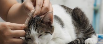
What to do if your cat has a swollen eye?
If a cat's eyes are swollen, the owner can provide first aid to his pet before going to the veterinarian. To alleviate the suffering of the animal, you need to moisten a cotton swab in strong tea leaves, chamomile decoction, a weak solution of hydrogen peroxide, boric acid, potassium permanganate or a solution of furatsilin. A wet swab is carefully applied several times around the eyelid. If there are crusts, they will become soggy and easy to remove.
The procedure is carried out several times a day to achieve a positive result. For a tumor under the eye of a cat, if it is accompanied by purulent discharge, tetracycline ointment helps. A small amount of the product is carefully placed under the lower eyelid. If none of the methods helps, and there is no improvement, then the animal should be urgently shown to a veterinarian.
It is forbidden to massage or rub the diseased organ of vision, so as not to increase the suffering of the pet.
Treatment
After interviewing the owner, examining the animal and conducting tests, the doctor will prescribe treatment procedures:
- In case of injury - Synthomycin for lubrication, drops (Iris, Tsipromed, Normax, Tobrex), wiping with Furacilin solution. If symptoms worsen, antibiotics are prescribed intramuscularly. Tavegil or Suprastin is given together with injections. The prescription of additional medications is decided by the doctor.
- To remove a foreign body, the cat must be wrapped in a blanket or blanket and carefully, using the edge of a napkin, pick up the speck. It is also recommended to use light eye drops to wash away sand and dust.
- Tumor caused by vitamin deficiency. For simple inflammation, apply ointment under the eyelid (Oxycort, Prednisolone). For phlegmatic inflammation - opening a purulent abscess, intramuscular antibiotics, rinsing with a solution of boric acid or chamomile decoction. Vitamin supplements are a must.
- For keratitis and conjunctivitis - eye drops (Novocain, Levomycetin, Sofradex), corticosteroids (Dexamethasone), ointments (Conjunctivin, Mizofen).
- Blepharitis - disinfection of the edges of the eyelids (brilliant green solution), suspension under the eyelid (oletriin, gentomycin, syntomycin), drops (Sorphadex). The crusts are removed with a cotton pad after wetting with Vaseline ointment.
- Treatment for cardiovascular and urinary pathologies depends on the identified disease. In oncology, the decision about the possibility of treatment is made by the doctor.
A tumor under a cat's eye is an alarming symptom that can lead to health problems for the pet. It is dangerous, so it is recommended to consult a veterinarian for professional help.
Possible causes of eye tumor in a cat
A tumor under a cat's eye can occur for several reasons. Cats are inquisitive and difficult to keep track of. An infection could have entered the animal's body; the tumor could have been caused by poor care or low immunity. The cause of the tumor can also be inflammation of the eyelids, the tissues around the eyes are hot and tense. Swelling around the eyelids may indicate problems with the excretory or cardiovascular system.
Injury
The cause may be a head hit on a hard object if the animal is active, runs and jumps a lot. The root cause of the damage may also be a fight with another cat. When injured, swelling appears under the eye. The animal behaves restlessly and the eye may become watery. This reason is difficult to determine, because there is no bruise after the injury. If the injury occurs in the presence of the owner, then a cold compress can be applied to the bruised area.
If a bruise occurs, a wound may appear; if it is not deep, it will heal on its own. In case of deep wounds and bleeding, it is necessary to urgently apply a bandage to the diseased organ of vision and show the pet to a doctor.
Foreign body
A foreign body, a midge, a speck or sand, could get into the eye. In addition to the tumor, purulent discharge or tears may appear. It is not recommended to pull out a foreign body on your own; it is better to contact a specialist at a veterinary clinic.
Conjunctivitis
The cause may be conjunctivitis, how to help, the eye is swollen and causes pain to the pet even with a slight touch. In addition to swelling, redness of the mucous membrane and tearing are observed. With this disease, purulent yellowish mucus is released. After sleep, the eyelids stick together, the animal hides from the light.
In this case, you should urgently contact a veterinarian to alleviate the pet’s suffering. The disease is caused by an infection that can also be transmitted to humans. The animal should be protected from communication with children; it should not be allowed into sleeping areas or personal hygiene items.
Tumor
Swelling in the eye area may indicate the presence of a neoplasm. At the same time, the cat is poorly oriented in space, because his vision decreases. The tumor will be hard to the touch, and the animal's temperature will rise. Only an examination at a veterinary clinic will allow you to make an accurate diagnosis and prescribe the necessary treatment.
Main causes of tumor
There are many factors that can provoke the appearance of a tumor. Before starting treatment for your pet, it is important to determine what caused such a symptom. Experts identify the following main causes of eye pathologies:
- Injury. A common factor that contributes to the formation of swelling is trauma. If a cat hits its head hard, it may develop traumatic swelling. Fights between a pet and another cat can also become the root cause of eye damage. Wounds of the eyelid can be superficial, deep or through. In this case, the swelling of the eyelid is accompanied by redness of the eyes or slight bleeding. If the wound is shallow, it will heal on its own and the swelling will subside. If there is deep damage to the eyelid, it is necessary to put a bandage on the pet’s eye and take it to the veterinarian.
- Foreign object. A foreign body entering the eye can also cause a tumor. Sand, thorns or some plant can get into the middle. In this case, the eye becomes hot to the touch and exudate oozes from it. Trying to remove the item yourself is not recommended. It's best to take your four-legged friend to the vet.
- Avitaminosis. With vitamin deficiency or metabolic disorders, cats sometimes experience inflammation of the eyelids. For simple inflammation (swelling appears and the edges of the eyelids become red), it is important to use an antibiotic ointment. Oxycort and prednisolone will help your pet recover quickly. Phlegmous inflammation manifests itself in the form of swelling of the eyelid and is accompanied by a purulent abscess. If a cat has such a tumor under the eye, it is recommended to immediately visit a veterinarian and open the abscess. The specialist may prescribe a course of injections, as well as further washing of the eyelid with chamomile or a 3% solution of boric acid.
- Conjunctivitis and keratitis. Inflammation of the eye in the form of conjunctivitis and keratitis is another reason for the formation of a tumor. In the first case, swelling appears around the eye, and redness is also noticed. With conjunctivitis, most often there is increased lacrimation and accumulation of mucous secretions in the corner of the eye. To treat the disease, eye drops are used (0.25% solution of novocaine or chloramphenicol, as well as sofradex or 1% solution of kanamycin).
- Oncology. The previously listed types of eye diseases respond well to treatment. If you quickly determine the cause of the swelling of the eye and undergo a course of procedures, your pet will soon recover. In some cases, a tumor under a cat's eye may indicate the presence of cancer. The pet loses vision and develops swelling under the eyes. Only a veterinarian must treat a malignant tumor.
- Blepharitis. Cats sometimes develop swelling under the eye due to inflammation of the eyelids - blepharitis. In addition to swelling of the eyelids, such a pathology is also accompanied by painful sensations. The eyelids become hot and tense when touched. Blinking provokes painful sensations in your pet, which is why most often he lies with his eyes closed.
- Heart and kidney disease. If a cat's eyelids are swollen, then in some cases this indicates a dangerous disease of the internal organs. In this case, the excretory and cardiovascular systems suffer. The general malaise of the cat should alert its owner. A veterinarian must diagnose and treat the disease.
Treatment of eye tumors
When examining the animal, the doctor determines whether the cornea and eyelid are swollen, whether there is discharge and what type it is, whether there is pain or redness. The sooner treatment is started, the faster the recovery will occur; every lost day increases the risk of complications.
In each case, the veterinarian will tell you what to do to make the cat feel good again. If a foreign body is detected in the eye, the doctor will remove it with sterile tweezers and treat the organ with Furacilin solution.
Treatment for a tumor caused by injury will depend on the severity of the disease. At the veterinary clinic, the eyes are washed and, if necessary, a 2% Novocaine solution and antimicrobial drops are instilled. In case of severe complications, even surgical intervention is possible. For a speedy recovery, it is recommended to feed your cat with vitamin A.
Redness of a cat's eye
For conjunctivitis, regular eye rinsing, drops and ointments are prescribed. For infectious diseases, if necessary, the doctor prescribes antibiotics and immune drugs. This will avoid complications and more severe diseases. Medicines are used only for their intended purpose and according to the instructions that come with them.
If a dense tumor is found in the eye area and the animal has a high temperature, an urgent biopsy is required. The test results will show whether the tumor is benign or malignant. If necessary, surgery to remove the tumor is prescribed.
Swollen eyes in a cat photo
It should be remembered that if a cat's eye is swollen and itchy, then there is a high probability of developing an allergic reaction. If pus is present, the process could be complicated by a bacterial infection. If any of these symptoms occur, it is best to take your pet to the vet, as cytology may be needed to make a correct diagnosis.
Prevention of eye diseases
To avoid eye diseases in a pet, the owner must prevent them. It is necessary to monitor the cat’s health, its behavior, nutrition and the state of the gastrointestinal tract. It is important to keep the entire house clean, including your cat's dishes and litter box. Dust and dirt can easily get into your pet's eyes and cause illness.
Your pet should be given a balanced diet to improve the immune system.
It is necessary to care for your pet, comb its fur and examine it regularly in order to identify the disease at an early stage.
To strengthen the immune system and protect against infectious diseases, cats need to be vaccinated in a timely manner. It is recommended to vaccinate from childhood so that adult cats can tolerate diseases more easily.
It is not recommended to self-medicate to prevent complications. Eyes are one of the most sensitive organs; with timely treatment of diseases, vision loss can be avoided.
All information posted on the site is provided in accordance with the User Agreement and is not a direct instruction to action. We strongly recommend that before using any product, you must obtain a face-to-face consultation at an accredited veterinary clinic.
What can cause a cat's cheek to swell?
The listed diseases have their own causes, clinical symptoms, methods of diagnosis, treatment and prevention, so a separate subsection is devoted to the description of each.
Abscess
The abscess has broken through
An abscess is a cavity that appears as a result of purulent melting of tissue. There are combat and dental ulcers.
Combat develops when an infection occurs during a fight. Not only the claws of cats conflicting over territory or females. The pet gets it from dogs or rodents. The infection enters the cheek with a splinter, or first injury occurs with sharp or prickly objects. Males are predisposed to fighting abscesses, since females are less likely to conflict with each other.
A swelling appears on the pet's cheek, which the owner notices when it has already formed. Most often, the abscess breaks out on its own during another fight and the owner notices a wound from which pus has already flowed out. The skin defect closes and scars without any treatment, or the owner applies an antiseptic spray or ointment.
Dental abscess most often develops in pampered pets when they eat soft, wet food that sticks to the gums. First, plaque forms from food debris and multiplied bacteria - permanent inhabitants of the mouth cavity. Calcium salts fall from saliva onto the surface of plaque. The mass hardens and tartar (dentolite) forms. The next stages of pathogenesis are gingivitis, caries, periodontal disease.
Be sure to read:
What causes growths on the pads of cats' front or hind paws?
Anaerobic conditions arise under the stone and suppuration develops, which resembles gumboil in humans. In addition to the fact that the cheek swells, the mouth smells unpleasant. The pet is depressed and refuses to eat. If the disease is not treated, the infection penetrates the internal organs and leads to the death of the cat.
After surgical opening of the abscess, surgical removal of tartar using ultrasound may be necessary.
It is impossible to prevent combat abscesses. Even castrati are not immune from being beaten by street cats.
Prevention of dental abscess involves using food of at least premium class, which includes specially structured dietary fiber that removes plaque from the gums.
If the owner notices the plaque too late and it has hardened, the problem is corrected by using veterinary food that contains an increased amount of structured fiber.
Veterinary nutrition for the treatment of tartar
The use of medicated food allows you not only to clean your teeth, but also prevents the development of urolithiasis, and also prevents obesity.
What should natural nutritionists do? Accustom your pet to brushing its teeth from kitten age. You cannot use paste intended for humans, otherwise the cat will resist subsequent procedures.
Acne
Acne, initial stage
First, comedones appear on the chin - blackheads. The reason is blockage of the sebaceous glands. The disease develops where food or water bowls are not washed. The occurrence of the disease is facilitated by stressful situations, as well as treating pets with human delicacies and table scraps.
With timely treatment, the process ends with the elimination of acne on the chin using external means. However, in advanced cases, the sebaceous glands on the face and other parts of the body become inflamed. Boils form.
The disease follows the same scenario as a combat abscess, however, it is characterized by a longer duration. If external remedies do not help, seek veterinary help. Anti-inflammatory corticosteroids and antimicrobial drugs - cephalosporins or amoxicillin derivatives - are prescribed.
Be sure to read:
The cat has a fever: what to do, reasons, what is the norm, how to bring it down at home, the best methods
Prevention involves regularly washing food and drinking bowls, stopping giving table scraps, and avoiding stressful situations.
Allergic reaction
A hypersensitive reaction to irritants is accompanied by swelling, as well as skin rashes, sneezing, coughing, shortness of breath, vomiting, diarrhea, and itching.
Stung by a bee
The cheeks and areas under the eyes swell under the influence of the following unfavorable factors:
- Insect bites: If a cat is stung by a bee, the bite site will immediately swell. The reaction to flea bites develops slowly. In some cases, the swelling disappears spontaneously. The treatment is carried out by the cat's owner. The pet is calmed and given Suprastin or another antihistamine. The owner of an allergic cat must have an ampoule of Dexamethasone with him and know how to use it, since individuals predisposed to the disease may develop anaphylactic shock.
- Anaphylaxis - develops in cats that are sensitive to vaccines or serums. If biological products are administered for the first time, you must wait and not leave the clinic for 15-20 minutes. A veterinarian will provide assistance. No less dangerous is repeated administration of the drug, to which the pet may react violently. If it turns out that the cat has a hypersensitive reaction to biological products, the veterinarian is informed about this, and he administers antihistamines before the procedure. At the request of the animal owner, a specialist will explain how to use Dexamethasone.
- Allergies to food and external irritants: occurs to dietary components, tobacco smoke, pollen, household chemicals, perfumes, accessories, toilet fillers, certain medications, flea products. In addition to swelling, conjunctivitis, skin rashes, and itching occur. To eliminate food allergies, use ready-made hypoallergenic food. Swelling that occurs in response to external irritants is relieved with antihistamines prescribed by a veterinarian.
Malignant neoplasm
A swelling on the cheek may be the result of a malignant tumor in the pet's mouth and cheek. Older cats most often develop fibrosarcoma or squamous cell carcinoma. Additional symptoms include drooling, bleeding, refusal of solid food, and bad breath. The cause of the disease is determined on the basis of a clinical examination, anamnesis, general blood test, x-ray, ultrasound, and histological examination.
Be sure to read:
Mastopathy in cats: causes, dangers, symptoms of the disease, treatment methods, prevention
The veterinarian prescribes surgical treatment, chemotherapy, and radiation. The prognosis ranges from doubtful to unfavorable.
Prevention of ophthalmic diseases in cats
Before getting a pet, think about the responsibility that falls on you. After all, the health of a pet depends on how strong its body is at an early age, what conditions it is in, what kind of nutrition it has and how well it is cared for.
To avoid unpleasant diseases and ensure a trouble-free existence for your animal, you must do the following for preventive purposes:
- Vaccinations. Timely vaccination will strengthen your kitten’s immunity, after which infectious diseases will no longer be so scary for an adult cat.
- Nutrition. When a cat’s diet is balanced, not only does its immune system improve, but also its appearance, playful mood, and digestion.
- Care. Daily brushing and examination of the cat will prevent any disease, because the disease does not begin suddenly, but with some preliminary incubation period.
If the cat owner does not have sufficient qualifications, then you should not self-medicate. After all, the diagnosis may turn out to be incorrect, and improper treatment can only worsen the disease. It is better to contact a veterinarian once again, since the vision of your pet is at stake, which should bring joy and comfort to your home, and not anxiety and regret.
First aid
It happens that it is not possible to immediately contact a veterinarian. It is then recommended to take measures to
providing first aid to alleviate the pet’s condition:
- ensure peace, eliminate stress factors;
- apply cold to the affected area - this slightly relieves swelling;
- do not touch your eyes unnecessarily, do not rub or massage them;
- Do not treat the animal yourself.
The main principle of first aid is to do no harm. If possible, it is necessary to reduce the pet’s suffering as much as possible and take it to a veterinary clinic.
Intraocular tumor in a cat
S. Simeon, L. Bugama, P. Coppen
Lymphoma is the second most common intraocular tumor in cats. Systemic disease most often manifests with intraocular symptoms. The article presents a clinical case of treatment of intraocular lymphosarcoma.
A 19-year-old neutered cat was brought to a specialized ophthalmology clinic for a consultation due to hemorrhage in the left eye.
Disease history
Ten days before admission, the cat fell from a great height while hunting. Three days later, the owners noticed a hemorrhage in the left eye. Previously, the animal had been treated for chronic renal failure for a year with benazepril (Fortekor, 0.5 mg/kg/day) and commercial diet food. The hemorrhage was treated with Beta-Septigen drops (atropine, gentamicin and betamethasone), but without effect - the hemorrhage did not disappear.
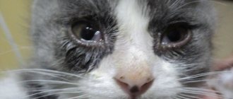
The patient's general condition is satisfactory. During the clinical examination, no abnormalities were detected. When remotely examining the animal, an adequate reaction was noted; no difficulties were noticed during movement. Ophthalmological examination of the left eye revealed the absence of the pupillary reflex and the protective blink reflex. There is a slight mucopurulent discharge from the eyes and an unexpressed, but generalized edema of the corneal endothelium. A hypopyon (accumulation of pus) is visualized in the anterior chamber of the eye. The pupil is deformed. The iris is hemorrhagic, with a thickened root. The Tyndall effect is present, but not pronounced (photo 1). The fluorescein test is negative. Measuring intraocular pressure using Tonopen revealed hypertension at 34 mmHg. Art. Biomicroscopy of the left eye confirms the disorders described above (mainly thickening of the iris and ventral infiltration). The fundus of the eye was not visualized during direct and indirect ophthalmoscopy.
A careful examination of the right eye revealed no abnormalities. The pupillary and protective blink reflex is present, intraocular pressure is within normal limits (10 mmHg). Biomicroscopy did not reveal any anomaly in the anterior chamber of the eye. When examining the fundus using indirect ophthalmoscopy, no abnormalities were found, including traces of hemorrhage on the retina.
| Photo 1. Left eye at the first consultation. The presence of uveitis in combination with mild conjunctivitis is noted |
Differential diagnosis
The identified unilateral lesions are characteristic of anterior uveitis with secondary hypertension, in combination with mild mucopurulent conjunctivitis. Deformation and thickening of the iris, as well as the absence of damage in the area of the right eye, indicate a tumor process in the left eye. In this regard, there was an assumption of iris melanoma or intraocular unilateral lymphosarcoma. The present picture is more typical for intraocular sarcoma.
Given the age of the animal, systemic arterial hypertension was considered as the cause of hemorrhage in the eye. However, hemorrhage in the anterior chamber of the eye is not considered a pathognomonic sign of arterial hypertension. In addition, no signs of hypertension were detected in the right eye. Therefore, this assumption is unconvincing.
Treatment
Enucleation was proposed followed by histomorphological analysis of the left eye, to which the owners gave their consent.
Conservative therapy and additional examination
Before surgery, treatment was prescribed with neomycin and polymyxin B (Tevemyxine collyre, three times a day), as well as an antihypertensive agent containing dorzolamide and timolol (Cosopt collyre, three times a day), which alleviated the animal's condition and eliminated conjunctivitis. Due to the need for anesthesia, and also taking into account the age of the animal, a clinical and biochemical blood test was carried out (Table 1). A clinical blood test, which focused on the quantitative content of formed elements and leukoformula, was normal. Changes in serum creatinine and urea levels and hyperkalemia were consistent with the course of chronic renal failure. Elevated alanine aminotransferase levels were not discussed during the ophthalmic consultation. The level of calcium in the blood was not determined due to the fact that paraneoplastic hypercalcemia in lymphoma in cats, unlike in dogs, has not been described. To identify metastases, a chest x-ray (frontal and lateral projections) was performed, but no abnormalities were detected.
Table 1. Results of biochemical blood tests in the preoperative period
| results | Norm | |
| ALT | 491 IU/L | 12–130 IU/L |
| Alkaline phosphatase | 129 IU/L | 14–111 IU/L |
| Urea | 0.85 g/L | 0.34–0.75 g/L |
| Creatinine | 39.6 mg/L | 8–24 mg/L |
| Glucose | 1.44 g/L | 0.7–1.6 g/L |
| Potassium | 5.8 mEq/L | 3.5 - 5.8 mEq/L |
Surgery
Dexmedetomidine (Dexdomitor, 10 mcg IM) was administered as a premedication, then the cat was oxygenated through a mask for five minutes.
Ten minutes after premedication, induction of anesthesia with isoflurane (Aerrane) via mask was started. Simultaneously with induction anesthesia, cephalexin (Rilexine, 15 mg/kg IV) was administered once. The animal was then intubated and placed under general gas anesthesia, which was maintained with a mixture of isoflurane and oxygen. Analgesia was achieved by injection of buprenorphine (Nemgesic) at 10 μg/kg IV. Infusion therapy included solutions of colloids (Volupen, 5.0 ml) and crystalloids (NaCl 0.9%, 4.0 ml/kg/hour) during surgery and during further hospital stay.
First, a lateral canthotomy with a length of 1.0 cm was performed, then two eyelids were fixed to the skin using a 4/0 nylon thread. The preparation of the eyeball and periocular structures (conjunctiva and muscles) was carried out using a blunt method. The optic nerve was fixed with hemostatic forceps, followed by a ligature of 2/0 braided absorbable suture (Vicril). Then the third eyelid with the additional lacrimal gland and the tarsal plate of the upper and lower eyelids were excised. A subcutaneous interrupted interrupted suture was placed using 3/0 braided absorbable suture (Vicril). The same, but quickly absorbable thread was used to sew the eyelids and the canthotomy site.
After isoflurane was stopped, the cat was administered atipamezole (Antisedan) at a dose of 60 mcg IV to quickly recover the animal from anesthesia. The cat was returned to the owners that evening with a prescription for a course of antibiotic enrofloxacin (Baytril) at a rate of 5.0 mg/kg/day orally, one dose for six days, and a recommendation to wear a protective collar.
Histomorphological analysis
Histomorphological analysis revealed pronounced inflammatory reorganization of the ciliary bodies, iris, part of the sclera and cornea, as well as complete obstruction of the iridocorneal angle. There are extensive areas of hemorrhage with necrosis, surrounded by an inflammatory infiltrate enriched with lymphocytes and plasma cells (photo 2). In the necrotic lesion, the presence of blast, round, unidentified and tumor cells with cytonuclear atypia is noted. The pattern of tumor cells is very similar to lymphosarcoma, but pronounced necrosis does not allow us to exclude achromatic (pigmentless) iris melanoma or metastasis to the eye of a round cell tumor (photo 3).
| Photo 2. Histological section of the left eye under a microscope (×2.5). Necrotic lesions and hemorrhages associated with the presence of a tumor (black arrows) | Photo 3. Histological section of the left eye under a microscope (×40). Large numbers of round, large, isolated and poorly differentiated tumor cells (arrows) |
Follow-up observation
The animal was delivered to the clinic 15 days after surgery. The patient's general condition is satisfactory, better than before enucleation of the eye. Postoperative wound without complications. Examination of the right eye revealed a small, slightly pigmented lesion in the temporal region of the iris (Figure 4). Intraocular pressure (10 mmHg), as well as the condition of the cornea, anterior chamber and fundus were normal. Based on the medical history, it was suggested that there was lymphoma in the iris. It is likely that the formation does not impede the circulation of aqueous humor and does not cause a painful reaction.
Owners were advised to consult their veterinarian to determine the location of the primary lymphoma and administer chemotherapy. No additional research methods were carried out to determine the localization of lymphoma. However, the owners accepted the offer of chemotherapy. The cat received an injection of L-asparaginase (Kidrolase) at a rate of 400 IU/kg IM a week after the lesion was detected. The eye damage has completely regressed. The animal died due to exacerbation of renal failure, two weeks after the start of chemotherapy. Most likely, death is not a consequence of therapy, since L-asparaginase is not nephrotoxic.
| Photo 4. Right eye 20 days after the initial consultation. Small formations were found in the temporal region of the iris |
Discussion
Establishing diagnosis
In cats, the most common causes of intraocular hemorrhage are considered to be systemic hypertension, uveitis and tumors with intraocular localization.
Suspicion of systemic hypertension
Systemic hypertension implies an increase in systolic blood pressure beyond the normal range (i.e., more than 170 mm Hg). Clinical manifestations of arterial hypertension are varied: chronic renal failure, heart failure (systolic murmur, gallop murmur, sinus tachycardia), nervous system disorders (disorientation, vestibular syndrome, convulsions, ataxia and paraparesis). Hyperthyroidism, hyperaldesteronism and diabetes mellitus lead to arterial hypertension much less often than is commonly thought (F. Magio, TC de Francesco, CE Atkins et al., 2000).
Morphofunctional lesions of the eye in arterial hypertension most often include uni- or (more often) bilateral vision loss, accompanied by a decrease in the pupillary reflex. The main disorder is partial or complete detachment of the retina. Often there are hemorrhages at various stages of development in the vitreous body, in the retina or under it. Hyphema (bleeding into the anterior chamber of the eye) and secondary glaucoma may be present. The diagnosis of arterial hypertension is confirmed using the Doppler method (F. Magio, TC de Francesco, CE Atkins et al., 2000). In our case, the development of systemic arterial hypertension could have been contributed to by chronic renal failure. However, the presence of hemorrhage only in the left eye, the absence of damage to the fundus of the right eye, and the satisfactory condition of the patient allowed us to exclude arterial hypertension as the cause of the symptoms in this cat. Therefore, blood pressure was not measured.
Suspicion of uveitis
During the first consultation, damage characteristic of uveitis was revealed in the left eye: conjunctival hyperemia, weak Tyndall effect, hyphema, edema of the corneal endothelium, hypopyon and iris deformation. Uveitis refers to inflammation of the iris and/or ciliary body (anterior uveitis) or choroid (posterior uveitis). In a cat with uveitis, you can also observe other signs of visual impairment, such as a decrease in intraocular pressure, precipitates on the cornea, miosis, pain reaction, and subsequently - posterior synechiae (adhesions), cataracts, lens luxation and secondary glaucoma in the chronic course of the disease ( C. M. H. Colitz, 2005).
Most uveitis in cats is idiopathic in nature (approximately 50%) and manifests itself only as inflammation. These are chronic lymphoplasmacytic uveitis (MC Davidson, MP Nasisse, RV Englich et coll., 1991). A significant proportion of uveitis is of a secondary nature: infectious, neoplastic, traumatic, including damage to the lens (the latter is relatively rare in cats) (KN Gelatt, 2007). In our patient, a cat who fell two days before the first consultation, the symptoms were attributed to injury. In this regard, treatment was prescribed aimed at relieving the symptoms of traumatic uveitis. However, it was not possible to achieve sufficient positive dynamics. Infectious and parasitic diseases are mainly systemic in nature and often lead to the development of uveitis in cats, which manifests itself bilaterally (CMH Colitz, 2005; MC Davidson, MP Nasisse, RV Englich et al., 1991). Uveitis can be caused by retroviruses, such as immunodeficiency viruses (FIV) and leukemia viruses (FeLV), or coronaviruses, which cause infectious peritonitis in cats. The protozoan Toxoplasma gondii also causes damage to the anterior and/or posterior segments of the eye (uveitis and chorioretinitis, respectively), which in this case is combined with respiratory, neurological or abdominal symptoms. Uveitis can develop with infection with Bartonella henslae and with rare mycoses caused by Criptococcus neoformans, Blastomyces dermatitis, Candida albicans or Cocidioides immitis.
To exclude an infectious cause of uveitis, there are a number of diagnostic methods: determination of anti-FIV and anti-FeLV in blood serum by the Eliza method; titration of Toxoplasma-specific IgM and IgG and PCR (polymerase chain reaction) to detect traces of DNA of Toxoplasma, coronaviruses, retroviruses, Bartonella in aqueous humor obtained by puncture. Aqueous humor can be plated on growth media to isolate filamentous fungi and yeasts (CC Powell, 2001).
In the case we described, uveitis is accompanied by the animal’s advanced age and a formation in the iris, which points toward an intraocular tumor.
Suspicion of intraocular tumor
Primary uveal melanoma is the most common intraocular tumor in cats (RC Riis, 2001; LW Williams, KN Gelatt, KN Gwin, 1981; M. Willis, 2001). Most often it affects one eye, mainly affecting the iris and ciliary body. Clinical signs include hyperpigmentation of the iris, impaired pupillary reflex; often a pigmented formation is found on the iris. Often the process is complicated by secondary uveitis. With the destruction of the ciliary bodies, severe hypotension occurs (LW Williams, KN Gelatt, RN Gwin, 1981; M. Willis, 2001). Obstruction of the iridocorneal angle by inflammatory cells or invasion by melanoma often leads to secondary glaucoma. Tumor growth into the orbit, conjunctiva or eyelid occurs in one third of cases. In cats, metastases appear in 62% of cases in regional lymph nodes, lungs and liver (M. Willis, 2001).
In our case, pigmentation of the iris was not noted, and therefore the presence of melanoma is unlikely. Melanoma can be completely excluded only after histomorphological analysis.
The second most common eye tumor is lymphoma (F. Delisle, C. Foumenteze, 1997). This tumor is secondary in nature, i.e. is a metastasis of primary lymphoma to the eye. Primary ocular lymphosarcoma has not been described in cats. Clinical manifestations of intraocular lymphoma are often the first manifestation of a systemic tumor (W. W. Carlton, 1976; M. Willis, 2001). A study by Australian scientists describes several rare cases of lymphoma localized only in the eye (4 out of 118) (LJ Gabor, R. Malik, PJ Gaufield, 1998). The study does not indicate that clinical signs of systemic disease are secondary (LJ Gabor, R. Malik, PJ Gaufield, 1998). All structures of the eye are sensitive to lymphosarcoma invasion (ML Castaing, 2002).
Lymphoma of the eye manifests itself both uni- and bilaterally, but in the latter case, one eye is affected before the other. In one study, 48 of 49 cats had unilateral lesions at the first consultation (KA Corcoran, R. Peifer, S. Koch, 1995), similar to the case we described.
With hematogenous dissemination, the uvea is the main site of adhesion of lymphomatous cells within the eye. In this case, the clinical picture is dominated by symptoms of uveitis, such as hypotony, precipitates in the cornea, changes in iris color, hyphema, synechiae, miosis and the Tyndall effect (ML Castaing, 2002). With diffuse infiltration of the iris, its thickening is observed, as in our case. In this case, a pigmented nodular formation is noted on the iris (WW Carlton, 1976; M. Willis, 2001; KA Corcoran, R. Peifer, S. Koch, 1995; KN Gelatt, 2007). Changes in the shape of the pupil are often observed. This is explained by the formation of synechiae with infiltration of the nerve pathways of the iris or the persistence of tumor cells in the ciliary body or iris. A case of D-shaped pupil deformation in a cat due to tumor invasion of eye structures was described (B. Nell, 1998).
Secondary glaucoma is often noted (ML Castaing, 2002; KA Corcoran, R. Peifer, S. Koch, 1995). This can be explained by obstruction of the iridocorneal angle by tumor, inflammatory cells and red blood cells, as well as the formation of posterior synechiae or destruction of the angle by lymphoma cells (WW Carlton, 1976; U. Dietrich, 2005). In our clinical case, there were no synechiae. As a histomorphological study showed, secondary glaucoma developed due to obstruction of the iridocorneal angle by cells.
With the development of lymphoma, corneal damage often occurs: keratitis, edema, neovascularization, hemorrhage and ulceration. Retinal damage in lymphoma (in our case, inaccessible to diagnosis due to the inability to visualize the fundus) includes hemorrhage, vascular dilatation, then degeneration and detachment (KA Corcoran, R. Peifer, S. Koch, 1995).
Our patient had a number of signs characteristic of intraocular lymphoma: a poorly pigmented formation on the iris, Tyndall effect, hyphema and secondary glaucoma. Further involvement of the second eye in the pathological process a few weeks later also confirms our assumption.
Additional Research
Lymphosarcoma of the eye is most often secondary. Consequently, its detection requires a search for the primary focus of lymphoma using additional methods, such as abdominal ultrasound, chest radiography and tomodensitometry. In our case, primary lymphoma could not be detected. The kidneys were examined using echography and a general examination was performed. The renal localization of the tumor should be taken into account, but in this case it is unlikely, because the animal had kidney disease for a long time and was slowly progressing.
Cytological examination
Cytological examination allows definitive verification of ocular lymphoma, because tumor cells persist most of the time in the aqueous humor. Direct puncture of the iris formation is also possible, but this method is dangerous due to serious complications, such as massive hemorrhage and rupture of the lens capsule.
Ultrasound examinations
Ultrasound can be performed as part of the differential diagnosis of uveitis and intraocular tumor. Sensors at 10–20 MHz allow you to visualize a formation in the ciliary body or iris without general anesthesia and assess the condition of the posterior chamber of the eye. This method is very important when it is difficult to visualize the formation during ophthalmoscopy, in particular when the anterior chamber is closed by hyphema or pronounced hypopyon, as well as when the transparency of the cornea decreases (E. Bentley, PE Miller, KA Diehl, 2003).
Histomorphological examination
In the case described, we chose to perform complete enucleation because intraocular hypertension led to blindness and pain. In addition, histomorphological analysis made it possible to definitively diagnose the nature of the formation in the iris, which could not be done using ultrasound or cytological analysis of aqueous humor. If both eyes were affected at once, then a needle biopsy of the aqueous humor would be preferable to enucleation at the first stage of the study.
Despite the characteristic clinical picture of ocular lymphosarcoma, without histomorphological analysis it is impossible to exclude achromatic (pigmentless) iris melanoma or metastasis of round cell carcinoma to the eye. Histomorphologically distinguishing lymphosarcoma from poorly differentiated melanoma can be difficult, which requires additional immunohistomorphological studies (BH Grahn, RL Peiffer, CL Culen et al., 2006). The authors report the likelihood of ocular metastasis from breast, lung, uterine and prostate carcinomas and multiple myeloma. Involvement of the second eye is not typical for non-pigmented melanoma, which is rare in cats and is characterized by unilateral localization. In addition, tumor response to L-asparaginase injection is confirmation of lymphosarcoma ex juvantibus (based on evaluation of the results of treatment).
Treatment
Lymphoma of the eye in a cat is a local manifestation of a systemic oncological disease, the priority treatment for which is chemotherapy. Corticosteroids given locally may temporarily cause regression of clinical signs. There are different treatment regimens and drugs used for mono- and polychemotherapy, which provides higher effectiveness. When using the developed treatment regimens, remission for all types of lymphomas is achieved in 65–75% of cases (G. Couto, 2001). The most commonly used drugs are vincristine, cyclophosphamide, L-asparaginase and doxorubicin. Cytosine arabinose, idarubicin and methotrexate are used less frequently (GK Ogilvie, A. Moore, 1997). Polychemotherapy is considered the most effective method. Initially S.M. Cotter developed a treatment regimen for COP, which includes cyclophosphamide, Oncovin (vincristine) and prednisolone. Research by A.S. Moore show that replacing vincristine and cyclophosphamide with doxorubicin in the maintenance phase can increase efficiency. However, the average time of remission is 281 days, in contrast to 83 days for cats receiving the COP treatment regimen (AS Moore, SM Cotter, AE Frimbenger, 1996). Doxorubicin appears to be preferred for the treatment of lymphomas in cats.
At the beginning of induction therapy, L-asparaginase alone or in combination with the described regimens probably leads to good results. The use of this hydrolase prolongs the time of remission (F. Delisle, C. Foumenteze, 1997; GK Ogilvie, A. Moore, 1997). Idorubicin may be of interest because it can be administered orally. In the described case, only one injection of L-asparaginase was used, which allowed regression of the disorders in the right eye.
The case we described is interesting due to the manifestation of unilateral ocular lymphoma in a cat at the age of 19 years. Intraocular lymphoma manifested itself with a clinical picture of uveitis and secondary glaucoma, which is often found in cats. The difficulty of clinical diagnosis and histomorphological examination for this type of neoplasm arises in the case of tissue necrosis.
Basic provisions
- Uveitis in cats is idiopathic, infectious, oncological and traumatic in nature.
- The optimal method for final tumor verification is histomorphological analysis.
- Lymphoma of the eye in a cat, sometimes primary, predominantly manifests itself bilaterally, but not synchronously.
- For ocular lymphoma, treatment is carried out systemically. Polychemotherapy is considered the optimal method.
SVM No. 6/2010
Rate material
Like Like Congratulations Sympathy Outrageous Funny Thoughtful No words