Struvite
The most common type of mineral found in dogs is magnesium ammonium phosphate hexahydrate, better known as struvite . This type of urinary stone accounts for about 50% of all urinary stones in dogs. Their prevalence in cats is about 30%. Miniature schnauzer, miniature poodle, bichon frize and cocker spaniel are the most susceptible dog breeds. Urinary tract infection is a major factor in the formation of struvite stones. The enzymatic action of certain bacteria in a dog produces urea, which increases its pH, making the urine more alkaline, and reduces the solubility of struvite crystals.
Cystitis increases the amount of organic residues in the urine, which undergo gradual crystallization.
Prerequisites for MBC disease
Urolithiasis can occur in any animal. The following factors often lead to its appearance:
- Wrong diet. Low-quality economy class feeds lead to the emergence of urolithiasis. Their composition is poorly balanced, they do not contain all the necessary nutrients, and various substances harmful to animals are often added to them. Because of this, metabolic disorders occur, failure of the gastrointestinal tract, kidneys, and genitourinary system. The animal gains excess weight, and the urine becomes more concentrated, salts and stones form in it.
- Lack of free access to drinking water. Cats, by nature, rarely drink. Lack of fluid in the body causes urine to become more concentrated and stones begin to form. Lack of fluid is especially dangerous if the animal eats only dry food. Metabolic disorders and dehydration occur.
- Stress. Strong emotional experiences can lead to exacerbation of diseases that previously did not make themselves felt.
- Excess weight. Obese cats need to drink a lot more fluid, which they rarely do. This leads to problems with the genitourinary system.
- Other chronic diseases that affect the general condition of the body and metabolism.
- Lack of vitamin A. This often happens if the animal eats natural food, but there are no vitamin and mineral supplements in its diet. A meager diet poor in vitamins leads to decreased immunity and metabolic disorders.
- A urinary tract infection that often occurs due to poor hygiene or hypothermia.
- Genetic predisposition. A cat is born with an abnormality of the urinary tract, or internal organs responsible for metabolism and hormone levels.
- Climate. The room where the pet is located is very hot. Dehydration begins and urine becomes more concentrated. Irregular urination. Many cats are too clean to go into a dirty litter box. They endure, the concentration of urine increases, which leads to the formation of stones and other unpleasant consequences.
- Hormonal imbalance. This is especially true only for sterilized animals, which after surgery due to hormonal changes become much less mobile, but at the same time eat a lot and quickly gain excess weight.
You might be interested in: Why does my cat have black plaque in her ears?
Calcium oxalate kidney stones
The percentage of calcium oxalate stones detected in dogs is about 35% of the total number of stones, while in cats this figure is higher and amounts to 50-70%. Urine stones recovered from a cat's kidneys or ureters were diagnosed as calcium oxalates in 70% of cases. The dog breeds most commonly affected by calcium oxalate include miniature and standard schnauzers, miniature poodles, bichon frizes, Lhasa Apso, Yorkshire terriers, and Shih Tzus. Of the cats, these are Burmese, Persian and Himalayan cats.
Older neutered cats and dogs are also highly susceptible to the formation of calcium oxalate salts. They are more common in animal kidneys than struvite stones. The reasons leading to the formation of oxalate stones have not yet been thoroughly studied, but there is some correlation between an increase in the concentration of calcium in the urine after eating certain foods and the formation of these urinary stones. In cats that have an accumulation of struvite, oxalate stones are practically not found and there is clearly a certain relationship in this. Results from epidemiological studies support the hypothesis that special urolithiasis diets to reduce struvite formation may inadvertently increase the likelihood of oxalate stone formation. Decreased urinary concentrations of natural crystal inhibitors and increased intake of mineral-rich foods may also play a role in the formation of calcium oxalate stones. The appearance of oxalate stones varies and depends on what form of oxalate is involved in its formation. The pictures show oxalate urinary stones , which form most often.
How to recognize the disease
Animals with stage 1 urolithiasis experience lethargy, apathy, and loss of appetite. The stones cause a frequent urge to urinate, but the cat does not manage to completely urinate immediately (a small amount of liquid is squeezed out at a time). The urine is cloudy with solid impurities that quickly precipitate. The appearance of the stones depends on the composition:
- Struvite (triple phosphates) are round, white, friable stones that crumble easily when pressed.
- Calcium oxalates are spiky brown or black crystals.
- Urates look like irregularly shaped sand grains of dark yellow or brown color.
- Cystines are hard, irregularly shaped sand grains of light (white or yellowish) color.
Four types of stones: 1) struvite; 2) oxalates; 3) urates; 4) cystines
At stages 2-3 of the disease, the animal begins to suffer from pain in the abdomen and groin - this can be recognized by the animal’s plaintive cries, constant licking of the perineum, hunched posture, tucked tail, walking on half-bent legs, and a constant desire to hide. The urine contains streaks of pus and blood.
The last stages of urolithiasis are accompanied by blockage of the urethra, causing complete urinary retention. With prolonged obstruction of the urinary ducts, pyelitis, pyelonephritis and apostematous nephritis develop. These complications are accompanied by fever, chills, severe weakness of the animal (cannot even stand on its paws), nausea, vomiting, paroxysmal pain (subsides and intensifies, but does not go away). The cat does not sleep, refuses food and water.
Veterinarians make a diagnosis not only based on the symptoms reported by cat owners, but also on the basis of diagnostic signs revealed during examination of the animal. Urolithiasis is indicated by:
- painful palpation of the back of the animal’s body;
- tense and enlarged lower abdominal wall (due to a full bladder);
- blood and urine tests revealing leukocytosis, accelerated ESR, increased concentration of urea, creatinine.
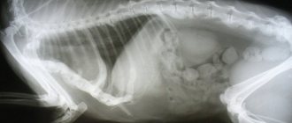
Since these symptoms coincide with a number of other diseases (malignant tumor, ascites), it is advisable to conduct ultrasound and x-ray examinations. Urolithiasis will be indicated by the presence of hyperechoic foci with clear acoustic shadows.
If a cat cannot go to the toilet (it constantly sits in the tray, but urine comes out in drops, while the animal meows loudly), then it must be checked for urolithiasis
Urats
The formation of urate stones in dogs occurs due to two different mechanisms. One of them is associated with high excretion of ammonium crystals in cases of pathology of portosystemic shunts. Dalmatian dogs have defects in the liver membranes of uric acid transport, and this also contributes to the formation of urate.
A retrospective study conducted to evaluate the clinical signs and results of surgical treatment in Dalmatians with urate stones found that blood in the urine was found in 85% of dogs, but crystals were found in only 54%. Most dogs affected by this are males. Contrast radiographs and ultrasound are the most useful tools for diagnosing urate.
Prevention of the formation of urate stones
The prevention of this type of crystal is much the same as the dissolution process described above. Dietary changes and urine alkalinization should be implemented. Urine should be checked regularly for the presence of urate crystals. If hyperurecemia persists despite dietary therapy, allopurinol at a dose of 10 to 20 mg/kg/day divided into two doses may be prescribed. Note that this dose is less than the dose used for pharmacological dissolution of stones.
Silicate stones
The mechanism of formation of silicate stones is completely unknown, but it may be associated with the consumption of silicates, silicic acids and magnesium silicate from food components. The formation of these stones is directly related to the consumption of large amounts of corn and soy gluten, in which the concentration of silicates is quite high. Great Danes, Old English Cattle Dogs, Golden Retrievers and Labradors are most susceptible to their formation. The average age of incidence is from 6 to 8 years.
Urinalysis and how it is performed
Let's look at a urine test and how it is carried out (more precisely, the types of tests):
- The simplest, cheapest and most effective method is microscopic examination of urinary sediment . In this case, you can find not only salt crystals and sand, but also casts of the urinary tubules of the kidneys, exfoliated epithelium and other traces of inflammatory processes in the organs of the urinary (as well as genitourinary) system.
- Another simple way to determine the properties of urine is to determine the density of urine. For this purpose, a device called a “urometer” is used. In terms of its design and principle of operation, this is a regular hydrometer. Normal cat urine density ranges from 1.020 to 1.040. With ICD, the density of urine increases significantly.
- In addition, in recent years, express diagnostics using express strips have become increasingly widespread. They are used by immersing them in urine for a couple of minutes. Using such strips, you can almost instantly find out the pH of urine, determine the presence/absence of blood in it, etc.
Prevention of urolithiasis in cats and dogs
Prevention of urolithiasis with struvite stones
Prevention of the formation of struvite stones depends on limiting the diet of protein, calcium, phosphorus and magnesium, and increasing the amount of salts that acidify the urine. Prompt treatment of urinary tract infection is also important and is a means of prevention.
Prevention of urolithiasis in calcium oxalate stones
Feeding protein- and sodium-restricted, alkalizing diets may reduce the recurrence of oxalate stones. Potassium citrate can be used to neutralize and slightly alkalinize urine. Sometimes high doses of B6 are used for prevention in combination with diuretics.
Prevention of urolithiasis in cystine stones
Eating a low protein diet and an alkalizing diet are highly successful in preventing the formation of cystine stones. The urine pH should be above 7.5. Potassium citrate and D-penicillamine are sometimes used for additional alkalization.
Prevention of urolithiasis with urate stones
The formation of urate can be prevented by using low-protein and alkalizing diets. Allopurinol is one of the drugs that helps prevent the formation of uric acid and is sometimes used to prevent urate. Allopurinol should not be given to cats. Urate stones can be prevented in 80% of dogs and 95% of cats.
Prevention of urolithiasis in silicate uroliths
Alkalinization of urine and low silicate diets are the only method to prevent their formation.
What it is?
This is what is called urolithiasis. As the name suggests, with this pathology stones form in the pet’s genitourinary system. Where do they come from and what is the mechanism of their occurrence? As a rule, the disease is closely related to the characteristics of your pet’s diet, its age and physiological state.
All stones are formed due to the precipitation from urine of salts and other compounds that are normally dissolved there. This happens when the concentration of some of these substances significantly exceeds the norm , as a result of which urine loses its “stability”, turning into a state of a supersaturated, unstable solution.
This often happens in pets whose owners feed them exclusively dry food . The fact is that in such cats, water-salt metabolism in the body can be seriously disrupted, which leads to many problems (including the formation of multiple stones in the kidneys and bladder).
About the relationship between nutrition and urolithiasis
In addition, stones in the genitourinary system are a “crown” feature of neutered cats . This is due to serious changes in their physiology after removal of the testes. The production of specific male hormones goes away, the pet’s body becomes very sensitive to excess weight gain (obesity is one of the predisposing factors for urolithiasis) and feeding characteristics. Remember that it is strongly not recommended to feed castrates exclusively with dry food, since this is an almost 100% guarantee of the formation of stones in the genitourinary system.
Another cardinal mistake of breeders is feeding their pets exclusively with fish . Of course, many sincerely believe that this food is ideal for feeding cats, but in fact this is not very true. The fact is that fish (sea fish in particular) contains a lot of phosphorus. Of course, this is very beneficial for the brain, bone tissue and teeth, but violating the ratio of phosphorus and calcium in the diet usually does not end well.
Finally, owners’ abuse of giving mineral and multivitamin preparations to their pets can lead to a similar outcome.
The desire to take care of your cat’s health is a good thing, but you still need to consult with a veterinarian before using any drug!
Important! If you don’t know what exactly is missing in your pet’s diet, and what, on the contrary, is in excess, you will not only not improve his health, but can also completely destroy him.
Treatment of urinary tract obstruction
Obstruction of the urethra by urinary stones is a very dangerous situation. A blockage in the urinary tract must either be cleared by removing the stone and emptying the bladder, or cystocentesis (puncture of a full bladder with a needle through the abdominal wall) must be performed if the stone cannot be removed.
Urinary stones lodged in the urethra can be dislodged and pushed back into the bladder using a technique called retrograde flushing.
The blockage can also be overcome using a technique called urinary flushing. (Fig. 1). After the stones blocking the canal are removed, the remaining stones in the bladder are removed surgically.
Figure 1 - Washing out urination
Prevention of stone recurrence
Once occurring, stones tend to form again if preventive measures are not taken. Factors that predispose to stone formation cannot be treated with surgery. Although the principles of prophylaxis are the same as for medicinal dissolution, prophylactic measures should be safer for continuous and long-term use.
In cases where the doctor does not give recommendations for prevention, stones in most cases recur.
Surgical treatment of urolithiasis in cats and dogs
The procedure for surgically removing urinary stones depends on where exactly they are located in the urinary tract. The procedure for removing them from the bladder is called cystotomy. When stones are located in the urethra and urethra, urethrotomy is performed. Sometimes, with complete obstruction of the urethra, a urethrostomy is performed to create complete and constant outflow and prevent possible obstruction. A perineal urethrostomy is often performed in cats with an obstruction.
Laser lithotripsy is the least invasive method for removing urinary stones , which has been tested and used relatively recently. This procedure requires advanced laser and endoscopic equipment and high technique. In some cases, the procedure is performed through the urethra, but sometimes a small incision is made into the bladder and an endoscope with a laser fiber is inserted through this canal into the urinary canal and then into the upper part of the animal's urethra. Laser lithotripsy is most useful for urethral obstruction with a small number of cystic stones.
Urinary stones that form in the kidneys can be removed using nephrotomy. Most veterinary specialists perform cystotomy quite professionally, however, urethrostomy, urethrotomy, and nephrotomy require special surgical training and not every specialist can do it. It should be performed by a surgeon with at least 5-7 years of experience in this field.
Rules for collecting samples from cats
To detect the presence of sand in the urinary system in cats, it is necessary to conduct a urine test. But the collection of this liquid must also be carried out in accordance with certain requirements. Here are the standard rules for collecting samples from cats:
- The most important nuance is that urine should be collected in the morning , since at this time its concentration is highest. In this case, the researcher will be able to identify even the initial stages of the appearance of tripel phosphate crystals.
- The second important rule is that urine should be collected before morning feeding. There is no point in taking samples after a hearty breakfast, since in this case the pH of the urine changes greatly.
- If for some reason urine had to be collected from the floor, a sterile disposable syringe should be used for this. In addition, you should definitely inform the receiving laboratory assistant about this circumstance!
- To collect and initially store samples, use special plastic containers for urine , sold at any nearest pharmacy. If for some reason this is not possible, the use of medicine bottles (glass only) is allowed, but not recommended. Before collecting urine in them, the vials must be thoroughly washed, boiled, and then rinsed thoroughly with settled or boiled water. This should be done to prevent “foreign” compounds from getting into the urine, the presence of which in the sample can distort the research results.
- The collected samples should be delivered to the clinic within half an hour. If for some reason it is impossible to do this immediately, storing samples in a household refrigerator for two hours is allowed. In the latter case, the urine is gradually (over half an hour) warmed to room temperature before testing.
Symptoms and first signs of ICD
The following symptoms and first signs of ICD are distinguished:
- Blood may appear in the urine, causing the urine to become either red or yellowish-pink. Hematuria (as this phenomenon is called) is characteristic of ICD with the formation of tripelphosphates. We have already written more than once that these stones/sand are distinguished by sharp, uneven edges.
- In severe cases, the animal does not urinate at all (dysuria or anuria). This condition is extremely dangerous for the life and health of the pet. It can develop either against the background of severe inflammation and swelling of the walls of the bladder/urethra, or for a more “prosaic” reason: sand completely blocks the lumen of the urinary canal, which is why urine does not come out.
- In addition, a characteristic sign of any type of ICD is prolonged and painful urination. The cat spends a long time pushing on the tray and constantly runs to the toilet, meowing pitifully as he does so.
But still, the most characteristic symptom of all types of urolithiasis is a very strong pain reaction. It is not uncommon for cats to die due to painful shock. Particularly dangerous are cases when “deposits” of tripel phosphates are formed directly in the renal pelvis. In severe cases, one wrong move is enough for the cat to literally begin to “break” from severe pain. For this reason, sick pets try not to move or jump too much.