Main signs of liver disease in cats
The liver is a vital organ that performs many functions:
- detoxification - processes metabolites, in particular ammonia, which is formed during protein metabolism;
- participates in digestion, produces bile;
- protein synthesis.
Impaired functioning of the organ leads to various disorders both in the gastrointestinal tract and other organs.
The clinical picture of liver diseases depends on the etiology and pathogenesis.
- In the presence of an inflammatory process or replacement of an organ with connective tissue, an increase in the size of the liver is observed - hepatomegaly, and sometimes an enlargement of the spleen is also observed.
- Dyspeptic disorders are manifested by nausea, vomiting, stool disturbances - both diarrhea and constipation can develop.
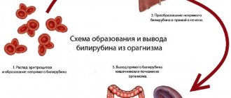
- A change in the color of the mucous membranes is observed with jaundice. Pathology is classified into the following types:
- hemolytic - not associated with diseases of the parenchymal organ, occurs as a result of massive destruction of red blood cells for various reasons;
- parenchymal develops against the background of severe damage to the liver structure, while the appearance of yellowness of the visible mucous membranes is caused by the loss of the ability of liver cells to capture free bilirubin from the blood;
- mechanical develops as a result of difficulty in the outflow of bile through the biliary tract, for example, in the presence of stones or a tumor in the liver or gall bladder.
- Ascites occurs with the development of portal hypertension - increased pressure in the portal vein. In this case, fluid exits into the abdominal cavity and accumulates. In addition, the formation of pathology is facilitated by the low content of albumin proteins in the blood, as a result of which the oncotic pressure of the blood decreases. The syndrome is manifested by an increase in the circumference of the cat's abdomen, and when the animal is positioned on its hind legs, ascitic fluid flows into the lower sections, as a result of which the abdomen takes on the shape of a pear.
- Some diseases are accompanied by the development of cholestatic syndrome, which is characterized by intense itching of the skin. This symptom is caused by irritation of skin receptors by bile acids released into the blood.
- With jaundice, there is a change in the color of urine towards dark shades, and becomes light in color.
- As a result of severe liver dysfunction, multiple hemorrhages develop on the skin. In severe cases, internal bleeding may occur.
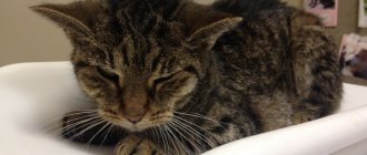
Symptoms of ascites in a cat
Brief characteristics of feline liver diseases
Hepatitis and hepatosis
Inflammation of the liver, which can occur in acute and chronic forms.
The causes of hepatitis are:
- Poisoning with poisons of plant and mineral origin.
- The action of toxic agents entering the blood as a result of infectious or allergic processes.
- Overdose of medications that can accumulate in the body.
Signs: jaundice, yellow discoloration of the mucous membranes, conjunctiva, and skin.
In some cases, the temperature may rise, loss of appetite, increased thirst, diarrhea or constipation may develop, while the feces have a grayish-yellow color, and in more severe cases of the disease, convulsions are observed.
The diagnosis is made based on clinical signs of the disease, laboratory tests of blood and urine for bilirubin.
Treatment of hepatitis (hepatosis) in cats depends on the cause that caused it. First of all, it is necessary to put the animal on a diet that does not contain fatty foods. On the first day of treatment, there is hunger, after which the cat is fed porridge; after a week, minced meat is gradually introduced into the diet.
Basic principles of therapy:
- the use of B vitamins and drugs containing choline (Essentiale, etc.);
- prescribing antispasmodic drugs to reduce pain and eliminate congestion in the liver (cholestasis);
- antibiotic therapy;
- drip infusion of saline in case of signs of dehydration;
- injections of glucose with vitamin C to relieve intoxication;
- antihistamines and prednisolone help eliminate allergic reactions.
Read more in the article: Hepatitis in cats: causes, symptoms and treatment.
Feline liver cirrhosis
Proliferation of connective tissue and changes in organ structure.
Reasons: hepatitis in the past, prolonged exposure to toxic substances in the body, lack of B vitamins and protein in the diet, infectious diseases of viral and bacterial origin.
Signs: loss of appetite, diarrhea, hemorrhages on the conjunctiva, jaundice, accumulation of fluid in the abdomen and an increase in its volume, hardening of the liver, palpable upon palpation, shortness of breath, disruption of the heart.
Diagnosis: made on the basis of anamnesis, clinical manifestations of the disease, laboratory tests of blood, urine and ultrasound.
Treatment: glucocorticoids, calcium and potassium preparations to restore hematopoietic function, choleretic and diuretics, vitamin therapy (mainly vitamins A, E, C, B).
Cholelithiasis
A rare disease of cats, characterized by the formation of stones in the gall bladder and liver ducts.
Causes: history of hepatitis, congestion in the liver, obstruction of the bile ducts, lack of vitamin A.
Signs: pain in the liver, jaundice, indigestion, foul-smelling feces, sometimes increased body temperature.
The diagnosis is quite difficult to make; laboratory blood tests and ultrasound are required.
Treatment consists of eliminating symptoms and prescribing painkillers and vasodilators. Surgery may be required.
Cholecystitis in cats
Inflammation of the gallbladder.
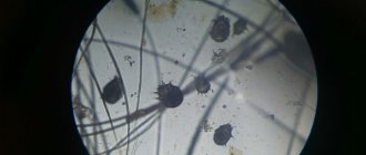
Causes: gallstones and giardiasis (an invasive disease caused by protozoa).
Signs: pain in the liver, increased body temperature, alternating diarrhea and constipation.
The diagnosis is difficult to make and is based on the history, clinical picture and blood tests.
Treatment: the diet includes easily digestible food, a heating pad on the abdominal cavity in the liver area (you cannot heat it during purulent processes), antibiotic therapy, choleretic drugs, and physiotherapy.
Most common liver diseases
The most common liver pathologies in cats include hepatitis, cirrhosis, lymphocytic, neutrophilic cholangitis, which are included in the general group of cholangitis syndrome in cats, cancer and other ailments.
Neutrophilic cholangitis
The pathology is characterized by damage to the bile ducts as a result of infection with pathogenic bacterial flora. Experts suggest that the source of infection is the intestines, but the etiology of the disease is not completely clear.
The disease usually manifests itself acutely - the animal vomits repeatedly, the mucous membranes and skin turn yellow, there is general malaise and signs of intoxication - decreased activity and appetite, a lethargic state.
Young individuals are susceptible to the disease. Treatment leads to complete recovery within one to one and a half weeks. Relapses are observed in rare cases; chronicity of the disease is not typical.
Lymphocytic cholangitis
Unlike neutrophilic, it is not associated with an infectious process. Experts suggest that a disorder of the immune response plays a major role in the pathogenesis of the disease. Most often, middle-aged and elderly animals are susceptible to pathology.
The resulting autoimmune complexes affect the bile ducts, resulting in a gradual increase in liver size, disruption of its function, and accumulation of ascitic fluid in the abdominal cavity. In the terminal stage, the pathological process also involves the liver parenchyma. There are high risks of transition of neutrophilic cholangitis to cirrhosis.
As a rule, animals respond well to treatment, but the course of therapy lasts several weeks. Recovery is often replaced by relapses of the disease and the acute course of neutrophilic cholangitis becomes chronic.
Hepatic lipidosis
Hepatic lipidosis is a disease caused by the deposition of lipids in hepatocytes. Fatty liver is manifested by dyspeptic disorders - nausea, vomiting, stool disorders develop, and the animal refuses to eat for a long time. Jaundice may appear.
Severe liver failure develops in the absence of adequate therapy. In this case, the cat exhibits signs of depression of the central nervous system - behavior changes, lethargy and increased salivation are noted, and an unusual tilt of the head may appear.
The causes of the disease are unknown. In many cases, the development of the disease is preceded by the pet's hunger or stress, intestinal diseases, or inflammation of the pancreas.
Poisoning
Food poisoning, as well as toxic substances, can occur, for example, while walking, when an animal eats food from the ground or from a trash can. Intoxication with salts of heavy metals, industrial compounds and other toxic substances often leads to the formation of hepatitis, cirrhosis and death of the pet.
Hepatitis
Hepatitis is an inflammation of the liver tissue, in which there is no violation of its architectonics. The etiological factor is infectious agents, as well as poisoning by toxic compounds.
Pathogenesis consists of infiltration of the liver with viral metabolites, bacterial waste products or toxins, resulting in local inflammation of the tissue. Massive poisoning can lead to the development of cirrhosis.
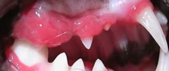
Symptoms are usually represented by yellowing of the mucous membranes and skin, nausea, vomiting, abnormal stool, and general poor health of the animal.
Yellow mucous membrane in a cat
Hepatosis
Develops due to the accumulation of lipids in the liver. Hepatosis can occur against the background of existing pancreatitis, cholecystitis and other ailments. Most often, the fatty form occurs, less often - the pigmented form, in which the metabolism of bilirubin is disrupted.
The pet experiences dyspeptic disorders, loss of appetite and weight, and a depressed state.
Cirrhosis
Cirrhosis is a chronic liver disease in which a structural disorder of the organ’s architecture occurs and the formation of regenerated nodes against the background of fibrosis, that is, replacement of the parenchyma with connective tissue. The disease is characterized by a severe course that cannot be treated. As a rule, it develops against the background of hepatitis, which can occur due to a lack of vitamins, poor nutrition, toxin poisoning, the presence of stones in the bile ducts, and helminth infestation.
Symptoms of cirrhosis in cats include signs of intoxication, loss of appetite, ascites, and jaundice. In the terminal stage, liver failure occurs as a result of toxic damage to the brain - the animal can become aggressive, restless, and gait is impaired.
Liver cancer
Cats are susceptible to cancer. Liver cancer often develops. The origin of atypical tumor cells can originate from both the hepatocytes themselves and the bile ducts and other structures of the liver. Most often, malignant liver tumors are metastases, and their introduction is possible both hematogenously and lymphogenously. The disease is characterized by a severe course, ending in death.
Symptoms of metastases
With lesions in the liver, pain in the right hypochondrium occurs, nausea and vomiting, weakness and fever, general malaise and other signs of intoxication. The patient's appetite decreases and there may be bowel irregularities. Signs of anemia appear - pale skin, decreased blood pressure. The patient loses weight. With severe liver dysfunction, an accumulation of fluid appears in the abdominal cavity - ascites.
Signs of the primary tumor also appear, which depend on its location. For example, an intestinal tumor is characterized by constipation, a pathology in the lungs is characterized by difficulty breathing and hemoptysis, and with tumors in the ovaries the menstrual cycle is disrupted.
Changes are detected in the blood. In a general blood test, signs of anemia appear - a decrease in the number of red blood cells and hemoglobin. ESR increases. In biochemistry, the level of liver enzymes increases - AST, ALT, alkaline phosphatase, GGT, bilirubin, and there may be blood clotting disorders. To find the source of the tumor, the doctor may order a test for tumor markers. Metastases in the liver are characterized by an increase in alpha-fetoprotein, a cancer embryonic antigen.
Ultrasound of the abdominal cavity, CT or magnetic resonance imaging, and scintigraphy will help clarify the diagnosis. If metastases are detected in the liver, a search for the primary tumor is performed. In unclear cases, diagnostic laparoscopy can be performed.
Diagnosis, treatment
When the first symptoms of liver disease appear in your pet, you should contact a veterinary clinic. The specialist first conducts an external and physical examination. At this stage, the presence of ascitic fluid, hepatomegaly, yellowness of the oral mucosa, eyes, and skin can be detected. Next, the veterinarian prescribes additional research methods.
- A clinical blood test in the presence of an inflammatory reaction in the body is characterized by leukocytosis and an increase in ESR.
- A biochemical blood test is aimed at determining the concentration of pigments, alkaline phosphatase and other criteria that may indicate the presence of liver disease.
- X-ray of the abdominal organs can reveal an increase in the size of the liver, changes in its contours, the presence of abscesses and other pathologies.
- Ultrasound accurately determines structural changes in the liver; in case of cirrhosis and other pathologies, it is possible to detect areas with increased echogenicity. In addition, the study allows you to estimate the size of the organ.
Treatment of the disease depends on the diagnosis. Determination of timing, dosage and choice of medication is carried out by a veterinarian.
For a bacterial infection, the veterinarian will prescribe a course of antibiotic therapy. The choice of drug, its dosage and duration of use is based on the type of pathogen.
If liver changes are detected due to autoimmune processes, it is advisable to prescribe glucocorticosteroids.
Cirrhosis does not respond to drug therapy; only relief of unpleasant symptoms is possible.
Therapy for helminth infestation involves taking special medications. In severe cases, surgery is possible.
An important component of treatment is diet, which helps reduce the load on the digestive organs and speed up the healing process.
CT diagnosis of liver hemangioma
WHAT DOES LIVER HEMANGIOMA MEAN?
Hemangioma of the liver (which means “vascular tumor” from the Latin “haema” - blood, “angio-” -vascular, “-oma” - tumor) is a benign neoplasm, consisting of multiple lacunae, well vascularized (with a large number of arteries ). The causes of liver hemangioma are unknown. As a rule, it is discovered accidentally during an ultrasound or computed tomography scan of the abdominal organs. According to statistics, hemangioma on the liver in adults occurs in approximately 5-7% (according to M. Prokop). Liver hemangioma in men is 5 times less common than in women. On microscopic examination, in the structure of the neoplasm one can see many arterial vessels with very slow blood flow, multiple blood clots (due to low blood flow speed), areas of connective tissue (fibrosis), as well as calcifications and manifestations of hyalinosis.
FEATURES OF HEMANGIOMA ON COMPUTED TOMOGRAPHY
Liver hemangioma is diagnosed using CT and MRI. Both of these studies must be performed with contrast. In this case, a special drug is injected into the vein, then scanning is carried out several times in a row in different contrast phases. For a reliable diagnosis, it is useful to scan in the portal-venous and delayed (after 10-20 minutes) phase. This vascular tumor can range in size from several (3-5) mm to several (3-5 or more) cm.
Get a CT scan of the liver in St. Petersburg
In most cases, hemangioma is characterized by the following CT signs.
1) On a native (without contrast) CT examination, the formation looks like a hypodense area in the liver parenchyma. The absolute values of the density of this area most often range between +20 and +40 units on the Hounsfield scale, while the density of unchanged liver parenchyma is +55...+65 units. The most typical location of a hemangioma is under the liver capsule. In approximately 10% of all observations, heterogeneity in the structure of the formation can be detected due to the presence of calcifications (according to M. Prokop).
This is what a typical cavernous hemangioma of the liver looks like on CT with contrast (right) in the arterial-parenchymal phase and with native CT (left). If on the left you can only notice a small (about 1 cm) low-density focus in the right lobe of the liver - on the periphery, then on the right there is characteristic peripheral contrast enhancement. Vascular lacunae are visible.
2) When contrasting in the arterial-parenchymal phase, a sharp increase in the density of the vascular tumor occurs due to the accumulation of contrasted blood. The density of the formation increases similarly to the density of blood in the aorta. In this case, the central part is contrasted more slowly, and in the arterial-parenchymal phase it usually remains hypodense. Sometimes multiple feeding arteries of different diameters can be identified along the edge of the tumor.
3) During the venous phase of contrast, the tumor acquires uniform density in the center and in the peripheral parts, its density characteristics are compared with those of the venous vessels of the liver (this is the so-called “blood pool” symptom). In general, contrasting of a vascular tumor lasts from several seconds to half an hour (depending on the degree of development of the vascular network in it and the speed of blood flow).
ERRORS IN DIAGNOSIS OF LIVER HEMANGIOMAS
Even with a three-phase CT scan, it is not always possible to reliably state that the detected tumor is benign (namely a liver hemangioma, and not something else). Differential diagnosis of vascular tumors must be carried out with the following formations:
1) Hepatocellular carcinoma. If large vascular tumors are detected, heterogeneity may be visible in their structure due to necrosis, fibrosis, and calcifications. Large nodular formations are contrasted heterogeneously, not over the entire section area, which is similar to the features of contrasting a cancerous tumor. It is possible to say for sure that a hemangioma or liver cancer has been identified if you pay attention to the characteristic “pattern” of lacunae in the arterial and venous phase, as well as to the nature of filling the formation with contrast - from the periphery to the center. However, in complex cases, a biopsy and histological examination of the tumor may be required to reliably confirm the diagnosis of hemangioma.
2) Metastases. When multiple lesions are detected in the liver, enhancing with the introduction of contrast along the periphery, secondary tumor nodes are the first thing a radiologist might think about. If you limit yourself to two-phase CT, you can come to a false conclusion that is unfavorable for the patient. If the differential series “metastases - multiple liver hemangiomas” occurs, a three-phase CT scan is required (with delayed scanning - after 10-20 minutes).
Metastasis or hemangioma of the right lobe of the liver? This image may confuse the radiologist because the contrast enhancement features (circular peripheral enhancement) are reminiscent of both cavernous hemangioma in the late arterial phase and metastasis.
LIVER HEMANGIOMA - WHICH DOCTOR SHOULD I CONSULT?
If a liver hemangioma is found on a CT or MRI, it is necessary to contact a surgeon to examine the patient clinically and prescribe the necessary additional examination methods. If the hemangioma does not cause compression of the bile ducts or vessels, there is no need to operate on it. In such cases, ultrasound or CT monitoring of the formation is prescribed at certain intervals. Keep in mind that these tumors usually grow very slowly and can never develop into cancer, that is, become malignant. Multiple liver hemangiomas also should not cause particular concern, provided that the diagnosis is accurate. Can liver hemangioma resolve? Sometimes such messages occur, but they are unlikely to be reliable. After all, any diagnosis is fraught with errors.
Get an MRI of the liver in St. Petersburg
DOUBT ABOUT YOUR DIAGNOSIS?
Sometimes even experienced doctors doubt whether the formation in the liver really is a hemangioma. How to distinguish liver hemangioma from cancer? Is it possible to confuse hemangiomas with metastases? Such questions are often asked not only by patients, but also by doctors.
A second opinion from a radiologist may be helpful in cases where the hemangioma has atypical features: for example, small hemangiomas often exhibit atypical enhancement (either homogeneous enhancement in the arterial phase or no enhancement at all). An atypical arterio-portal shunt may also be detected near the formation, which results in a wedge-shaped hypodense area (a local defect in the blood supply to the liver). An experienced specialist, as a result of reviewing a CT or MRI, can determine the nature of the formation or recommend a research method that most fully contributes to its correct identification.
Vasily Vishnyakov, radiologist
Read more about Second Opinion
Read more about telemedicine
Pavel Popov
Candidate of Medical Sciences, Member of the European Society of Radiologists
Mr. Cat recommends: prevention of liver diseases in cats
To avoid the occurrence of liver diseases in your pet, you need to monitor your pet’s diet - the menu should include all the necessary nutrients, and the portions and amount of food consumed should correspond to the age, breed, and weight of the pet. In addition, you should undergo routine vaccination and preventive administration of anthelmintic drugs in a timely manner.
Such pathologies occur in old animals (8-10 years of life). Liver cancer in cats is usually a metastasis, a complication of the development of tumors in other parts of the body. In addition, due to the high compensatory properties of the liver, cancer in it is not immediately detected. In this regard, surgical treatment often does not give a favorable outcome, and chemotherapy often only slows down the development of tumors.
Symptoms of liver disease in cats and how to treat them
The liver is one of the most important organs that helps protect against harmful substances in the cat's body, such as allergens, poisons and toxins. In some cases, the influence of negative factors is so strong that the liver cannot cope with its functions. As a result, diseases arise that require immediate treatment. In this article we will look at liver diseases in cats, their symptoms and methods of treating each of them.
Types of Liver Tumors
Since the liver is a filtering organ through which all blood passes, neoplasms in it can appear from any organ.
The most common types are:
- neuroendocrine tumor;
- bile duct carcinoma;
- hepatocellular carcinoma;
- mesenchymal sarcoma.
Typically, a liver tumor in a cat is a metastasis; a little less often it is of a hemolymphatic nature (develops from blood vessels and lymph). Cases of primary cancer when a neoplasm develops from hepatocytes are extremely rare.
Often liver metastases appear from the spleen, gastrointestinal tract or pancreas.
In cats, as a rule, there is a bile duct adenoma, a benign tumor that is relatively easy to remove. Less common is bile duct carcinoma, a malignant neoplasm that is inoperable.
Symptoms of the disease
The main problem of this pathology is that in the early stages there are practically no serious signs, the symptoms are nonspecific, and usually only general weakness is observed.
Only at stages 2 and 3 of tumor development, the following symptoms gradually develop:
- Lack of appetite.
- Reducing body weight.
- Lethargy.
- Vomiting and nausea.
- Intense thirst.
- Diarrhea.
- Frequent urination.
- Ascites (edema of the abdomen).
- Abdominal enlargement.
- Internal bleeding (anemia, ascites).
- Yellowness of the mucous membranes and skin.
- Convulsions (a sign of intoxication).
These signs develop in stages, starting from mild ones (no appetite) and ending with harbingers of imminent death (convulsions).
Death occurs as a result of intoxication of the body with ammonia, which the liver should normally process into uric acid.
In addition, when the tumor increases in volume, the following surgical complications are possible:
- Internal hemorrhage due to liver rupture.
- Obstruction of bile outflow due to tumor pressure on the bile duct.
- Obstruction of blood circulation due to pressure of the tumor on the surrounding vessels (portal and inferior vena cava).
Diagnostics
First, the veterinarian collects an anamnesis and, if a tumor is suspected, palpates the animal’s right hypochondrium. A large tumor protrudes beyond the lower edge of the ribs and is easily palpable. Blood and urine must be sent for laboratory diagnostics, which can provide detailed data on the condition of the liver.
In addition, if appropriate equipment is available, radioisotope studies, MRI and CT, and ultrasound of the chest can be performed. Prognosis and treatment are determined on the basis of all studies, since none of them alone can give a complete picture.
Liver cancer treatment
In the treatment of liver cancer, none of the methods is highly effective:
- Chemotherapy. Its low effectiveness is due to the fact that the liver is a wastewater treatment plant for the body. Delivery of the drug to cancer cells is possible only if this ability of the liver is reduced (it needs to be “killed”). Chemotherapy is advisable only as an auxiliary treatment method after surgery to prevent the formation of metastases in other organs.
- Surgical treatment is more effective, but the problem is due to the fact that the liver has a complex histological structure. Violation of its structure at the cellular level (which occurs during a cut) leads to serious consequences, poisoning of the body and death. As a result, the tumor is removed along with the removal of a certain lobe of the liver (an incision is made along the border of the lobes).
As you can see, of all treatment methods, only surgical intervention makes sense. In addition, cats are saved by the strong segmentation of the liver (7 lobes, this is more than in humans). The method is segment resection, bisegmental resection or economical liver resection.
For a benign tumor of only one segment of the liver, surgical treatment can provide a favorable prognosis. In this case, the average life expectancy is 3-4 years, which is a very high figure for cats (their average life expectancy in general is 8-10 years).
With a malignant tumor of several lobes of the liver, the average life expectancy is six months, no more than 10-15% of animals survive up to a year, less than 5% survive up to 3-5 years. Unfortunately, metastases are often recorded in other organs (usually in the lungs and kidneys), which usually greatly reduces the survival rate of animals.
What is cancer, what causes it and how does it occur in cats?
By its nature, cancer is an autoimmune process that occurs against the background of a malfunction of the immune system and genetic code molecules. A malignant tumor develops from a small number of cellular structures in which qualitative changes in DNA have occurred. Such mutant cells begin to grow and divide uncontrollably, their structure and functional characteristics change.
As the disease progresses, the mutated cells rupture the primary tumor in which they were formed and spread through the lymph and blood throughout the body, settling in the cells of other organs. In new locations, mutated cellular structures begin to divide and destroy new organs. This form of cancer is called metastatic.
The location of new mutant cells leads to new tumors. Thus, cancer in cats in the lungs easily turns into bone cancer. It is worth noting that benign neoplasms do not have the ability to metastasize, and therefore are not so dangerous.
During the first stages of cancer development in an animal’s body, the immune system is able to fight and suppress the negative effects of toxic substances secreted by tumor cells. But as the pathology grows, the body stops coping. This condition is characterized by the development of anorexia in the animal, severe dyspeptic disorders, regular vomiting and diarrhea, and total hair loss.
Each malignant tumor has its own lifespan. As its cycle ends, the cancerous tumor decomposes, causing bleeding, necrotization processes and massive ulcerations in the affected areas of the internal organs.
Quite often, veterinary specialists find it difficult to make an accurate diagnosis in the final stages of the disease. For example, cancer of the small or large intestine is accompanied by severe bleeding, which is also inherent in ulcerative colitis during the acute stage.
How long an animal diagnosed with a malignant neoplasm will live depends directly on the stage of development of the disease and the treatment methods used to eliminate the danger.
Scientists around the world have been arguing about the causes of cancer for many decades. During scientific debates, a version was even put forward about the viral nature of oncology. In fact, this is nothing more than a myth, since cancer cannot be contracted even by transplanting affected tissues into healthy ones. Such proof tests have been carried out in many developed countries - Japan, the United States, France, Great Britain.
Types of cancer in cats
Cancers are classified in veterinary medicine in the same way as in human medicine.
The common nomenclature is TNM, which is used to describe the anatomical features of the spread of degenerative processes. So the letter of the Latin alphabet T means the size and location of the primary focus of the pathological process. This is a necessary indicator for identifying solid types of tumors - carcinomas or sarcomas. The letter N (translated from Latin as nodus or lymph node. This designation is used to determine and record the presence of mestastases in the area of regional lymph nodes (located in close proximity to the tumor), the absence or presence of metastases in the regional lymph nodes and the degree of their damage. Letter M - metastasis is used to characterize the presence or presence of metastases in the body.
There are a huge number of types of malignant tumors in cats. But they are all divided into several main stages, namely:
- initial stage - characterized by the formation of a small nodule, does not have pronounced symptoms;
- second stage – there is active growth and development of the pathological process in the tissue structures of the organ, nearby cells and tissues are affected;
- third stage - characterized by the end of tumor growth, but progressive destruction of the entire organism;
- the fourth stage is the process of decomposition of a cancerous tumor, poisoning the body with toxic substances from the tumor. During this period, almost the entire body of the animal is affected and most often the fourth stage ends with the death of the pet.
There are many types of cancer, but the most commonly diagnosed types are:
- Skin cancer in a cat . One of the most dangerous and diverse diseases in the field of symptoms. It represents about 20% of the total number of diagnosed malignant neoplasms in pets. Factors that provoke skin cancer are excess ultraviolet rays, chemical burns and others. Because there are several layers of the dermis, skin cancer is divided into cutaneous lymphoma, mast cell tumors, melanocytic and sarcomas, and squamous cell carcinomas.
- Lungs' cancer . A disease such as lung cancer in cats is also one of the most frequently diagnosed in veterinary medicine. It is worth noting that this disease is rarely registered independently, but more often with metastases. In the first stages of the disease, any symptoms are completely absent. The appearance of a cough and characteristic shortness of breath are symptoms of the final stages, and in such cases it is rarely possible to save the animal.
- Liver cancer in cats. Oncology, more often diagnosed in older animals aged 7-12 years. Just like lung cancer, it is rarely an independent disease. More often it develops as a result of metastasis from other organs affected by the tumor. As a result of the special compensatory properties of the liver structures, diagnosing cancer in them is very problematic. The most common malignant tumors in the liver are neuroendocrine tumors, bile duct carcinomas, mesenchymal sarcoma, and hepatocellular carcinoma.
- Blood cancer in cats. Lymphoma or lymphosarcoma is another name for blood cancer. It is a malignant process of proliferation of lymphocytes and immune cells throughout the body. Lymphoma is diagnosed as often as skin cancer, and accounts for about 30% of the disease diagnosis statistics. The reasons for the development of lymphosarcoma, like other neoplasms, remain questionable, but there is a predisposition that lymphoma develops against the background of damage to the cat’s body by the leukemia virus, increasing the chances of developing pathology tenfold. Blood cancer has the following types - multifocal lymphoma, alimentary lymphoma, mediastinal lymphoma, renal, bone, spinal and nasal lymphomas.
- Colon cancer in cats. The pathology is rarely diagnosed in domestic cats, but always has an unfavorable prognosis and outcome. As a rule, intestinal cancer manifests itself as blockage and obstruction, as well as various digestive disorders. There are many reasons for the development of a malignant intestinal tumor, but they are all based on assumptions and not on exact facts. Intestinal cancer develops in overwhelming cases in old animals after 12 years.
- Stomach cancer in cats . A dangerous disease that is difficult to diagnose. Symptoms of the first stages of the disease are not immediately noticeable and are similar to infectious or chronic diseases. Quite often, stomach cancer is confused with gastritis in the acute stage or ulcerative manifestations. When an accurate diagnosis is established, as a rule, it is already too late to take measures to eliminate the disease.
- Kidney cancer in cats . It is rarely presented as an independent formation, and more often occurs as a result of the transfer of malignant mutated cells from another focus of pathology. Since the kidneys are a filtration organ, disturbances in their functioning are noted almost immediately. Over time, the progression of cancer in one or both organs of the urinary system leads to problems with urination, vomiting and eruption of gastric contents mixed with blood. All this indicates the beginning of poisoning of the body with its own waste products. The kidneys cannot cope with the job and begin to fail. Often secondary metastasis and renal carcinomas are fatal.
- Uterine cancer in a cat . A rarely diagnosed malignant neoplasm that occurs in cats over 8 years of age. It has been proven that in cats that are sterilized at an early age, the risks of developing pathology are reduced. Uterine adenocarcinoma is particularly aggressive, capable of growing and developing most rapidly. Gives metastases to the ovaries, bladder, kidneys and adrenal glands. In the last stages of uterine cancer, the pulmonary structures, the diaphragm, and even the brain are affected.
- Brain cancer in a cat. A practically untreatable manifestation of malignant neoplasms. A brain tumor is one of the most secretive in the area of symptoms. When characteristic signs of pathology appear, the process goes too far and the chances of recovery with treatment are very small. The most dangerous symptom of brain damage as a result of a tumor is convulsions and seizures, as well as paralysis. Swallowing may be impaired and normal breathing becomes difficult. When the respiratory center is compressed, the animal dies from asphyxia.
Oncology in cats
Like other types of domestic animals, cats are prone to cancer. Moreover, the majority of neoplasms identified in cats are malignant in nature, so this pathology requires complex treatment in a modern veterinary clinic by an experienced veterinary specialist.
A malignant tumor in a cat occurs as a result of a malfunction of DNA molecules, which are a repository of information and are present in every cell of the cat. The development of cancer in a cat begins with a small group of cells in which the DNA has changed, as a result of which these cells begin to divide abnormally quickly, their structure and functions that they usually performed change. Subsequently, these mutant cells are carried through the blood and lymph flow throughout the cat’s body, settling in the tissues of the organs. Once in the organ, “daughter” neoplasms are formed. Malignant tumors often metastasize, while benign tumors do not metastasize.
A malignant tumor, as it grows, increases in size and begins to grow into the healthy tissue of the affected organs, causing inhibition of their function.
Types of tumors in cats.
The types of tumors depending on the affected organ are as follows:
- Tumor of the basal layer of the skin.
- Mast cell cancer.
- Lymphoma. This type of tumor is often caused in cats by the leukemia virus.
- Cancer of the gastrointestinal tract.
- Tumor of the liver and pancreas.
- Bone cancer.
- Sarcoma.
- Carcinoma (breast, lung, nose). Mammary cancer is most common in older cats.
Stages of development of malignant neoplasms.
Malignant neoplasm has four stages of development.
Stage 1 (initial) - at this stage, the neoplasm in a cat is represented by a primary limited tumor nodule. There are no metastases.
Stage 2 – a malignant neoplasm grows deep into the organ affected by the tumor, the size of the tumor reaches 6 centimeters. Metastases spread from the tumor to regional lymph nodes.
Stage 3 – the number of metastases increases. The tumor becomes less mobile upon palpation.
Stage 4 (terminal) – the malignant tumor affects large areas of organs in the cat’s body, which ultimately leads to death.
Etiology. The reasons that cause the appearance of cancer in cats have not been precisely determined to date. At the same time, scientists in the field of oncology have collected certain material on the factors that could give impetus to the appearance of a cancerous tumor in animals. Among these factors that can cause cancer are:
- Hereditary predisposition.
- Feeding low quality dry food containing a large number of preservatives and other chemicals.
- Frequent and prolonged exposure to sunlight sometimes leads to the development of skin cancer.
- Reduced immunity due to inadequate feeding, a sedentary lifestyle, poor living conditions, worms in the cat, etc.
- Traumatic damage to certain organs and tissues, including microtrauma.
- Hormonal imbalance that occurs in a cat as a result of the use of medications that suppress the reproductive cycle (breast cancer is almost always the result of an excess of certain hormones).
- Systematic stressful situations (frequent changes of environment, moving from one owner to another, frequent shouting, living next to an aggressive dog, etc.).
Clinical picture. The clinical picture of cancer in a cat depends on the organ affected by cancer, the stage of the disease and the general condition of the cat. For many years, the neoplasm in a cat cannot be externally manifested in any way. Symptoms of the disease in an animal may be vague and unclear.
In cases of liver cancer, veterinary specialists note general weakness, fatigue, decreased appetite in a cat, a change in the consistency and color of feces, and a sharp decrease in weight. The cat begins to develop jaundice (visible mucous membranes, the cornea of the eyes and the skin acquire a yellowish tint). Disease symptoms characteristic of liver disease in cats appear.
Stomach cancer in cats. During a clinical examination, the veterinarian notes that the cat has general weakness, drowsiness, poor appetite, and a painful abdomen on palpation. Increased body temperature. There is a change in the color and consistency of fecal matter, sometimes we find blood clots in the fecal matter, the cat suddenly loses weight, and anemia develops.
Bowel cancer. During a clinical examination, a veterinarian finds the same clinical signs in a sick cat as for stomach cancer. Unlike stomach cancer, fresh, scarlet blood appears in the stool, a foul odor emanates from the feces, and bloating occurs. A fetid odor is felt from the oral cavity, and rumbling sounds are heard on auscultation of the intestines. Depending on the affected part of the intestine, diarrhea (diarrhea in a cat) or, conversely, constipation may occur.
Lungs' cancer. When conducting a clinical study, veterinary specialists at an early stage notice that the cat has a hysterical, frequent, dry cough with the appearance of sputum, in which patches of pus are visible; later, blood clots appear in the sputum. During a clinical examination, the veterinarian notes that the cat has shortness of breath that is not associated with physical exertion. The cat experiences fluctuations in body temperature (it rises by one degree, then drops one degree below normal).
Mammary cancer. According to statistics, mammary gland cancer in cats accounts for more than 60% of all malignant tumors. During a clinical examination, a veterinarian uses palpation to note the appearance of small compactions at first; later, as the cancerous growths grow, they take the form of lumps and entire ridges of tumors. The tumors are then opened. Forming non-healing wet, foul-smelling ulcers that are very painful on palpation.
Kidney cancer . With kidney cancer, pet owners note the periodic appearance of blood clots in the urine (blood in the urine in cats). During a clinical examination of such a cat, a veterinary specialist notes a depressed state, the cat’s appetite is reduced, and the cat develops aching pain in the kidney area. The cat's temperature rises slightly in the evening.
Skin cancer. During a clinical examination, a veterinarian finds non-healing ulcers in any part of the cat’s body, swelling, and bumps. These lesions in cats are especially common in the area of the lips, nose, inner surface of the ears, around the genitals, on the pads of the paws and between the toes.
Diagnosis of cancer. Cancer in a cat can be diagnosed only in modern veterinary clinics, where there is ultrasound, MRI, X-ray, CT, by taking a biopsy of pathological material and conducting a histological examination and laboratory blood test results.
Cancer treatment. The method of treating a particular form of cancer in a cat is determined by a veterinary specialist at the clinic. At the same time, performing a surgical operation to remove a tumor in a cat is possible only in the early stages of the development of a neoplasm, when cancer cells through the blood and lymph flow have not had time to spread throughout the body and there are no metastases.
Chemotherapy is used to treat cancer in cats, as in humans For a sick cat, veterinarians prescribe special medications that inhibit the active growth of cancer cells and prevent their division and functioning. One of the ways to treat cancer is radiation therapy , in which the tumor is exposed to radiation. For all forms of neoplasm, immunotherapy is used, which helps increase the body's resistance and strengthens the protective barrier.
Forecast. The prognosis for neoplasms depends on the stage at which the cat’s tumor was detected and the level of immunity. At the first stage of the disease, the prognosis is generally favorable.
Prevention. To date, science has not developed methods aimed at preventing the development of cancer, both in humans and in animals. In order to reduce the risk of developing cancer in animals, veterinary experts recommend taking measures aimed at strengthening the cat’s immunity. Prevent worms (worms in cats). Nutrition should be balanced in macro, microelements and vitamins. Reduce the cat's exposure to the street during periods of solar activity, when there is strong ultraviolet radiation. Sterilizing a cat before her first heat (preventing mammary cancer).
Symptoms of cancer in cats
It is important to recognize the symptoms of cancer in cats in a timely manner, which will allow you to contact a specialist and give the animal a chance for recovery. If you notice characteristic signs of the development of oncology, you must immediately contact a qualified veterinarian at a veterinary hospital.
Signs of oncology depend on the location and degree of neglect of the pathological process. If the digestive system is damaged, dyspeptic disorders will be observed; with cancerous lesions of the skin, itching and hair loss occur. The main signs of stomach and intestinal cancer in cats are:
Cancer of the lower jaw in a cat is characterized by the appearance of symptoms such as an abscess directly on the bone or tongue, which breaks through over time. The animal initially experiences severe discomfort, and then pain. The pet loses the ability to chew food and swallow it normally. As a result, this leads to anorexia.
With breast cancer, characteristic signs develop in the area of the cat's nipples. A malignant tumor is characterized by unusually rapid growth, large size and pain. Ulcerations often appear, which bleed and cause pain to the animal.
Oncology in cats is manifested by various disturbances in vital processes. So, with cancerous tumors in the kidney area, edema develops throughout the body, the cat does not eat or drink, but at the same time swells, as fluid leaks from the cells into the subcutaneous tissue. The act of urination becomes difficult, urine becomes rich in color, and a coma may develop due to kidney failure.
Diagnosis, treatment and prevention
Correct diagnosis of a liver problem affects the course and outcome of treatment, so it is important to notice the first symptoms of the disease in time.
The following methods are used for diagnosis:
- general and biochemical blood test;
- urine and stool analysis;
- X-ray and ultrasound of the abdominal organs;
- liver biopsy.
Based on examination and visual examination of the animal, a diagnosis is made. Treatment is prescribed by a veterinarian; you cannot independently determine the symptoms and treatment of liver diseases. For this, tablets, homeopathic preparations and intramuscular injections can be used. The main condition for effective treatment is proper nutrition. If the animal has no appetite, then you can feed it through a syringe or tube.
Basic preventive measures to prevent the development of liver diseases in cats:
- Selection of quality feeds with good timing.
- The use of a balanced diet, taking into account the required amount of fats, carbohydrates, proteins, minerals and vitamins.
- Minimizing the possibility of drug overdose and toxic substances entering the cat’s body.
- Prevention of infectious diseases.
- Carrying out timely vaccination of the animal.
- Prevention of helminths every 4 months.
The cat's health depends on the attentiveness and care of the owner, so it is important to promptly consult a specialist at the first signs of illness.
Diagnosis of cancer in cats
In order to determine cancer in a cat, it is necessary to conduct special instrumental studies and laboratory tests. Diagnosis of malignant and benign tumors consists of ultrasound diagnostics, computer diagnostics, endoscopy (if intestinal or stomach cancer is suspected).
It is important to carry out cytological studies and histology, which makes it possible to determine the degree of neglect of the pathology and select chemotherapy drugs.
A biopsy is performed to identify histological changes. Often, a surgical biopsy is performed by opening the abdomen and removing particles of diseased tissue. The advantages are quick access to organs located in the peritoneum, and the disadvantages are general anesthesia.
Treatment of cancer in cats
Treatment of cancer in cats requires a comprehensive approach and depends on the degree of development of the disease. Therapy consists of surgery, radiation and chemotherapy. The choice of treatment depends on the type of cancer cells, the location of the cancer, and the presence of metastases. To treat cancer in pets, surgery is often used.
In most clinical cases, excision of the tumor leads to recovery. But not in all cases it is possible to use a scalpel and eliminate metastases. Includes radiotherapy and chemotherapy treatment. Radiation therapy helps prevent cancer relapses after surgery.
The sick animal must be immobilized during radiation therapy using general anesthesia. The animal should not move for several minutes. As a rule, radiation therapy is combined with chemotherapy. This makes it possible to prevent further growth of the tumor. The most commonly used treatments are radiation and chemotherapy without surgery for stomach cancer in cats.
Chemotherapy involves the treatment of malignant tumors using poisonous and toxic substances that destroy mutant cells. This type of therapy includes taking pills or using IVs. In humans, chemotherapy causes a large number of side effects, but in cats the treatment is more relaxed. Isolated cases of loss of whiskers or fur in cats after a course of chemotherapy have been described.
A veterinarian should advise whether a cat with cancer should be euthanized, based on the stage of the disease and the severity of the process. If a cancerous tumor has given a large number of metastases, and necrosis processes have begun in the main focus of the pathology, it is more advisable to carry out euthanasia, saving the animal from suffering.
Features of treatment
The method of getting rid of a tumor largely depends on its size and shape.
Small tumors (up to 5-6 cm in volume) do not require special action. They are monitored and ultrasonic controlled after 3 months from the moment of detection. In the absence of any dynamics, ultrasound diagnostics are prescribed every 6-12 months.
The need for surgical removal is determined individually. This course of events leads to:
- rapid growth of liver hemangioma - more than 50% of the original size per year;
- volume more than 5-6 cm;
- bleeding of atypical tissue;
- aggressive manifestation of symptoms;
- inaccurate determination of the benignity of the tumor.